BIOCHEMISTRY FOR TEACHERS - F.F. BOYECHKO - 1985
BASIC CHEMICAL COMPONENTS OF CELLS
PROTEINS — THE BASIS OF VITAL ACTIVITY IN ORGANISMS
Among all organic substances, Proteins occupy a special place: they form the structural foundation of living organisms and perform a wide range of vital Functions.
Protein substances have been known to humans since ancient times, yet it was only at the beginning of the 18th century that scientists established that substances found in plant juices and animal tissue extracts share a common nature and, much like egg white, coagulate upon heating. The chemistry of protein substances was first investigated by the Italian researcher J. Beccari. In 1728, he isolated a protein — gluten — from wheat flour and examined some of its properties. Later, similar protein substances were isolated from various PLANT AND ANIMAL Tissues and fluids. Detailed studies of the Elemental Composition of these substances showed that they are similar not only in external appearance and certain properties, but also in their composition.
The properties of protein substances were also studied during that period by the French chemist A. Fourcroy. He thoroughly investigated Blood Serum proteins and named three of their components: gelatin, albumin, and fibrin. The researcher used the term "albumin" to distinguish the main component of blood serum from egg white, which shares similar properties in that it also coagulates upon heating and turns white. Similar substances that coagulated upon heating to form a white precipitate were isolated from numerous animal and plant sources and came to be known as proteins.
Later, in 1839, the Dutch chemist G. Mulder, based on his research into the primary constituents of protein substances, named them proteins (derived from the Greek "protos" — first, most important). Both names have remained in Protein Chemistry to this day.
Within the Organism, proteins represent the most abundant and heterogeneous group of organic substances of paramount importance. In particular, the most critical functions and characteristic Specific features of Living organisms are intrinsically linked to proteins. These include movement, irritability, the capacity for growth and self-reproduction, Digestion and The excretion of metabolic products, ADAPTATION TO ENVIRONMENTAL conditions, heredity and Immunity phenomena, sensory perception, and more. Proteins are involved in every single one of the thousands of reactions taking place within The Cell, constituting the bulk of its active matter.
Research in the biological sciences confirms that proteins play a vital role in shaping the unique Structure of living organisms, although the reproduction of living systems and hereditary phenomena are determined by the molecular structure of other cellular Biopolymers — Nucleic Acids.
The Characteristic Features of proteins as the foundation of life are largely determined by the STRUCTURE AND FUNCTIONS of their molecules. Proteins possess A number of unique characteristics that set them apart from other key biopolymers found in living systems. These primarily include high polyfunctionality and the capacity for diverse Physical and Chemical transformations, structural diversity and The ability to form supramolecular complexes and structures, catalytic activity and intermolecular interaction capabilities, high species Specificity, and more.
Chemical composition of Proteins
Investigations into the Chemical Nature of proteins began with The Study of their elemental composition. In 1810, the French scientists J. Gay-Lussac and L. Thénard analyzed the elemental composition of protein substances and established that they contain the following basic elements: carbon, hydrogen, oxygen, and nitrogen. Additionally, small amounts of sulfur, phosphorus, and other elements were detected in the composition of certain proteins.
Later, the German scientist L. Michaelis investigated the elemental composition of blood serum proteins, while the French researcher J. Boussingault analyzed the elemental composition of plant proteins. These and subsequent studies demonstrated that the proportion of major elements in proteins of both plant and animal origin is remarkably consistent, averaging: 50–55% carbon, 6.5–7.3% hydrogen, 15–17% nitrogen, 21–23% oxygen, and 0.3–2.5% sulfur.
Since the nitrogen content in various proteins remains the most stable—approximately 16%—it became the standard basis for quantifying proteins in diverse plant and animal products (the Kjeldahl method). To determine this, The amount of nitrogen obtained from analyzing specific products is multiplied by a factor of 6.25, based on the assumption that protein nitrogen content is 16% (100 : 16 = 6.25). This method is widely used to measure protein levels in various Organs and tissues of plant and animal organisms, as well as in food products.
However, even a thorough examination of the elemental composition of proteins did not yet provide insight into their molecular structure.
The structure of proteins began to be studied in the first half of the 19th century. In the course of these investigations, scientists focused their attention on Amino Acids, which for a long time were viewed not as Building Blocks of protein molecules, but rather as the byproduct of strong chemical Reagents acting on protein substances.
The first amino acid from protein hydrolysate of gelatin was isolated in 1820. This amino acid has a sweet taste, which is why it was named glycocoll (from the Greek glykys meaning sweet and the Latin colla meaning glue). The Russian chemist N. Lyubavin was the first to propose METABOLISM/2.html">THE CONCEPT OF the Amino Acid Composition of proteins.
Applying the method of Protein Hydrolysis, a whole range of Other Amino Acids was isolated. By 1900, more than 10 different acids had been isolated from proteins. Today, it is known that proteins are composed of 20 different amino acids.
Amino acids: the fundamental structural units of proteins
Nearly 200 different amino acids exist in nature. However, only 20 of them consistently make up the majority of proteins. Another 10 amino acids occur very rarely in proteins. All of these are frequently referred to as Proteinogenic Amino Acids. The remaining Amino acids are part of physiologically active substances (Hormones, Coenzymes, Antibiotics) or are found in a free state within animal and plant organs and tissues, where they perform vital biological functions.
The natural synthesis of amino acids occurs continuously in plant organisms. Some amino acids are also synthesized in the bodies of humans and animals. A number of amino acids—particularly proteinogenic ones—cannot be synthesized by the body and must be obtained through diet, as they cannot be replaced by other amino acids. These are known as Essential Amino Acids. They include valine, leucine, isoleucine, Methionine, Threonine, Lysine, phenylalanine, and Tryptophan. Additionally, Arginine and Histidine are considered conditionally essential.
A deficiency of essential amino acids leads to the disruption of numerous biochemical processes, primarily the synthesis of various BIOLOGICALLY ACTIVE SUBSTANCES. Furthermore, the proportion of essential amino acids
in food products is of great importance to the organism. Proteins with an optimal balance of essential amino acids possess the highest biological value. In such cases, the body's protein requirements can be met with a minimal intake.
Amino acids are derivatives of organic acids in which one or more hydrogen atoms are replaced by amino groups. In proteinogenic amino acids, the amino group is typically located adjacent to the a-carbon atom, meaning they are all a-amino acids. Their general formula (with the exception of Proline and hydroxyproline) can be represented as follows:
Class="center">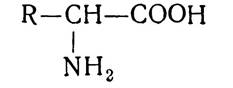
Table 3. Physical Properties of amino acids
Amino acids |
Relative molecular weight |
Solubility in 100 g of Water at 25°C |
Melting point, °C |
Isoelectric point pH |
|
Full name |
Letter Abbreviations |
||||
Ala Ala А |
89,06 |
16,51 |
297 decomp |
6,0 |
|
Arginine |
Arg Arg Р |
174,14 |
15,0 |
207 |
10,76 |
Aspartic acid |
Asp Asp Д |
133,06 |
0,50 |
251 |
2,77 |
Valine |
Val Val V |
117,09 |
8,85 |
315 |
5,96 |
Histidine |
His His Н |
155,09 |
4,29 |
7,59 |
|
Gly Gly G |
75,05 |
24,99 |
292 |
5,97 |
|
Glutamic acid |
Glu Glu Е |
147,08 |
0,84 |
248 |
3,22 |
Isoleucine |
Ile Ile І |
131,11 |
4,12 |
280 decomp |
6,02 |
Leucine |
Leu Leu L |
131,11 |
2,19 |
33 |
5,98 |
Lysine |
Lys Lys К |
146,13 |
Good |
224 |
9,74 |
Methionine |
Met Met М |
149,15 |
3,35 |
283 |
5,74 |
Proline |
Pro Pro Р |
115,08 |
16,23 |
228 |
5,68 |
Ser Ser S |
105,06 |
5,02 |
228 |
5,68 |
|
Tyr Tyr У |
181,09 |
0,05 |
310 decomp |
5,66 |
|
Tryptophan |
Trp Trp W |
204,11 |
1.14 |
298 |
5,89 |
Threonine |
Thr Thr Т |
119,08 |
20,05 |
253 |
5,16 |
Cys Cys С |
121,12 |
Very good 3,00 |
258 decomp |
5,07 |
|
Phenylalanine Asparagine Glutamine |
Phe Phe Г Asn Asn N Gln Gln Q |
165,09 |
283 decomp 207 |
5,91 5,41 5,65 |
|
All amino acids are built on the same principle and differ from one another solely in the chemical nature of their side chains (radicals). These radicals can be residues of acyclic and cyclic Hydrocarbons, their derivatives, various heterocycles, etc. Amino acids containing more than one amino or carboxyl group have also been isolated from proteins. When an amino acid contains two amino groups, one is located at the a-position and the other at the ω-position, i.e., at the carbon atom at the opposite end from the α-amino group. Amino acids containing an NH group instead of NH2 have been isolated from protein hydrolysates.
Amino acids isolated from proteins are colorless crystalline substances, the vast majority of which are readily soluble in water and sparingly soluble in organic Solvents. The basic physical constants of amino acids are presented in Table 3.
Amino acids, like a number of other Organic compounds, are optically active. They have the ability to rotate the plane of polarized light passing through their solutions. This property is characteristic of all compounds whose molecules contain an asymmetric carbon atom (bonded to four different atoms, groups of atoms, or radicals). Each amino acid can exist in two optically active forms: a dextrorotatory form, designated by the plus sign (+), and a levorotatory form, designated by the minus sign (-).
The configuration of amino acids is determined in comparison with glyceraldehyde. As is known, glyceraldehyde exists in two forms: the D-form and the L-form.

If THE POSITION OF the hydrogen atom at the a-carbon atom of The amino acid is analogous to the position of hydrogen at the same carbon in D-glyceraldehyde, the amino acid belongs to the D-series. Conversely, if the position of the amino acid's hydrogen atom corresponds to the L-form of glyceraldehyde, the amino acid belongs to the L-series.

The D-form of glyceraldehyde always rotates the plane of polarized light to the right (+), while the L-form rotates it to the left (-). However, for a number of compounds, including amino acids, the designations "D" and "L" do not correspond to the direction of optical rotation; rather, they characterize not the optical activity of the amino acids, but their membership in a group of related compounds that share the same spatial configuration at the a-carbon atom.
The Biological Significance of D- and L-amino acids varies. Specifically, proteins of plant and animal origin predominantly contain L-amino acids. Some of these are dextrorotatory and others are levorotatory, which is why the signs (+) or (-) are added next to the letters "D" and "L".

The optical antipodes of natural amino acids are D-amino acids, which are obtained primarily through chemical synthesis. Recently, D-forms of Certain amino acids have been discovered both in the free state and as components of specific antibiotics and microorganisms. In ruminants, the source of amino acids is microbial protein from the rumen. Some of these amino acids belong to the D-series and cannot be utilized for building animal protein molecules. Furthermore, the enzyme D-Amino Acid Oxidase has been found in certain organs of higher vertebrates, showing significantly higher activity than the L-a-amino acid oxidase. This enzyme catalyzes the Oxidative Deamination of amino acids. D- and L-amino acids also differ in taste: D-amino acids are sweet, whereas L-amino acids are bitter or entirely tasteless. These two forms of amino acids also behave differently in the bodies of humans and animals, as the organism's enzyme systems are specifically adapted to assimilate L-amino acids.
Under certain conditions, the configuration of the H and NH2 groups around the asymmetric a-carbon atom of Amino acids can change, causing a portion of the L-amino acids to convert into the D-form. This process is called racemization, and mixtures of D- and L-amino acids are termed racemic mixtures. Using special techniques, one of the optical isomers can be isolated from these mixtures in pure form. The Racemization of Amino Acids also partially occurs in the intestines of higher animals and humans. Specifically, evidence indicates that some L-amino acids are partially converted into D-amino acids under The Influence of racemase Enzymes. This pathway of conversion has thus far been proven for alanine and glutamic acid.
Amino acids are amorphous compounds containing two functional groups with opposite properties: a carboxyl group, which confers acidic properties, and an amino group, which determines the Basic Properties of amino acids. In aqueous solutions or in the crystalline state under normal conditions, the carboxyl and amino groups of amino acids can interact with each other: a hydrogen ion dissociates from the carboxyl group of the Amino Acid and attaches to the lone electron pair of the nitrogen:
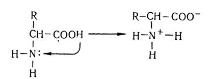
Thus, the amino acid molecule is converted into a dipolar ion (zwitterion). As a result of this intramolecular interaction, a neutral compound—an internal salt of the amino acid—is formed. Consequently, amino acid solutions are neutral in most cases and do not affect indicators (except for amino acids containing multiple amino or carboxyl groups in their molecules).
In solutions, amino acids behave as amphoteric electrolytes (ampholytes): depending on the pH of the medium, they can exhibit either acidic or basic properties. For instance, in an acidic medium, due to the excess of H+ ions, the dissociation of the carboxyl group is suppressed, causing the amino acid to acquire a positive charge and behave as a cation:

In an alkaline medium, a hydrogen ion is split off from the NH3 group, resulting in its neutralization and causing the amino acid to acquire a negative charge, i.e., to behave as an anion:

The pH of the medium at which the net overall charge of an amino acid is zero is called the isoelectric point. In this state, the amino acid molecule is electrically neutral and cannot migrate in an electric field toward either the anode or the cathode.
It should be noted that the isoelectric point for different amino acids depends on the presence of ionizable amino and carboxyl groups in their structure (Table 3).
The degree of ionization of carboxyl groups is slightly higher than that of amino groups. Therefore, the isoelectric point of Monoaminomonocarboxylic Acids lies within a pH range of about 6,0. Aqueous solutions of these amino acids, In addition to dipolar ions, contain a small amount of anions and hydrogen ions (H+).
The presence of amino and carboxyl groups in amino acid molecules ensures their participation in Chemical Reactions typical of these groups. They can form derivatives characteristic of amines and carboxylic acids. Like other carboxylic acids, amino acids form salts, amides, acyl halides, and esters.
Thus, amino acids react with alkalis to form salts:

With alcohols, they form esters:

This reaction is used to separate amino acids via fractional vacuum distillation of their esters.
Amino acids can react with nitrous acid, yielding a hydroxy acid and releasing free nitrogen:

This reaction forms The basis of the Van Slyke method for the Quantitative determination of amino acids.
Amino acids also react with formaldehyde:

This reaction underlies formol titration, a method used for the quantitative Determination of Amino acids according to Sørensen.
The ninhydrin reaction is quite characteristic of the amino group. It is used for the quantitative determination of low amino acid concentrations. This reaction is typical for all Amino Acids and peptides containing a free amino group.
With inorganic acids, amino acids act as bases and form salts:

Due to the presence of various functional groups in their molecules, amino acids can react with one another to form: a) peptides, and b) diketopiperazines:

Amino acids are often classified by the structure of their carbon Skeleton and the number of amino and carboxyl groups (see endpaper I).
Another widely used Classification is based on the polarity of their side chains (R-groups). According to this criterion, amino acids are divided into four classes: nonpolar, uncharged polar, negatively charged polar, and positively charged polar.
Studies on the amino acid composition of various plant and animal organs and tissues have shown that free amino acids are very rare in nature. Instead, amino acids are obtained through protein hydrolysis, as well as chemical and microbiological synthesis.
There are Three types of protein hydrolysis: acidic, alkaline, and enzymatic. Acid hydrolysis is most commonly used in laboratory and industrial settings. Hydrolysis yields a mixture of the amino acids that made up the protein. Individual amino acids are then isolated from the hydrolyzate using Specific Reagents for precipitation, conversion into amino acid esters, fractionation on special resins, or through Electrophoresis and Chromatography. However, hydrolysis yields primarily those amino acids that are present in proteins in relatively large amounts, rather than all amino acids.
Amino acids are isolated from hydrolyzates as sparingly soluble salts or Other Compounds. In this manner, glycine is isolated from gelatin, glutamic acid from casein and cereal gluten, tyrosine from Silk Fibroin, cystine and cysteine from wool, and histidine from blood proteins, among others.
Synthetic Methods are widely employed to produce a variety of amino acids. These include the amination of α-halocarboxylic acids, the cyanohydrin method, the alkylation of aminomalonic ester, and syntheses utilizing methyl nitroacetate.
In addition, modern methods have been developed for the Chemical synthesis of specific amino acids. These include the synthesis of methionine from methyl mercaptan and acrolein, tryptophan from 3-indolyl aldehyde and nitroacetic ester, glutamic acid from acrylonitrile, and lysine from cyclohexane or 1-nitro-1,3-butadiene and nitroacetic ester, among others. The application of diverse chemical methods has made it possible to synthesize Almost all amino acids. The synthesis of methionine and glutamic acid is widely used on an industrial scale. Research institutions continue to work on the industrial production of several other amino acids.
Microbiological synthesis of amino acids is gaining widespread importance year by year. It is based on the ability of certain microorganisms to synthesize a specific amino acid from simple substances—nitrogen and carbon sources—and excrete it into the environment. The industrial production of amino acids such as lysine, glutamic acid, and tryptophan via microbiological methods has already been established.
Sometimes chemical synthesis methods are combined with microbiological ones. Chemical synthesis typically yields racemic mixtures, from which a specific isomer is subsequently isolated using microorganisms.
The high growth rates of industrial Amino Acid Production are primarily driven by their extensive use in animal husbandry. The amino acid composition of feed plays a crucial role in ensuring a wholesome, balanced diet for farm animals.
Balancing animal diets for the most essential amino acids—methionine, lysine, and tryptophan—makes it possible to reduce feed protein consumption by 20–25% and correspondingly increase both the utilization rate of dietary nitrogen compounds and animal productivity.
Amino acids are also widely used in medicine. In particular, amino acid mixtures and protein hydrolysates are required for parenteral (extra-gastrointestinal) Nutrition. Certain amino acids serve as treatments and preventatives for a range of diseases. Methionine, for example, is used in Liver disorders, anemia, and Burns; histidine for gastric ulcers; tryptophan for Gout; and glutamic acid for Nervous system disorders. Amino acids occupy a central position in Nitrogen metabolism, serving as precursors for numerous biologically active substances that play vital roles in the metabolic processes of living organisms.
Structure of Proteins
Among the many problems in biochemistry, the Study of the structure of natural biopolymers—proteins—is one of the most important and exceptionally complex. It encompasses several key questions:
1. Investigating the quantitative and qualitative amino acid composition of protein molecules. 2. Examining the types of bonds within the protein molecule. 3. Determining The sequence of amino acids in protein molecules. 4. Exploring the conformation of protein molecules, etc.
Scientists from many countries have worked on solving these problems for a long time. Numerous assumptions and hypotheses regarding the structure and constituent Components of the protein molecule have been put forward. Initially, researchers attempted to elucidate Protein Structure by studying its enzymatic degradation products—peptones, which for some time were considered the fundamental Structural components of protein molecules.
In the late 19th century, Russian scientist A. Ya. Danilevsky hypothesized that amino acids should be considered the primary building blocks of protein molecules. While studying protein degradation products, he noticed that upon adding copper (II) sulfate to products of incomplete alkaline protein hydrolysis, the mixture turned a violet-blue color. The same coloration was observed when copper (II) sulfate was added to biuret solution. Biuret solution is formed by splitting off an ammonia molecule from two urea molecules:

Since A. Ya. Danilevsky believed that the biuret reaction was caused by alternating —CO—NH— structures (groups) within the biuret complex, he suggested that this very complex forms the structural basis of all proteins.
Thus, Danilevsky was the first to point out the presence of a bond between the α-carboxyl and α-amino groups in proteins, which was later named the peptide bond. Therefore, Danilevsky is considered the discoverer of the peptide bond, although his works do not yet contain a clear Definition of the polymeric structure of protein molecules.
Further research into protein structure was conducted by the German scientist A. Kossel, who proposed the hypothesis of so-called protein cores. His compatriots E. Fischer, E. Abderhalden, and W. Hofmeister thoroughly studied the products of complete protein hydrolysis, their Qualitative and quantitative composition, and the ratios of amino acids in hydrolysates. Based on experimental research, E. Fischer confirmed Danilevsky's idea regarding the presence of a keto-imide type bond between the α-carboxyl and α-amino groups of adjacent acids formed through the elimination of water. This assertion was based on the facts that, firstly, amino acids are the primary degradation products upon protein hydrolysis, and secondly, native proteins contain a small number of amino groups, whereas products of alkaline, acidic, or Enzymatic hydrolysis contain them in large quantities. Furthermore, this type of bond allows for The formation of a vast number of amino acid Condensation products.
Structures arising from the formation of peptide bonds between amino acid residues were named peptides. Depending on the number of amino acid residues participating in the polycondensation reaction, the resulting compounds are termed di-, tri-, tetra-, or Polypeptides. The name of a peptide is derived from the names of its constituent amino acid residues, starting from the (N)-terminal amino acid. The suffix "-ine" of amino acids whose carboxyl groups participate in the condensation reaction and are transformed into acyl residues is changed to "-yl". For example, the tripeptide formed by the condensation of the amino acids alanine, valine, and methionine is called alanyl-valyl-methionine. In structural formulas of peptides, the amino group is always written on the left, and the carboxyl group on the right. The STRUCTURE OF THE aforementioned tripeptide can be represented as follows:
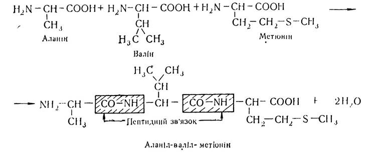
The resulting tripeptide possesses free carboxyl and amino groups and can undergo further condensation with other amino acid residues to form polypeptides. A large number of peptides are synthesized in the Human and Animal organism as a result of various metabolic processes. Many of these exhibit high biological activity and participate in Intermediary Metabolism.
Through the persistent efforts of scientists, the complex and vital problem of protein chemistry was solved: The Nature of amino acid bonding within the protein molecule was clarified, and the peptide theory was established, retaining its significance to this day. It was precisely by elucidating the nature of amino acid bonding in the protein molecule that it became possible to determine the Fine Structure of individual proteins and study protein Conformations.
Investigations of interatomic distances within —CO—NH— peptide bonds, which form the backbone of the polypeptide chain, as well as Bond Angles among hydrogen, oxygen, nitrogen, and carbon atoms, helped reveal several Structural Features of the polypeptide chain and peptide bond. Specifically, it was established that in the polypeptide chain, carbon and nitrogen atoms lie approximately in the same plane, while hydrogen atoms and side chains (radicals) are oriented at an angle of 109°29' to it.
A characteristic feature of the polypeptide chain structure is that the main chain, built from (—NH—CH—CO—) units, is surrounded by various side chains containing highly reactive functional groups such as phenyl, thiol, amide, and hydroxyl groups.
The ability of certain functional groups to ionize causes the protein molecule, depending on their quantitative ratio, to acquire a net positive or negative charge, as well as a specific pH value in aqueous solution. Functional groups also determine a number of other Characteristic Properties of protein macromolecules, such as the spatial configuration of the polypeptide chain, biological activity, and the Nature of the biopolymer's chemical interactions.
X-Ray Diffraction Analysis of the Peptide bond Structure showed that due to the delocalization of p-electrons from the carbonyl Carbon and Oxygen atoms with the lone pair electrons of the imino nitrogen atom, an electron density redistribution occurs:

As a result, the carbon-nitrogen bond length decreases from 1.47 nm to 1.32 nm. Thus, the peptide bond occupies an intermediate position between a single bond (length 1.47 nm) and a double bond (C—N distance of 1.25 nm). To explain this, it was hypothesized that the structure exhibits mesomerism between two Resonance forms—neutral and polar:
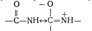
At the same time, a slight elongation of the carbon-oxygen bond from 1.21 to 1.24 nm was observed, rendering it partially single-bonded. Mesomerism, or resonance, is a factor that enhances the stability of chemical compounds, and it is precisely this phenomenon that explains the robustness of the peptide bond. Mesomerism also accounts for the planarity of the amide bond in peptides (all four atoms of the amide bond lie in a single plane) as well as its stability and rigidity.
Studies on bond angles and the mutual orientation of atoms in the peptide group established that substances containing amide groups, with the exception of certain cyclic compounds, possess a planar amide group in the trans-configuration, which is more stable than the cis-configuration.
A characteristic feature of the trans-configuration is that the four atoms of the peptide bond and the two α-carbon atoms lie in the same plane, while the carbonyl oxygen and the imino hydrogen are in a trans-position relative to each other (Fig. 5).
Due to the stability and rigidity of the peptide bond, free rotation around it is hindered or virtually non-existent. The backbone of the polypeptide chain can be visualized as a series of stably fixed planes separated by CH—R groups, relative to which rotation is unhindered.
The phenomenon of resonance largely determines the reactivity of PROTEINS AND PEPTIDES. Peptide bonds undergo tautomeric rearrangements yielding an enol form of the peptide bond, which is characterized by high reactivity.

Experimental data have fully confirmed the polypeptide theory of protein molecular structure. Methods of X-ray diffraction analysis, infrared and ultraviolet spectroscopy enable the visualization of the polypeptide chain with a specific sequence of amino acid residues. Confirmation of the polypeptide theory and irrefutable proof that the monomeric units in a protein molecule are linked by peptide bonds are the successes achieved in the synthesis of proteins and peptides—Insulin, Ribonuclease, oxytocin, vasopressin, and a number of others exhibiting the biological activity of native proteins.
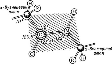
Fig. 5. Peptide bonds and angles between them.
The atoms shown in the shaded area lie in the same plane.
Peptide Synthesis. Naturally occurring peptides
Peptide synthesis is a rather complex and laborious process. It comprises a series of consecutive stages: blocking all groups that should not participate in the reaction, Synthesis of the polypeptide chain, removal of the blocking reagents, etc.
Peptide synthesis has been pursued by a large group of scientists since the late 19th century. However, it was not until 1907 that the German chemist E. Fischer and his pupil E. Abderhalden successfully synthesized peptides containing D- and L-amino acid residues using optically active haloacyl halogenanhydrides. At that time, E. Fischer synthesized the famous octadecapeptide: L-leucyl-(triglycyl)-L-leucyl-(triglycyl)-L-leucyl-(octaglycyl)-glycine. The synthesis of this polypeptide was a significant achievement for its time and was considered unsurpassed for a long time. It was only in 1916 that Abderhalden accomplished the synthesis of a polypeptide with one more amino acid residue—a nonadecapeptide.
E. Fischer developed a number of methods that made it possible to obtain polypeptides with any sequence of α-amino acid residues. The most frequently used method was the successive condensation of α-halocarboxylic acid chloride anhydrides with α-amino acids:

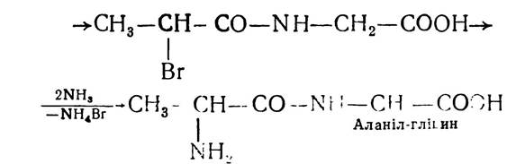
This method, with some refinements, is still used today for peptide synthesis. L-amino acids are employed as esters because the carboxyl group of the amino acids is blocked, and the α-amino group enters into the reaction:

Subsequent saponification of the resulting ester yields the free dipeptide.
Among other methods for obtaining polypeptides, the method involving amino acid Acyl phosphates and the method of carboxyl anhydride condensation in aqueous solutions deserve attention. Carboxyl anhydrides are formed through the action of phosgene on an amino acid According to the reaction:

The carboxyl anhydrides then undergo condensation to form polypeptides:


This method is used to synthesize a number of biologically active polypeptides, such as insulin, adrenocorticotropic hormone (ACTH), and others.
In total, E. Fischer and his students synthesized more than 125 peptides varying in composition and relative molecular weight. By comparing the Properties of the synthesized peptides with the products of complete protein hydrolysis—peptones—E. Fischer concluded that the similarity between them increases with chain length. Polypeptides, much like peptones, gave a positive biuret reaction, had a bitter taste, and, most importantly, were cleaved by Proteolytic Enzymes, although they still differed from native proteins in certain parameters. This indicated that although Proteins are built from a large number of amino acid residues linked by peptide bonds, they possess a significantly more complex structure and specific features compared to polypeptides obtained via chemical synthesis.
Significant progress in peptide synthesis was achieved In the second half of the 20th century, when biologically active Peptide Hormones were successfully synthesized. During this period, oxytocin and vasopressin were synthesized, followed later by bradykinin, ACTH, and other peptides. Triumphs in the field of polypeptide synthesis included the synthesis of the pancreatic hormone insulin and the synthesis of ribonuclease in 1968.
The synthesis of these biologically active substances required intense efforts by entire research teams over several decades. For example, synthesizing insulin—which consists of 51 amino acid residues—required 5,000 chemical reactions, while the synthesis of ribonuclease required 11,000.
Polypeptide synthesis was significantly streamlined after the 1968 invention of an automated, program-controlled synthesizer, which enabled automated solid-phase synthesis of polypeptide chains in a relatively short time. Synthesizing a polypeptide containing 60–65 elementary units takes just a few days, while synthesizing the nonapeptide bradykinin takes 27 hours. The synthesis of the hormone insulin was achieved in 10 days. This method was also used to synthesize the polypeptide chain of bovine ribonuclease, which consists of 124 amino acid residues. The core Principle of the method is that the first amino acid is anchored via its carboxyl group to a specially treated resin, bonding with it through covalent linkages. In the presence of a condensing agent, dicyclohexylcarbodiimide, the anchored amino acid is acylated by the next amino acid, whose amino group is blocked. This yields a resin-bound, protected dipeptide attached to the resin via a covalent bond at its terminal carboxyl group. In the second step, after acidifying the medium, the blocking agent—which breaks down into carbon dioxide (IV) and isobutylene—is removed, and the dipeptide is then acylated with the subsequent amino acid. Upon completion of the polycondensation process, the polypeptide is released from the resin-peptide complex and purified from foreign impurities.
To date, a large number of peptides have been obtained through chemical synthesis, and many have been isolated from natural sources—about 120 in total—though the functions of many remain not fully understood.
Many low-molecular-weight peptides are components of the human and animal organism, participating in intermediary metabolism due to their biological activity. In particular, the tripeptide Glutathione ($\gamma$-glutamylcysteinylglycine) is widespread in plant and animal tissues:
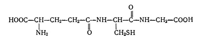
This tripeptide plays a crucial role in redox reactions, owing to its ability to transition between an oxidized form (GS—SG) and a reduced form (GSH), and vice versa. Glutathione forms part of the active centers of enzymes; moreover, its reduced form acts as a coenzyme for certain enzymes that catalyze the isomerization of various compounds.
Animal and human Muscles contain carnosine ($\beta$-alanylhistidine) and anserine ($\beta$-alanyl-1-methylhistidine), which are of great importance for metabolic processes. Soviet scientist V. S. Gulevich demonstrated that in DISEASES ASSOCIATED WITH dystrophic processes in Muscle tissue, carnosine levels decrease significantly.
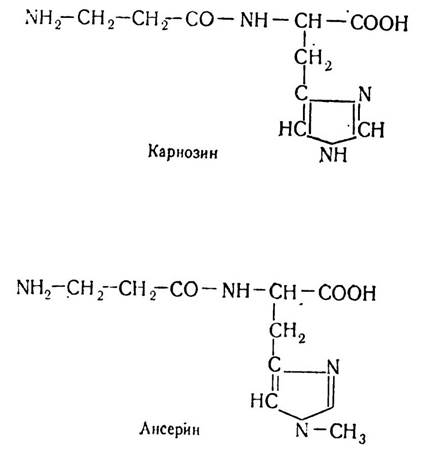
Carnosine helps accelerate carbohydrate breakdown in muscles and, in the form of phosphate, is integrated into the Energy Metabolism of muscle tissue. Anserine is found in the muscles of birds and fish, occurring most abundantly in skeletal muscles, particularly those characterized by high contraction frequencies. Evidence suggests that muscle performance depends on the presence of anserine and carnosine.
Peptide hormones are also of vital importance, notably the Posterior Pituitary Hormones—oxytocin and vasopressin—which are composed of nine amino acid residues. The former stimulates the contraction of muscle Cells surrounding the alveoli of the Mammary Glands and the smooth muscle of the Uterus. Vasopressin increases blood pressure and helps reduce diuresis.
The human and animal body contains physiologically active peptides such as bradykinin, kallidin, and angiotensin, which are generated from biologically inactive Plasma Proteins. All of them are physiologically active: kallidin and bradykinin stimulate muscle contractions, while angiotensin produces a vasoconstrictive effect. A number of antibiotics—such as gramicidin, mycobacillin, and others—also belong to the class of polypeptides, existing as cyclopeptides with widespread medical Applications.
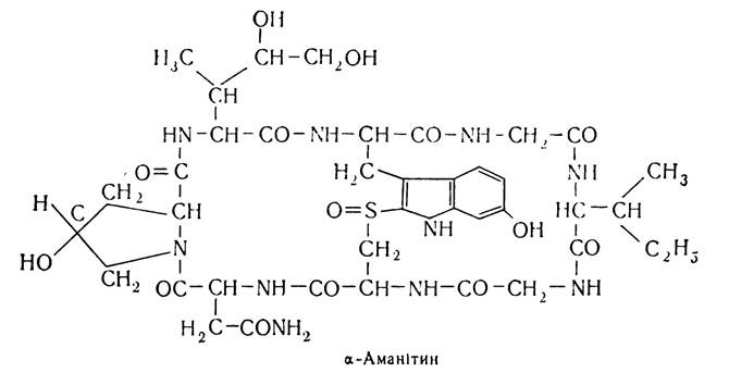
Among plant-derived cyclopeptides, $\alpha$-amanitin, isolated from the death cap mushroom (*Amanita phalloides*), is noteworthy for its high toxicity. Its structure is as follows:
Types of bonds in protein molecules. Research has established that, in addition to peptide bonds, protein molecules contain hydrogen, disulfide, ionic, and Hydrophobic bonds.
A Hydrogen bond forms between covalently bonded hydrogen atoms, which possess a partial positive charge, and negatively charged, covalently bonded atoms of other elements.
In protein molecules, a hydrogen bond is most commonly formed between the hydrogen atom of an imino group (NH) of one peptide bond and the electronegative oxygen atom of a carbonyl group (CO) of a second peptide bond. The presence of this bond is due to the fact that the mesomeric form of the peptide bond has an electron surplus near the oxygen atom and an electron deficit near the nitrogen atom, which leads to enhanced electronegativity of the carbonyl group and electropositivity of the imino group. Consequently, a peculiar competition for the hydrogen atom arises between the oxygen atom of one peptide bond and the nitrogen atom of another.

Unlike other bonds, a hydrogen bond is relatively weak. Its formation energy is approximately 6.0 kJ, whereas the bond energy between carbon atoms is about 250 kJ. For this reason, Hydrogen Bonds form and break easily under normal conditions. Nevertheless, the large number of such bonds within a protein molecule creates quite robust structures, which is of paramount importance for the Formation of the protein molecule's conformation.
A hydrogen bond can be intrachain—in which case it connects separate turns of a single helix—or interchain, connecting different polypeptide chains. In native proteins, this type of bond can occur not only between the hydrogen and oxygen of peptide groups, but also between the side Functional groups of the polypeptide chain, such as the residues of tyrosine, serine, threonine, aspartic acid, and glutamic acid.
A disulfide bond is a relatively strong covalent bond formed between cysteine amino acid residues within a polypeptide chain as a result of the removal of two hydrogen atoms. This type of bond can link cysteine residues within a single polypeptide chain to form disulfide bridges, or it can connect cysteine residues from different polypeptide chains, thereby stabilizing their specific folding during the formation of the tertiary structure.
The stability of a significant number of proteins is attributed to the presence of Disulfide Bonds in their structure. In particular, they are abundant in proteins that make up connective and integumentary tissues, as well as in a number of proteins with high biological activity, such as Collagen, Elastin, ribonuclease, and insulin.
At the sites where disulfide bonds are localized, mechanical stress arises that can partially disrupt Hydrogen bonds and contribute to the destabilization of Helical structures. As a result, polypeptide chains contain linear, uncoiled regions in addition to helical ones. Disulfide bonds ensure the biological activity of numerous proteins; therefore, when these bonds are disrupted, the protein loses its biological activity. Specifically, disulfide bonds play a crucial role in ensuring the biological activity and structural stabilization of the insulin protein molecule.
The Lysozyme molecule contains disulfide bonds that link different Regions of the polypeptide chain, imparting a characteristic spatial conformation that ensures the biological activity of this biopolymer.
An ionic bond arises in protein molecules when their composition includes amino acid residues capable of ionization, such as aspartic acid, glutamic acid, lysine, arginine, and others.
As is well known, in aqueous solutions proteins exist as dipolar ions, or zwitterions, in which the carboxyl group carries a negative charge and the amino group carries a positive charge.
As a result, electrostatic interaction forces arise between the carboxyl groups 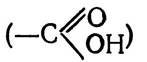 and amino groups (NH2) as they come close to each other, leading to the formation of salt bridges:

An ionic bond can form between individual segments of a polypeptide chain or between polar groups of different polypeptide chains. Although native proteins contain a large number of amino acids with polar side chains, only a small fraction of them actually participate in ionic bonding. This is because some of the polar groups interact with the solvent, as the protein molecule tends to adopt a conformation in which the maximum number of polar groups is exposed on its surface.
Hydrophobic bonds arise due to Van der Waals forces between nonpolar side-chain residues of amino acids such as alanine, valine, leucine, isoleucine, phenylalanine, tryptophan, etc. A schematic representation of a hydrophobic bond is shown below:

The formation of nonpolar (hydrophobic) bonds is driven by the tendency of nonpolar molecules to associate in aqueous solutions. When nonpolar amino acid residues come into close proximity, hydrophobic interactions occur, accompanied by a specific orientation of water molecules surrounding the nonpolar groups. All of this restricts the translational motion of water molecules and leads to a decrease in Entropy. Water molecules are effectively squeezed out from the regions where hydrophobic interactions take place, while the protein molecule adopts a conformation that places hydrophobic groups in its interior, leaving hydrophilic groups On the surface. Under these conditions, the radical–water system transitions into the more stable radical–radical system.
The hydrophobic interior of the protein molecule, by excluding water, remains "dry" and anhydrous. This phenomenon helps proteins perform their specific functions. In Hemoglobin, for example, the iron atom is located deep inside the protein molecule. Protected from water in this manner, it is able to bind oxygen. Although hydrophobic bonding is the weakest of all nonconvalent interactions, the high cooperativity of hydrophobic interactions results in fairly stable structures. This type of bonding plays a crucial role in the formation and stabilization of the tertiary and, potentially, quaternary structures of proteins.
Current Concepts of Protein Structural Organization
Studying the quantitative and qualitative amino acid composition of proteins and the bonds between amino acids in their molecules does not yet provide a complete picture of protein structure. It is essential to determine
the sequence of amino acid residues in the polypeptide chain and the spatial conformation of the protein molecule.
According to current data, proteins exhibit four levels of structure: primary, secondary, tertiary, and quaternary.
Primary Protein Structure. Primary protein structure refers to the sequence of amino acid residues linked together by peptide bonds within a polypeptide chain. Each polypeptide chain of a protein molecule, like any peptide, has two terminal amino acids:

Amino acid residues in polypeptide chains are conventionally numbered starting from the N-terminus and ending at the C-terminus.
Since protein molecules consist of a large number of amino acid residues, investigating their Primary Structure presents certain difficulties. However, research pace has accelerated significantly over the past decade. This is primarily due to improved methods for isolating highly purified protein preparations, as well as the development and widespread adoption of precise analytical research techniques. Individual stages of primary structure analysis have now been automated.
Protein molecules may consist of one, two, four, or even more polypeptide chains.
Polypeptide chains within a protein molecule can be linked by covalent bonds, specifically disulfide bridges (S—S) formed between two cysteine residues. In addition, a disulfide bond can also form between two cysteine residues located within the same polypeptide chain.
Along with disulfide bonds between polypeptide chains, other noncovalent bonds (ionic, hydrogen, and hydrophobic) can form between polypeptide chains or between different regions of the same polypeptide chain. These interactions are vital for the stabilization of higher-order protein structures.
The number of polypeptide chains in a protein molecule is determined by the number of N-terminal amino acids per protein molecule. This is achieved using the dinitrophenyl method, or Sanger method, originally developed by the English biochemist Frederick Sanger. It was first applied to study the Introduction/19.html">Primary structure of the protein insulin. The method is based on treating the protein in a mildly alkaline medium with 2,4-dinitrofluorobenzene, which reacts with the N-terminal amino acid of the peptide chain to form a dinitrophenyl derivative: a DNP-protein (peptide) derivative.

In the next step, the dinitrophenyl protein (peptide) derivative is subjected to acid hydrolysis (6 N HCl), which cleaves all peptide bonds except the one formed between the 2,4-dinitrophenyl group and the α-amino group of the N-terminal amino acid. Under these conditions, free amino acids and the dinitrophenyl derivative of the N-terminal amino acid are produced:

Subsequently, qualitative and quantitative analysis of the N-terminal amino acids is performed using Specific methods (chromatography, electrophoresis, colorimetry, spectrophotometry). Thus, the number of N-terminal amino acids determines the number of polypeptide chains in the protein molecule.
Other methods, notably the dansyl and phenylthiohydantoin methods, are also used to identify N-terminal amino acids.
In addition to chemical methods, enzymatic techniques are employed to determine N-terminal amino acid residues.
If a protein consists of multiple polypeptide chains, disulfide bonds must first be cleaved via oxidation or reduction.
When studying the primary structure of insulin and ribonuclease, performic acid was used to cleave the disulfide bonds.
However, this method also has drawbacks. Performic acid partially destroys tryptophan and does not always ensure The oxidation of all disulfide bonds within the protein molecule.
To cleave disulfide bonds, reduction methods using reagents such as mercaptoethanol, thioglycolic acid, and others are frequently employed.
Once the disulfide bonds in protein molecules are disrupted, individual polypeptides are isolated. Ion-exchange chromatography and gel electrophoresis are used for this purpose.
The next stage of the work involves determining the sequence (order) of amino acid residues in the isolated polypeptide chains. To achieve this, selective partial hydrolysis of The polypeptide chains is first carried out using proteolytic enzymes or Hydrochloric acid to yield smaller fragments (peptides). In shorter peptides, it is significantly easier to determine the arrangement of amino acid residues.
The Use of enzymes results in the Cleavage of peptide bonds between specific amino acids. To prevent enzymes from contaminating the protein under study, insoluble enzyme preparations immobilized on special carriers are utilized.
Trypsin exhibits a highly specific action on polypeptide chains. As a rule, it cleaves peptide bonds formed by the carboxyl groups of lysine and arginine. Trypsin belongs to Endopeptidases and hydrolyzes a peptide bond only when another peptide bond is adjacent to it. Consequently, free amino acids are not released during the hydrolysis of peptides by trypsin.
Other proteolytic enzymes (Pepsin and Chymotrypsin) are less specific. For instance, pepsin cleaves peptide bonds formed by the amino group of tyrosine, and occasionally those of glutamic acid, glycine, and alanine. Chymotrypsin hydrolyzes bonds in a polypeptide formed by the carboxyl groups of amino acids such as methionine, phenylalanine, tyrosine, and several others.
The resulting mixture of peptides is subjected to preliminary fractionation based on charge and size, and is subsequently purified from various impurities. These processes are carried out using electrophoresis and ion-exchange chromatography.
The isolated peptides (fragments) are used to study the Amino Acid Sequence within each of them.
The sequence of amino acid residues in these peptides is determined starting from the N- and C-terminal amino acids. To identify N-terminal amino acids, alongside the dinitrophenyl method, the phenylthiohydantoin method, or Edman Degradation, has been applied over the past decade. The principle is that the protein (peptide) is treated with phenylisothiocyanate, leading to the formation of a phenylthiocarbamyl derivative (PTC-derivative) of the protein (peptide), which undergoes cyclization under the influence of hydrochloric acid. During this process, the N-terminal amino acid is cleaved off as a phenylthiohydantoin derivative. This process can be schematically represented as follows:
Phenylthiohydantoin is identified using chromatographic methods.
The main advantage of the Edman degradation over other methods is that the protein (peptide), shortened by a single amino acid, can be isolated in an intact form and subjected to a fresh Treatment with phenylisothiocyanate. This once again yields a phenylthiohydantoin containing the next N-terminal amino acid. Because this process can be repeated many times, the sequence of amino acid residues in the protein polypeptide chain can be fully determined.
An instrument (sequenator) has already been developed in which all operations—Treatment of the protein (peptide) with phenylisothiocyanate, hydrolysis of the resulting product, and isolation of phenylthiohydantoin—are automated.
After determining the primary structure of individual peptides, their arrangement within the polypeptide chain is established. This is achieved by comparing peptides obtained from the hydrolysis of the same protein using trypsin, chymotrypsin, and pepsin.
Continuous improvements in Methods for determining the sequence of amino acid residues in protein molecules have made it possible to elucidate the primary structure of nearly 1,000 different proteins. The first protein whose primary structure was determined was insulin, a pancreatic hormone that plays a crucial role in regulating Carbohydrate Metabolism. It is a simple protein with a relative molecular mass of 6,000. It is built from 16 amino acids, contains 51 amino acid residues, and consists of two chains: chain A contains 21 amino acid residues, and chain B contains 30 amino acid residues.
The primary structure of insulin was investigated during the 1940s and 1950s by the English biochemist F. Sanger. For these and other studies related to protein structure, F. Sanger was awarded the Nobel Prize in 1958.
Following insulin, the primary structure of another enzymatic protein, ribonuclease, was deciphered. It consists of 124 amino acid residues.
In the 1970s, under the leadership of Soviet scientists Academicians Yu. A. Ovchinnikov and A. E. Braunstein, the primary structure of the enzyme aspartate aminotransferase was elucidated in our country. This protein contains 412 amino acid residues. The structure of the polypeptide chains has also been fully deciphered for such enzymes as pepsinogen, pepsin, chymotrypsin, carboxypeptidase, Glutamate dehydrogenase, aldolase, lysozyme, Papain, a number of proteins belonging to Cytochromes, as well as hemoglobin, Myoglobin, and others.
Advances in studying protein primary structure have made it possible to produce them via chemical synthesis. Notably, all such synthesized proteins exhibit their characteristic biological activity.
The study of protein primary structure and the chemical synthesis of some proteins indicate that the amino acid sequence is a specific characteristic of each individual protein. It is predetermined within the polypeptide chain and genetically encoded. Since proteins are composed of 20 different amino acids, theoretically around 2·1018 protein isomers can exist in nature. Analysis of amino acid sequence data across various proteins has revealed several interesting and important regularities, such as THE PRINCIPLE OF structural similarity. Its essence lies in the fact that identical peptide motifs occur in the polypeptide chains of different proteins or in different regions of the polypeptide chain of the same protein. For instance, the primary structures of insulin and ribonuclease (Table 4) contain three identical tripeptides and one tetrapeptide in different regions of their polypeptide chains.
Table 4. Structural similarity of insulin and ribonuclease
Peptide motifs |
Insulin |
||
Chain A |
Chain B |
Ribonuclease |
|
Identical tripeptides |
Ala-Ser-Val 8 9 10 Val-Cys-Ser 10 11 12 |
Val-Glu-Ala 12 13 14 |
Ala-Ser-Val 122 123 124 Val-Cys-Ser 57 58 59 Val-Glu-Ala 54 55 56 Asn-Tyr-Cys-Asn 24 25 26 27 |
Identical tetrapeptides |
Asn-Tyr-Cys- 18 19 20 Asn 21 |
||
A characteristic pattern is a certain similarity in the Primary Structure of Proteins that perform similar biological functions. An example is the group of enzymes that catalyze the dehydrogenation reactions of various substrates. This feature is also characteristic of hydrolase enzymes, which have a similar arrangement of amino acid residues near the active center.
At the same time, it has been established that proteins are characterized by species specificity, which is determined by the specific primary structure of certain regions of the polypeptide chain. For instance, the protein hormone insulin in various animal species has a non-identical composition of chain A fragments at positions 8–10. The study of the amino acid sequence in the insulin molecule of various animal species has shown that the changes observed in the insulin molecule when passing from one animal species to another are characteristic only of certain amino acids. Thus, in polypeptide chain A, alanine and threonine can substitute for each other at position 8, Serine and Glycine at position 9, and valine, isoleucine, and threonine at position 10 (Table 5).
Differences in primary structure are also found in individual proteins of the same species, which is caused, in particular, by Mutations. It is known, for example, that the hemoglobin of a healthy human differs from that of a patient with Sickle-Cell Anemia solely in that the glutamic acid residue in the |3-polypeptide chain is replaced by a valine amino acid residue:

Table 5. Amino acid Changes in the A polypeptide chains of insulin molecules from various animal species and humans
Species |
Position of amino acid residues in chain A |
||
8 |
9 |
10 |
|
Bull |
Ala |
Ser |
Val |
Pig |
Thr |
Ser |
Ile |
Ram |
Ala |
Gly |
Val |
Horse |
Thr |
Gly |
Ile |
Sperm whale |
Thr |
Ser |
Ile |
Sei whale |
Ala |
Ser |
Thr |
Dog |
Thr |
Ser |
Ile |
Rabbit |
Thr |
Ser |
Ile |
Human |
Thr |
Ser |
Ile |
More than 100 abnormal (pathological) Hemoglobins have already been studied, and in almost all cases, the substitution of a single amino acid in the α- or β-polypeptide chain of the given protein has been established. Characteristically, the changes detected in abnormal hemoglobins often involve hydrophilic amino acids. Studying the primary structure of a number of proteins makes it possible to elucidate the molecular mechanisms underlying The Development of many Hereditary diseases.
Secondary structure of proteins. The secondary structure of proteins refers to the spatial configuration of the polypeptide chain. The determination of Protein secondary structure was preceded by prolonged and extensive studies on the structure of a number of amino acids and peptides using X-ray diffraction analysis. This made it possible to establish certain Regularities of the peptide bond, as well as the probable Conformations of Peptide chains. Based on X-ray diffraction data, American scientists L. Pauling and R. Corey proposed a model of protein secondary structure in the form of an α-Helix (Fig. 6).
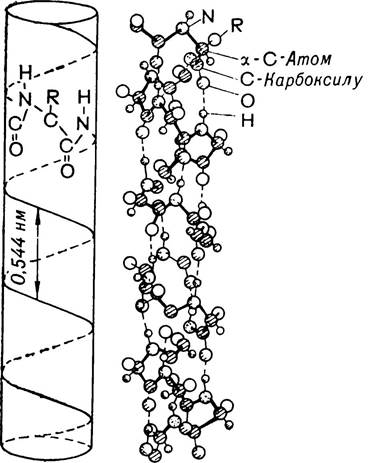
Fig. 6. Model of the polypeptide chain according to Pauling and Corey (α-helix).
It represents a helical, twisted structure. In nature, Two Types of helices can potentially exist: right-handed and left-handed. However, data obtained from X-ray diffraction Analysis of proteins indicate the presence of a right-handed helix in them. This may be related to the fact that proteins consist exclusively of L-series amino acids.
The spatial configuration of the α-helix is characterized by the following parameters: one turn of the α-helix contains 3.6 amino acid residues, and the pitch of the helix (the distance between turns) is 0.54 nm. The rise angle of the turn is 26°, and the height per amino acid residue is 0.15 nm. The identity period, i.e., the length of the segment of the helix that completely repeats, is 2.7 nm and includes 18 amino acid residues.
Hydrogen bonds play an important role in stabilizing the secondary structure of proteins. In the α-helix, they form between the carbonyl oxygen and the imino hydrogen of adjacent turns of the polypeptide chain. It should be noted that although the energy of hydrogen bond formation is small, their large number ensures sufficient stability and rigidity of the α-helix. However, in an aqueous environment, the α-structure may be less stable because water competes with the protein by forming hydrogen bonds.
The stability of protein structure is also determined by other factors, in particular the side groups (radicals) of amino acid residues located in different regions of the polypeptide chain. Thus, studies with polyalanine, whose radicals are small in size and uncharged, have shown that it spontaneously forms an α-helical structure in aqueous solution at pH 7.0. A polypeptide based on another amino acid—lysine, i.e., polylysine—does not form an α-helix at this same pH value, but instead exists as a disordered coil. In a neutral environment, lysine radicals carry a positive charge, which prevents them
from approaching each other due to repulsive forces that hinder the formation of intra-chain hydrogen bonds by the polypeptide. Similar experiments were conducted with other polyamino acids. This made it possible to establish that a number of amino acids—alanine, valine, leucine, methionine, phenylalanine, tyrosine, tryptophan, histidine, and some others—favor the formation of the α-helix, especially when they are located adjacent to each other in the polypeptide chain. Conversely, amino acids such as lysine, arginine, tyrosine, serine, threonine, aspartic acid, and glutamic acid promote the destabilization of the α-helix. Certain amino acids, such as proline and hydroxyproline, do not fit into the helical structure. In these regions, the direction of the polypeptide chain changes by 103°, and the helical structure is disrupted.
This is obviously one of the reasons why polypeptide chains in the molecules of certain proteins are not entirely helical. Such cases are quite rare. Each protein is characterized by a specific degree of polypeptide chain helicity (Table 6).
Table 6. Degree of helicity of polypeptide chains
(according to Davies, 1965)
Protein name |
Degree of helicity, % |
Paramyosin |
100 |
Myoglobin |
75 |
Hemoglobin |
75 |
Serum albumin |
50 |
45 |
|
Lysozyme |
35 |
Tobacco mosaic virus (subunit) |
30 |
Pepsin Ribonuclease |
28 17 |
Chymotrypsin |
11 |
Thus, the secondary structure of a protein should be understood as The ratio of helical regions of the polypeptide chain to linear, irregular, amorphous transitions in which Hydrogen bonds are disrupted.
Studies of the structure of various proteins have shown that, in addition to the α-helix, another ordered conformation of the polypeptide chain can exist, known as the β-structure. For its formation, polypeptide chains must be extended to a certain extent and arranged in a parallel manner. The stabilization of the β-structure, like that of the α-helix, occurs through hydrogen bonds. It is believed that the β-structure can exist in two varieties. The first is called the antiparallel pleated sheet. It is formed by polypeptide chains whose amino acid residues are directed in opposite directions. The second variety of the β-structure is called the parallel pleated sheet. It is formed when the orientation of the amino acid residues in the polypeptide chains coincides.
The β-structure is characteristic of Fibrous proteins—fibroin (silk protein), keratin (Connective Tissue protein), Myosin (muscle protein), and several others.
Thus, the α-helix and β-structure represent the second level or order of the Structural organization of a protein molecule, i.e., the secondary structure, in the stabilization of which hydrogen bonds play an important role.
Tertiary Structure of proteins. This level of structure is characterized by a specific spatial conformation of the helical and linear regions of polypeptide chains. In other words, tertiary structure is the folding of the secondary structures of polypeptide chains to form globules of various shapes.
Each protein is characterized by its own tertiary structure, upon which its specific biological properties depend. The tertiary structure is largely determined by the primary structure of proteins, i.e., the sequence of amino acid residues in their molecules, as well as the size, shape, and polarity of amino acid side chains. It should be noted that polypeptide chains do not simply fold to form near-spherical structures, but rather fold through a series of precisely defined states, resulting in a unique conformation of the protein molecule. During the formation of the tertiary structure, Polypeptide chains fold in such a way that the maximum number of hydrophilic groups of amino acid residues is positioned on the outside, facing the aqueous environment, while hydrophobic groups are located inside the structure (globule). The conformation of the resulting globule is significantly influenced by such factors as ambient pH, Ionic strength of the solution, Temperature, and the interaction of protein molecules with other substances, etc.
A minor disruption of the tertiary structure of a protein generally leads to the loss of its biological properties. Hydrogen, disulfide, ionic, and hydrophobic bonds play a vital role in stabilizing the Tertiary Protein Structure. They come into play once the polypeptide chain acquires the specific conformation characteristic of the native protein molecule. General Principles concerning the forces that stabilize the protein globule structure were established back in 1944 by Soviet scientists. For instance, disulfide bonds can form spontaneously as two sulfhydryl groups of cysteine residues come into close proximity. These bonds are covalent and, compared to other bonds, are characterized by high strength. However, the presence of disulfide bonds is not obligatory for all proteins. There are known proteins that lack these bonds, such as hemoglobin, myoglobin, tobacco mosaic virus protein, Escherichia coli proteins, and several others. At the same time, the destruction of disulfide bonds in proteins where they are present leads to the loss of their specific biological properties and reduces the Stability of the protein molecule.
It is believed that among non-covalent bonds, hydrophobic bonds are of the utmost importance for stabilizing the tertiary structure of proteins. They are formed when the polypeptide chain contains residues of such Amino acids as valine, leucine, isoleucine, phenylalanine, and tryptophan.
A series of studies conducted in recent years indicate that hydrogen and ionic bonds also play an important role in stabilizing the tertiary protein structure.
Today, the tertiary structure of many dozens of different proteins has been elucidated. The tertiary structure of the myoglobin protein was first deciphered by the English researcher J. Kendrew in 1957.
Myoglobin is a protein with a relatively small relative molecular mass of 17,500. Its molecule is built from a single polypeptide chain containing 153 amino acid residues. More than 70% of the myoglobin polypeptide chain is helicized. Non-helicized regions contain a large number of proline residues. In myoglobin isolated from various animal species, the conformation of the polypeptide chains is similar, although they differ slightly from one another in amino acid composition.
The English scientist M. Perutz established the tertiary structure of a more complex protein—hemoglobin, which consists of four subunits. Each subunit is structurally similar to myoglobin. Myoglobin and hemoglobin share similar functions: hemoglobin binds and transports oxygen to the body's tissues, while myoglobin serves as an oxygen reserve in animal muscles. The structure of both proteins includes an iron-porphyrin complex.
In the second half of the 1960s, the tertiary structure of the enzyme lysozyme, which is built from a single polypeptide chain, was elucidated in considerable detail. Furthermore, researchers successfully determined the positions of all 129 amino acid residues forming the polypeptide chain of this protein. It was found that when the lysozyme polypeptide chain folds, it forms a specific-shaped cleft that houses the active center interacting with the corresponding substrate.
The structures of such proteins as ribonuclease, aldolase, Lactate dehydrogenase, Carbonic anhydrase, papain, pepsin, trypsin, chymotrypsin, and others have been determined. The studies carried out in this regard once again demonstrate that the tertiary structure is quite complex, unique, and specific for each protein molecule.
Quaternary Structure of proteins. The application of X-ray diffraction analysis and Electron Microscopy has made it possible to establish that proteins whose molecules are built from a single polypeptide chain exhibit only primary, secondary, and tertiary structures. Proteins that consist of two, four, or a greater (even) number of individual polypeptide chains with their own tertiary structure possess a quaternary structure. Such polypeptide chains are called protomers or subunits, and the proteins composed of them are called oligomers. Thus, the mutual spatial arrangement of protomers (subunits) in a protein molecule constitutes its quaternary structure.
The protomers that make up The quaternary structure of a protein may have identical or different amino acid compositions. It is believed that proteins with a relative molecular mass exceeding 50,000 are built from multiple polypeptide chains (protomers).
A classic example of a protein with a quaternary structure is hemoglobin, which consists of four protomers (two α- and two β-polypeptide chains). The protomers built from α-chains contain 141 amino acid residues each, while those from β-chains contain 146 residues each. By combining with one another, these protomers form the hemoglobin molecule (Fig. 7). The relative molecular mass of hemoglobin is 68,000, and that of each protomer is 17,000. Characteristically, hemoglobin undergoes slight Conformational Changes in its quaternary structure during the uptake and release of oxygen.
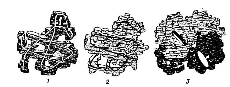
Fig. 7. Structure of the α-chain (1), β-chain (2), and the intact hemoglobin molecule (3) according to X-ray diffraction analysis data.
Oxygen binding is accompanied by the compression of its molecule due to the approximation of protomers. Oxygen release produces the opposite effect.
Tobacco mosaic virus protein also exhibits quaternary structure, with a relative molecular mass of approximately 40 million. The molecule of this protein comprises nearly 2,200 identical protomers with a relative molecular mass of 17,500, arranged in a helix. Quite recently, the quaternary structure of turnip yellow mosaic virus protein has been elucidated; it has a relative molecular mass of about 5 million and consists of 180 identical subunits.
The quaternary structure of proteins underlies the architecture of a wide range of enzyme proteins and is directly related to the existence of Isoenzymes. For example, if a protein-enzyme molecule consists of four type A and B protomers, the enzyme can exist in five isoforms: AAAA, AAAB, AABB, ABBB, BBBB, meaning that five isoenzymes with varying degrees of catalytic activity may exist. The formation of isoenzymes is a crucial regulatory process for enzyme systems. Enzyme isoforms are characteristic of lactate dehydrogenase and several Other Enzymes.
The quaternary structure of proteins is stabilized and maintained in its Native State through non-covalent bonds formed between various functional groups located on The surface of protomers (protein globules). Under the influence of various physical or Chemical factors, proteins dissociate into individual protomers, thereby losing their biological activity. Factors causing this dissociation may include organic solvents, urea, changes in ambient pH, and others. Eliminating the dissociating factor causes the protomers to reassociate, restoring the protein's Quaternary Structure and its biological activity, which demonstrates the close interrelation between protein Structure and function.
Functions and Role of Protein in the Organism
A vast Diversity of proteins exists in nature. Living organisms contain 1 • 1012 proteins, of which about 5 million distinct proteins are found in the human and animal body. Each of these proteins performs specific functions that determine its involvement in vital physiological processes. Some proteins ensure movement and Muscle contraction, others mediate immune or catalytic processes, determine the structure of organs and tissues, and promote growth, development, and self-reproduction.
One of the most essential Protein Functions is the structural (plastic) function, whereby proteins form the structural basis of organisms, are constituent parts of all organs and tissues, and participate in building the framework of cells and their Organelles—membrane structures, Mitochondria, Ribosomes, and Cytoplasm. Proteins are major components of Biological Membranes, performing diverse functions within them, such as structural, regulatory, and catalytic. The specificity and role of membranes are determined primarily by their protein component. In higher animals and humans, proteins participate in forming vascular walls, integumentary, muscular, and Connective Tissues, and constitute the organic matrix of Bone tissue, Cartilage, ligaments, and tendons. Proteins also play a vital role in forming various structures in plant organisms. All PLANT CELL AND tissue apparatuses that sustain life are built from diverse protein compounds.
Proteins account for an average of 10–21% of the wet body mass of humans and animals and about 50% of their dry mass. In individual organs and tissues, their content may vary within wide ranges, which is associated with the structural features and functions of these organs and tissues.
In particular, protein content is high—exceeding 85% calculated on a wet weight basis—in cells that are in a physiologically active state.
The average protein content in individual organs and tissues, calculated on a wet weight basis, is as follows (%):
Muscles |
18–23 |
7–9 |
Leaves |
1.0–3.0 |
|
Liver |
17–19 |
Adipose tissue |
6–0 |
Roots |
0.5–3.0 |
16–18 |
Plant seeds |
10–13 |
Vegetables and fruits |
0.3–1.6 |
|
12–15 |
Stems |
1.5–3.0 |
The plastic function of proteins in the organism is due to their ability, along with other biological molecules (CARBOHYDRATES, Lipids, nucleic acids), to form supramolecular complexes that ensure the formation of diverse subcellular structures in tissues and organs.
In humans and animals, Proteins perform a catalytic function, acting as specific regulators and catalysts for numerous biochemical processes. All enzymes—biological catalysts that govern the direction and rate of the chemical reactions underlying metabolism—are proteinaceous in nature. Enzymes catalyze reactions that drive the Chemical transformations of various substances necessary to generate the energy required for metabolic processes and vital activity. More than 17,000 proteins with enzymatic activity are currently known, and over 200 of them have been isolated in crystalline form.
Thousands of different chemical reactions occur simultaneously within a cell with the aid of enzymes, ensuring the Synthesis and Breakdown of diverse compounds at normal temperatures and pressures, efficiently and without the use of chemical reagents. For instance, a single molecule of the enzyme catalase can catalyze the decomposition of 5 million molecules of hydrogen peroxide in one minute. Thus, proteins form the foundation not only of the organism's structure, but also of metabolism—one of the most defining characteristics of life.
Proteins play a key role in regulating metabolic processes. This regulatory function is carried out with the participation of hormones, which are secreted by Endocrine glands and control the level of metabolic activity in the body. Most hormones are proteins or products of Protein metabolism. These include insulin, theelin, thyrotropin, thyroxine, adrenocorticotropic hormone, oxytocin, vasopressin, and others. Products of protein metabolism also include Central Nervous System mediators such as histamine, serotonin, γ-aminobutyric acid, and others. Additionally, certain peptide hormones participate in the synthesis and Regulation of enzyme Activity, thereby establishing a balanced system that allows the organism to actively respond to changing environmental conditions. Hormonal REGULATION OF METABOLISM occurs through effects on cell membrane permeability, the modulation of enzyme activity, and influence over Translation and Transcription processes, among others.
Proteins also play a crucial role in regulating genome activity. Considerable attention is devoted to studying the structure, functions, and characteristics of genome-regulatory proteins. It is believed that the Chromatin of cell nuclei contains specific proteins capable of associating with DNA through ionic bonds and Intermolecular Forces, thereby stabilizing the DNA molecule. However, since the destabilization of its structure is necessary to ensure template activity, the ability of DNA to replicate is restored only when the bonds between the DNA and protein molecules are weakened—in this case, acting as regulators of genome activity.
Proteins play an essential role in supporting GROWTH AND REPRODUCTION—the "Replication of one's own kind"—as well as the transmission of hereditary traits. This is especially true for complex proteins known as Nucleoproteins, which consist of biopolymers such as Nucleic Acids and histone proteins.
The protective function of proteins is of great importance, realized through γ-globulins, which are associated with the body's immune responses. Antibodies produced by the organism in response to adverse factors—such as pathogenic Bacteria, Viruses, and toxins—are proteinaceous in nature. By binding to microorganisms and toxins (Antigens), antibodies inactivate them, inhibit their pathogenic effects, and neutralize toxic substances. Proteins produced by the organism in response to toxic action are called IMMUNOGLOBULINS (Ig). There are 5 classes of immunoglobulins (IgG, IgM, IgA, IgD, IgE).
Another vital protective function of proteins is their involvement in blood clotting. This shields the body from excessive blood loss during various traumas and injuries to organs and tissues. Blood clotting is mediated by the protein fibrinogen, which, upon blood vessel damage, is converted into fibrin, precipitates, and blocks the lumen of the vessel, thereby stopping the bleeding.
Movement is one of the most important manifestations of life in living organisms. Various Forms of mechanical movement—such as muscle contraction and relaxation, and the functioning of Internal Organs (heart, brain, lungs, Stomach)—rely on contractile proteins such as Actin, myosin, and Tropomyosin. These proteins drive the mechanochemical processes that convert chemical energy into mechanical energy. They transform the chemical energy of high-energy compounds into the mechanical energy of muscle contraction, which forms the basis of mechanical movement. Soviet biochemists V. O. Engelhardt and M. N. Lyubimova proved that the contractile protein myosin is enzymatic: it accelerates the hydrolysis of ATP—the primary energy store—yielding ADP and inorganic phosphate. The energy released in this process is utilized by the muscles. Thus, myosin performs a dual function: it regulates both the release of energy and its utilization during muscle work.
Proteins facilitate the energy supply for various intracellular processes because they can transform the energy derived from nutrient oxidation or sunlight into electrical energy, leading to the generation of membrane potentials in Mitochondria and Chloroplasts. This energy is subsequently used to perform various types of work or is transported along membranes as a transportable form of energy.
Proteins drive the mechanisms of self-assembly and self-regulation in biological systems (such as complex enzyme systems, cytoplasmic structures, and Viral Particles) at THE MOLECULAR LEVEL, owing to their ability to recognize specific molecules amidst a vast array of others.
Equally vital to the organism is the transport function of proteins, which facilitates the distribution of energy-rich and biologically active compounds such as fats, lipids, and Fat-soluble Vitamins. The Essence of this transport function lies in the ability of proteins to form temporary complexes with various substances and deliver them to different tissues. One prominent transport protein is hemoglobin, which transports oxygen to various organs and tissues and carbon dioxide (IV) in the opposite direction. Hemoglobin is found in the erythrocytes of higher animals and humans, accounting for 90% of their content. In some lower organisms, such as Mollusks and crustaceans, erythrocytes are absent, and hemoglobin is dissolved directly in the cytoplasm. In squids and cephalopods, Oxygen transport is carried out by hemocyanin, a copper-containing protein that gives the blood of these animals a blue color.
Serum albumin binds and transports Fatty acids, Steroid Hormones, various metabolic products, anions, and cations. This protein binds 50% of calcium and transports Cu2+ ions from the intestine to the liver. Another notable transport protein is ceruloplasmin, which transports copper from the liver to cellular organelles.
The protein transferrin transports iron ions, while Lipoproteins ensure The transport of lipids and fat-soluble vitamins. Specifically, low-density lipoproteins daily transport 25–50 g of endogenous triglycerides, while others handle the transport of Cholesterol, carotene, carbohydrates, and Phospholipids. Transport proteins function at both the macro-structural level (circulatory and lymphatic systems) and the micro-structural level (cells and subcellular structures).
A critically important function of proteins is signal transmission across cell membranes, known as the signaling or receptor function. The reception of signals and their transmission to intracellular centers form the basis of yet another fundamental property of life: irritability. Here, stimuli—whether chemical or mechanical—induce specific conformational changes in proteins, serving as the cell's primary reaction to external stimuli. This principle underlies the functioning of The Nervous System and the brain.
The Mechanism of reception involves receptor proteins capturing stimulus energy, converting this energy via specialized protein molecules into specific information, and transmitting that information to the central nervous system. Receptor proteins are localized within the cellular membrane apparatus and serve as The Site of Action for numerous hormones. For instance, a receptor protein associated with adenylate cyclase alters its conformation under the Influence of the peptide hormone Glucagon. This leads to the activation of the coupled adenylate cyclase, an enzyme that stimulates cAMP synthesis and accelerates Glycogen phosphorolysis. This process proceeds via protein kinase reactions and The conversion of inactive phosphorylase B into active phosphorylase A. An example of a photoreceptor protein is opsin, which can form a complex with the aldehyde form of vitamin A—retinal. As a result, the protein undergoes a conformational change that triggers nerve impulses and enables Vision.
Choline receptor proteins play a vital role in perceiving sound waves and converting them into nerve impulses. These proteins also modulate the permeability of cell membranes.
Proteins are essential components of human and animal diets; their deficiency or absence in nutritional rations leads to Metabolic Disorders and various diseases. They cannot be replaced by any other substances in nutrition. Furthermore, proteins are widely utilized across various sectors of the national
economy. Natural protein fibers include wool and silk. Proteins are also employed in The production of gelatin, leather goods, plastics, and medical protein preparations such as hormones, antiserums, and blood substitutes.
As we can see, proteins sustain the vital processes of living organisms, which has spurred rapid research developments in this field. Once merely a biochemical sub-problem, it has evolved into one of the most crucial frontiers of modern biology and medicine. The multifaceted and vital nature of protein research stems from its connection to unraveling the fundamental laws governing living matter, understanding the highest forms of its existence, decoding the essence of life's phenomena, and gaining conscious control over them. Therefore, it is safe to assert that the progress and achievements of many fields in biology, medicine, and agriculture largely depend on the advancement of protein chemistry and biochemistry.
Last update: 06/08/2026
Editorial and Educational Adaptation: This material has been compiled based on the primary/original source text. The project team performed an editorial review, corrected technical inaccuracies, structured sections, and adapted the content for an educational format.
What was processed:
- elimination of formatting defects (OCR errors, structural breaks, corrupted characters);
- editorial organization of content;
- standardization of terminology in accordance with academic sources;
- verification of factual statements against the original source text.
All mentions of the author, publication year, and origin of the primary text have been preserved in accordance with the source.