Human Anatomy and Physiology (with Age-Related Features of the Child's Body) - Sapin M.R., Sivoglazov V.I. 2002
Structure of the Human Body
Cells
Cell Division (Cell Cycle)
Organismal growth, Cell proliferation, and reproduction occur through Cell Division. Mitosis and Meiosis are the primary Methods of cell division in The Human Body. Although the processes occurring during these types of division are similar, they yield different results. Mitotic cell division leads to an increase in cell number, driving the growth of the Organism. This process ensures cell renewal as cells wear out and die. (It is currently known that epidermal cells live for 3–7 days, while red Blood Cells live up to 4 months. Nerve and Muscle cells (fibers) persist throughout a person's entire life.) Thanks to mitotic division, daughter cells receive a chromosome set identical to that of the parent cell.
During meiosis, which occurs in Germ Cells, division results in new cells with a single (haploid) set of Chromosomes, which is essential for the transmission of Genetic information. When a germ cell fuses with a cell of the opposite sex (during Fertilization), the chromosome number doubles, becoming a complete, double (diploid) set.
Meiosis is a specialized form of cell division in which a single nucleus divides to produce four daughter nuclei, each containing half the number of chromosomes of the parent nucleus. Meiosis involves two consecutive (meiotic) cell divisions. As a result, a single (haploid) set (1n) is formed from the double (diploid) number of chromosomes (2n). Meiosis occurs only during the division of germ cells, maintaining a constant chromosome number across generations, which ensures the transmission of hereditary information from one cell to another. During reproduction (division), all cells undergo changes that occur within the framework of the Cell Cycle.
The Cell cycle refers to the processes that occur in a cell during its preparation for division and during the division itself, which results in one cell (the parent cell) dividing into two daughter cells (Fig. 4). The cell cycle is divided into the preparation of the cell for division (interphase) and mitosis (The process of cell division).
During interphase, which lasts approximately 20–30 hours, the mass of the cell and all its structural components, including centrioles, doubles. Replication of nucleic acid molecules takes place. The parental DNA strand serves as a template for the synthesis of daughter Deoxyribonucleic Acids. As a result of replication, each of the two daughter DNA molecules consists of one old and one new strand. During the preparatory period for mitosis, the cell synthesizes the Proteins necessary for cell division (mitosis). By the end of interphase, the Chromatin in The Nucleus is condensed.
Mitosis (from the Greek mitos, meaning thread) is the period during which the parent cell divides into two daughter cells. Mitotic cell division ensures the equal distribution of Cellular Structures and nuclear material (chromatin) between the two daughter cells. The duration of mitosis ranges from 30 minutes to 3 hours. Mitosis is divided into prophase, metaphase, anaphase, and telophase.
Class="center">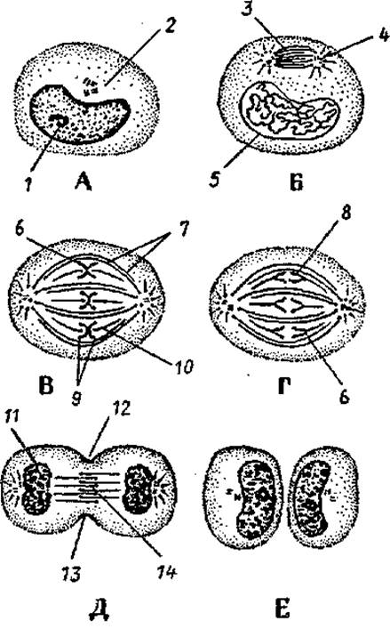
Fig. 4. Stages of mitosis. Shown are chromatin Condensation to form chromosomes, mitotic spindle formation, and the equal distribution of chromosomes and centrioles between the two daughter cells.
A — interphase, B — prophase, C — metaphase, D — anaphase, E — telophase, F — late telophase.
1 — nucleolus, 2 — centrioles, 3 — mitotic spindle, 4 — aster, 5 — nuclear envelope, 6 — kinetochore, 7 — polar (non-kinetochore) microtubules, 8, 9 — chromosomes, 10 — kinetochore microtubules, 11 — nuclear formation, 12 — Cleavage furrow, 13 — Actin filament bundle, 14 — midbody (residual body)
During prophase, the nucleolus gradually disassembles, and the centrioles migrate to opposite poles of the cell.
During metaphase, the nuclear envelope breaks down, and spindle fibers extend toward the poles while maintaining connection with the equatorial region of the cell. The structures of the Endoplasmic reticulum and the Golgi apparatus break down into small vesicles, which, along with Mitochondria, are distributed into both halves of the dividing cell. At the end of metaphase, each chromosome begins to split longitudinally into two new daughter chromosomes.
During anaphase, the chromosomes separate from each other and migrate toward the cell poles at a rate of up to 0.5 µm/min.
During telophase, the chromosomes that have reached the cell poles decondense, reverting to chromatin, and RNA METABOLISM/31.html">Transcription (production) begins. The nuclear envelope and nucleolus re-form, and the membrane structures of the future daughter cells rapidly take shape. A cleavage furrow appears at the cell's equator and deepens, dividing the cell into two daughter cells.
Review and Self-Assessment Questions:
1. Name the Structure/83.html">Structural elements of a cell.
2. What Functions does a cell perform?
3. List the membrane-bound and non-membrane-bound Organelles of the cell and state their functions.
4. What elements make up the Cell Nucleus, and what functions does it perform?
5. What types of cell junctions exist?
6. What is the cell cycle, and what periods (phases) are distinguished within it?
7. What is meiosis, and how does it differ from mitosis?
Last update: 10/08/2026
Editorial and Educational Adaptation: This material has been compiled based on the primary/original source text. The project team performed an editorial review, corrected technical inaccuracies, structured sections, and adapted the content for an educational format.
What was processed:
- elimination of formatting defects (OCR errors, structural breaks, corrupted characters);
- editorial organization of content;
- standardization of terminology in accordance with academic sources;
- verification of factual statements against the original source text.
All mentions of the author, publication year, and origin of the primary text have been preserved in accordance with the source.