PLANT MORPHOLOGY - T. A. Sautkina - 2012
CHAPTER 8. POLLINATION AND FERTILIZATION IN ANGIOSPERMS. EMBRYO AND ENDOSPERM DEVELOPMENT
8.2. The Process of Double Fertilization in Angiosperms
The time interval between Pollination and Fertilization varies among different angiosperm species. The shortest interval has been observed in members of the Asteraceae family, ranging from 15–30 minutes, as in kok-saghyz (Taraxacum kok-saghyz), to 1.5 hours in most species. In some members of the Betulaceae family, this period ranges from 1 to 4 months, and in Fagaceae (some oak species), from 12 to 14 months.
Most commonly, the pollen tube is directed toward the micropylar pole of the ovule, although it may also enter the ovule through other regions. When the pollen tube enters through the micropyle, this mode of entry is termed porogamy; through the chalaza, chalazogamy; and through the lateral Zones of the integuments and nucellus, mesogamy. Porogamy is considered the most ancient type of pollen tube entry, characteristic of both angiosperms and gymnosperms. Chalazogamy and mesogamy are specialized modes of entry discovered and described in A number of woody plants (Casuarina, birch, walnut). Regardless of how the pollen tube enters the ovule, it meanders through the ovule Tissues until it reaches the micropylar pole near the micropyle.
The pollen tube enters the embryo sac through one of the synergids, which degenerates in the process. The tip of the pollen tube positions itself near the egg apparatus and bursts. The Contents of the pollen tube are discharged in the immediate vicinity of the egg Cell. The Cytoplasm of the pollen tube merges with the cytoplasm of the embryo sac located near the egg apparatus. The vegetative Nucleus disintegrates, while the two sperm Cells lie in the cytoplasm near the egg cell for some time before beginning to move apart. One sperm cell moves toward the egg cell, while the second moves toward the polar nuclei. The exact mechanism of sperm movement within the embryo sac remains incompletely understood. S. G. Navashin believed that sperm cells exhibit enantiomorphism, meaning they are mirror-symmetric. It is precisely due to this mirror Symmetry that the sperm cells actively repel one another and move within the cavity of the embryo sac.
E. N. Gerasimova-Navashina held a different view. According to her hypothesis, the sperm cells enter the embryo sac in a state of unfinished mitotic division, and it is this residual mitotic activity that drives their independent locomotion. Other hypotheses have also been proposed.
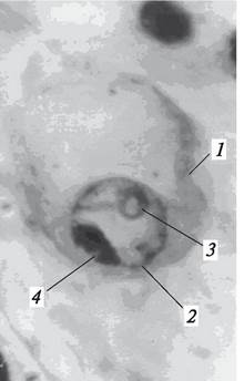
The fertilization process begins with the fusion of a sperm cell with the egg cell. This fusion occurs as follows: the sperm penetrates the cytoplasm of the egg cell, where it sheds its own cytoplasm, which then mixes with the egg cell's cytoplasm. Subsequently, the naked sperm nucleus moves toward the egg nucleus, penetrates it (Fig. 194), and fuses with it. Only after the sperm nucleus has fused with the egg nucleus can the fertilization of the egg cell (amphimixis) and The formation of the zygote be considered complete. The diploid chromosome number is thus restored in the zygote nucleus.
Class="center">Fig. 194. Stage of double fertilization: sperm cell within the egg nucleus: 1—egg cell; 2—egg nucleus; 3—nucleolus of the egg nucleus; 4—sperm cell
There are three recognized types of egg cell fertilization: premitotic, postmitotic, and intermediate (Fig. 195). In the premitotic type, the nuclei of the Gametes fuse while in a resting state. Division of the zygote nucleus begins only after it emerges from this dormant state. In the postmitotic type, the fusing egg and sperm nuclei are in an active state, meaning THE PROCESS OF mitotic division has already initiated within them and culminates in nuclear fusion, leaving the zygote nucleus effectively in prophase of mitosis. In the intermediate type, fusion occurs when one of the nuclei—most frequently the sperm nucleus—has begun dividing, while the egg nucleus remains quiescent. Division of the zygote nucleus continues after nuclear fusion is complete.
Fig. 195. Types of egg-sperm fusion (fertilization types according to Gerasimova-Navashina, 1961, with modifications): A—premitotic; B—intermediate; C—postmitotic: 1—egg cell; 2—egg nucleus with nucleolus; 3—sperm cell in the egg cytoplasm; 4—onset of sperm mitosis preceding fusion with the resting egg nucleus; 5—fusion of the sperm with the egg nucleus (amphimixis); 6—zygote formation; 7—onset of mitosis in the zygote nucleus; 8—entry of the sperm in a mitotic state into the egg nucleus; 9—onset of mitosis in the egg nucleus; 10—fusion of the sperm and egg nucleus in a mitotic state, zygote formation; 11—sperm and egg nucleus in a resting state; 12, 13—onset of fusion between dividing sperm and egg nuclei, zygote formation

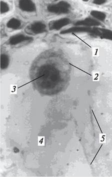
Following fertilization, the shape of the fertilized egg cell changes. While the unfertilized egg cell is pear-shaped, the zygote becomes spherical (Fig. 196). The egg cell contains a single nucleolus, whereas the zygote nucleus may contain several nucleoli. Cytologists believe that the number of nucleoli in the zygote nucleus can indicate the number of sperm cells that penetrated the egg cell.
Fig. 196. Zygote within the cavity of the embryo sac: 1—degenerating nucellar cells; 2—zygote; 3—zygote nucleus; 4—embryo sac cavity; 5—cytoplasmic strands of the embryo sac
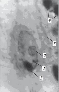
The second sperm cell approaches the polar nuclei or the central nucleus (the Nucleus of the central cell) of the embryo sac and begins to fuse with them (Fig. 197). This fusion process can take various forms. If the polar nuclei are separate, the sperm may first fertilize the micropylar nucleus and subsequently fuse with the chalazal one. Alternatively, the order of fusion may be reversed: the sperm may enter the chalazal nucleus, after which the fertilized nucleus fuses with the micropylar one. If a unified central nucleus of the embryo sac has formed prior to fertilization, the fusion of the sperm with the central nucleus proceeds similarly to the fertilization of the egg cell. Thus, following the fertilization of the polar nuclei or the central nucleus of the embryo sac, a polyploid primary endosperm nucleus or a polyploid (triploid in the simplest case) primary endosperm cell containing a primary nucleus is formed. In certain variant types of embryo sacs, the primary endosperm nucleus is highly polyploid. For instance, the embryo sac of Peperomia muna contains from 7 to 13 polar nuclei, upon fertilization of which a very large primary endosperm nucleus is formed, containing from 8n to 14n.
Fig. 197. Stage of the double fertilization process: sperm cell in the central nucleus of the embryo sac: 1—central nucleus of the embryo sac (nucleus of the central cell of the embryo sac); 2—nucleolus of the central Cell Nucleus of the embryo sac; 3—sperm cell; 4—degenerating nucellar cells; 5—cytoplasmic strands of the embryo sac
The phenomenon of fertilization in angiosperms was discovered by Professor S. G. Navashin of Kyiv University in 1898 and was termed by him the process of double fertilization. This discovery served as a major impetus for investigating this phenomenon in angiosperms, particularly those of significant practical importance.
Following fertilization, the zygote and the primary endosperm nucleus enter a period of dormancy before their subsequent development begins. The zygote develops into the embryo
of the new Organism, while the primary endosperm nucleus (or primary cell) gives rise to a specialized nutritive tissue known as the endosperm. The process whereby an embryo develops from a zygote formed through fertilization is termed amphimixis. However, in a number of plants, the seed embryo develops without fertilization—apomictically.
Last update: 07/08/2026
Editorial and Educational Adaptation: This material has been compiled based on the primary/original source text. The project team performed an editorial review, corrected technical inaccuracies, structured sections, and adapted the content for an educational format.
What was processed:
- elimination of formatting defects (OCR errors, structural breaks, corrupted characters);
- editorial organization of content;
- standardization of terminology in accordance with academic sources;
- verification of factual statements against the original source text.
All mentions of the author, publication year, and origin of the primary text have been preserved in accordance with the source.