Anatomy of the Vessels and Nerves of the Trunk (Angioneurology) - Chornokulskyi S. T. 2011
Autonomic (Vegetative) Nervous System
Peripheral Division of the Autonomic Nervous System
The Phenomenon of Cutaneous Sensory Disorders in Visceral Diseases
The phenomenon of cutaneous sensory disorders (hyperesthesia, paresthesia, pain) in visceral diseases (HEAD's zones)
Pathological processes in visceral Organs are known to be accompanied by changes in sensitivity (hyperesthesia, paresthesia, pain) in specific Skin areas. This is the so-called phenomenon of referred pain (radiating to the skin of the left scapular region, the left arm, and the fifth digit of the hand in angina pectoris; to the interscapular skin region in gastric ulcer; and to the right inguinal skin region in appendicitis). These are dermatomes that correspond to the Spinal Cord segments (down to the nuc. intermediocentralis) receiving impulses from the diseased organ. It is at the level of the second neuron's body that numerous associative connections occur between the axons of the receptor neuron and the sensory nuclei of the dorsal horns of the spinal cord. Specifically, connections with the nuclei proprii are transmitted to the Cerebral Cortex via the ascending Pathways of the cutaneous analyzer and are perceived as pain, whereas those with the substantia gelatinosa are perceived as paresthesias and hyperesthesias. Such skin segments are termed Head's zones. By influencing the skin of these zones (via acupuncture or physiotherapeutic Procedures), one can thus affect the Internal Organs, which has been a part of the arsenal of Chinese and Tibetan medicine since ancient times.
Class="center">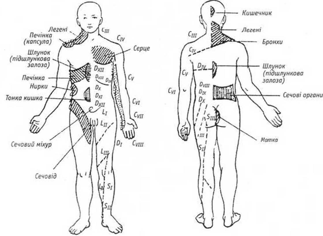
Fig. 66. Skin areas (Head's zones) in visceral pathology
However, it must also be taken into account that the afferent link of the interoceptive analyzer, In addition to visceral nerves (mediated by receptor Neurons of the g. spinale), is supplied by the vagus and phrenic nerves. Therefore, hyperesthesia and pain in the skin of the occiput during colitis are associated with associative connections of the nuc. solitarius of the 10th cranial nerve with the sensory nuclei of the dorsal horns of the cervical spinal cord, while painful facial skin areas are linked via connections of the 10th cranial nerve with the nuc. spinalis n. trigemini. Reverse connections are also possible, where skin irritation is reflected as pain in the internal organs.
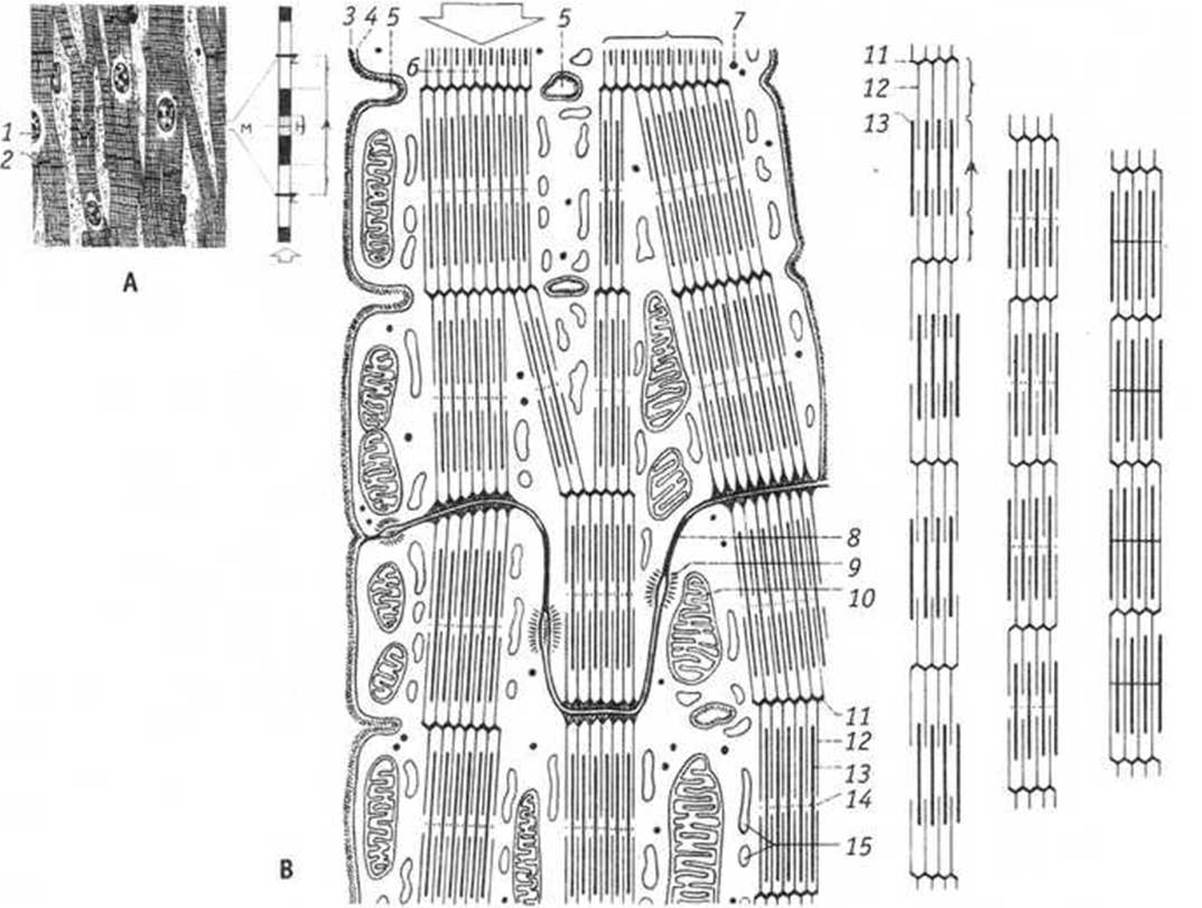
Cardiac Muscle under: A - light Cell/15.html">Microscopy;
B - Electron microscopy (after W. Kahle et al.)
A. 1 — cardiomyocyte Nucleus
2. Intercalated discs (disci intercalati).
B. 3. Basement membrane.
4. Plasma Membrane.
5. T-tubules (tubules).
6. Myofibrils.
7. Glycogen.
8. Desmosome.
9. Gap junction (nexus).
10. Mitochondrion (sarcosome).
11. Myofibril attachment site.
12. Actin filaments.
13. Myosin filaments.
14. Interfibrillar protein network.
15. Sarcotubular network
Last update: 11/08/2026
Editorial and Educational Adaptation: This material has been compiled based on the primary/original source text. The project team performed an editorial review, corrected technical inaccuracies, structured sections, and adapted the content for an educational format.
What was processed:
- elimination of formatting defects (OCR errors, structural breaks, corrupted characters);
- editorial organization of content;
- standardization of terminology in accordance with academic sources;
- verification of factual statements against the original source text.
All mentions of the author, publication year, and origin of the primary text have been preserved in accordance with the source.