Human Biochemistry, Volume 2 - Murray R. 1993
Special Topics
Contractile and Structural Proteins
Muscle
Muscles serve as the primary biochemical transducers of potential (chemical) energy into kinetic (mechanical) energy. Muscle tissue ranks first in volume among all human Tissues, accounting for slightly less than 25% of total body weight at birth, over 40% in middle-aged adults, and just under 30% in the elderly.
The efficient conversion of chemical energy into mechanical work requires several conditions to be met: 1) a continuous supply of chemical energy must be ensured. In vertebrate muscles, chemical Energy is stored in molecules of ATP and creatine phosphate; 2) mechanisms must exist to regulate mechanical activity—specifically, the velocity, duration, and force of contraction in the case of muscles; 3) the conversion process must remain under operator control, a function fulfilled here by The Nervous system; 4) for the energy-transducing "machine" to be used repeatedly, a mechanism is required to restore the system to its initial state.
A muscle can be compared to a machine that can "pull" but not "push"; consequently, every muscle must be subject to the antagonistic action of another muscle group or some other force, such as gravity or elastic recoil.
Three Types of Muscles exist in the vertebrate body: skeletal, cardiac, and smooth. Skeletal and cardiac muscles exhibit cross-striations under microscopic examination, whereas smooth muscles lack such striations. While skeletal muscles are under voluntary nervous control, cardiac and smooth muscles function involuntarily.
Class="center">
Fig. 56.1. Structure of striated muscle. (Drawing by Sylvia Colard Keene. Reproduced, with permission, from Bloom W., Fawcett D. W. A Textbook of Histology, 10th ed. Saunders, 1975.)
Striated Muscle consists of multinucleated Cells (muscle fibers) enclosed by an electrically excitable membrane, the sarcolemma. Microscopic examination of an individual muscle Cell, which may extend the entire length of the muscle, reveals a bundle of numerous parallel myofibrils embedded in an intracellular fluid known as sarcoplasm. This fluid contains Glycogen, high-energy compounds (ATP and phosphocreatine), and glycolytic Enzymes.
The sarcomere is the functional unit of a muscle. Sarcomeres are arranged sequentially along the axis of the fibril, repeating every 1500–2300 nm (Fig. 56.1). Electron Microscopy of a myofibril reveals an alternation of dark and light bands (A and I bands). The central region of the A band (the H zone) appears less dense than the rest of the band. The I band is bisected by a very dense and narrow Z line. These structural details of the muscle are illustrated in Fig. 56.2.
The striations visible in muscles under a Light Microscope result from a high degree of Organization, wherein most muscle cells align such that their sarcomeres lie in parallel (Fig. 56.1).
Examination of myofibril cross-sections using an Electron microscope demonstrates that each myofibril is composed of Two Types of longitudinal filaments. The first type ("thick" filaments)
are confined to the A band, are composed primarily of the protein Myosin, measure approximately 16 nm in diameter, and form a hexagonal array in cross-section (Fig. 56.2). The second type of filaments ("thin" filaments) occupies the I band, extends into the A band, but does not reach its H zone (Fig. 56.2). The diameter of thin filaments is approximately 6 nm. They contain the Proteins Actin, Tropomyosin, and troponin. Within the A band, thin filaments are arranged around each thick (myosin) filament in a second hexagonal array. Thus, each thin filament occupies a symmetrical position between three thick filaments, and each thick filament is symmetrically surrounded by six thin filaments (Fig. 56.2).
Thick and thin filaments interact via cross-bridges spaced at 14 nm intervals along the thick filament. As shown in Fig. 56.2, the cross-bridges, or "heads," of the thick filaments exhibit opposite polarities at the two ends of the filament. These polar ends are separated by a 150 nm central segment (the M band) devoid of projections.
During Muscle contraction, the lengths of the thick and thin filaments remain unchanged, but the H zone and I bands shorten; consequently, the interdigitating filaments must slide past one another. The tension developed during muscle contraction is proportional to the degree of filament overlap and, hence, to the number of cross-bridges. The "HEAD" of each cross-bridge is connected to the thick filament by a flexible fibrous segment that can bend to adjust the spacing between the filaments.

Fig. 56.2. Filament arrangement in striated muscle. A. Relaxed muscle. B. Contracted muscle.
Fresh muscle fibers consist of 75% Water and contain over 20% protein by mass. The two major muscle proteins are Actin and myosin.
Monomeric (globular) actin, or G-actin, is a globular protein with a Molecular Weight of 43,000, accounting for 25% of the total muscle protein mass. At physiological Ionic strength and in the presence of magnesium, G-actin undergoes non-covalent polymerization to form an insoluble double-helical filament termed F-actin (Fig. 56.3). The F-actin filament has a diameter of 6–7 nm with repeating structural units every 35.5 nm. Neither G- nor F-actin possesses catalytic activity.
Striated muscles contain four additional proteins that make minor contributions to the mass of muscle tissue yet perform vital Functions. Tropomyosin is an elongated, strand-like molecule composed of two chains, α and β, which lies alongside F-actin within the groove between the two polymer strands (Fig. 56.3). This protein is present in all muscles and similar structures. A characteristic feature specific to striated muscles is the presence of a troponin system comprising three distinct proteins. Troponin T (TnT), like the other two troponin components, binds to tropomyosin (Fig. 56.3). Troponin I (TnI) inhibits the interaction between F-actin and myosin and also binds to other troponin components. Troponin C (TnC) is a calcium-binding protein whose Primary and secondary structures, as well as function, are entirely analogous to those of calmodulin, a protein widely distributed in nature. Both TnC and calmodulin bind four Calcium Ions per protein molecule and have a molecular weight of 17,000. The thin filament of striated muscle is composed of F-actin, tropomyosin, and the three troponin components: TnC, TnI, and TnT (Fig. 56.3). Tropomyosin and the troponin complex repeat at intervals of 38.5 nm.
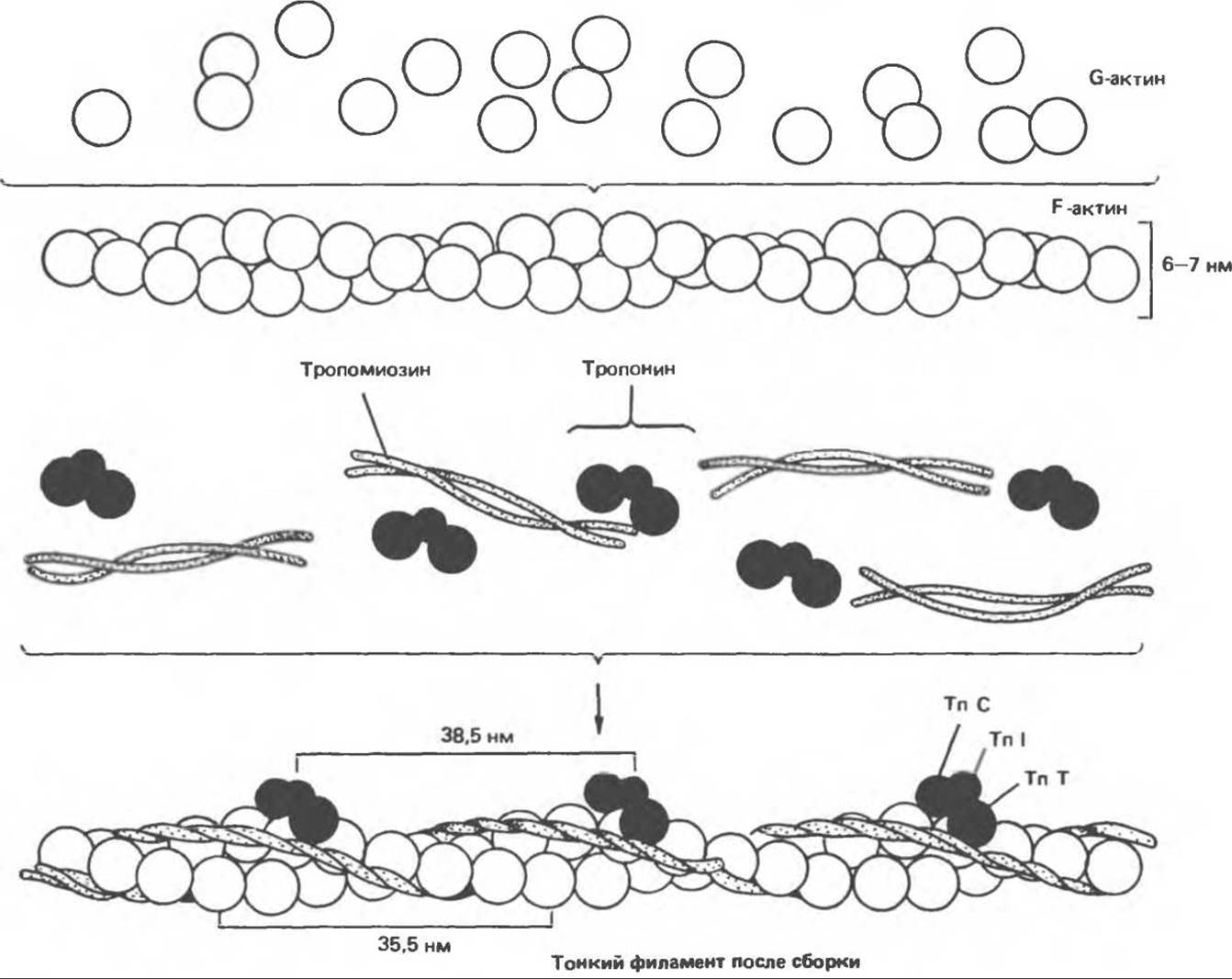
Fig. 56.3. Schematic representation of the thin filament. The spatial configuration of the three major protein components—actin, tropomyosin, and troponin—is shown.
Myosin constitutes 55% of muscle protein by mass and forms the thick filaments. It is an asymmetric hexamer with a molecular weight of 460,000. Myosin comprises a fibrous region consisting of two intertwined helices, each terminating in a globular "head" at one end (Fig. 56.4). The hexamer includes one pair of heavy chains (molecular weight 200,000) and two pairs of light chains (molecular weight 15,000–27,000). Skeletal Muscle myosin exhibits ATP-hydrolyzing (ATPase) activity and binds to the insoluble molecule F-actin.
Most knowledge regarding myosin has been derived from studying the products of its partial Hydrolysis. Treatment of myosin with Trypsin yields two fragments known as meromyosins. Light meromyosin (LMM) consists of aggregated, insoluble α-helical fibrils (Fig. 56.5). It lacks ATPase activity and does not bind to F-actin.
Heavy meromyosin (HMM) is a soluble protein with a molecular weight of 340,000 that contains both fibrous and globular domains (Fig. 56.5). It possesses ATPase activity and binds to F-actin. Hydrolysis of HMM with Papain produces two subfragments, S-1 and S-2. S-2 has a fibrous structure, exhibits no ATPase activity, and does not bind F-actin.
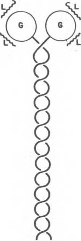
Fig. 56.4. Diagram of a myosin molecule with two intertwined a-helices (fibrillar domain), globular region (G), and light chains (L).
S-1 has a molecular weight of 115000, exhibits ATPase activity, and in the absence of ATP binds to actin, capping it with "arrowheads" (Fig. 56.6). Although both S-1 and LMM themselves possess ATPase activity, The addition of F-actin increases this activity by 100–200 fold. It has been shown that F-actin dramatically accelerates the release of the reaction products of myosin ATPase—ADP and inorganic phosphate. Thus, although F-actin itself does not affect ATP hydrolysis, its ability to stimulate product release from the ATPase reaction results in a significant increase in the overall rate of catalysis.
a-Actinin is a protein molecule found in the Z-line region, to which the ends of F-actin molecules of thin filaments attach (Fig. 56.2).
Molecular Function of Muscle

Fig. 56.5. Enzymatic Cleavage of myosin. HMM — heavy meromyosin; LMM — light meromyosin; S-1 — fragment 1; S-2 — fragment 2.

Fig. 56.6. Formation of "arrowhead" structures upon binding of actin filaments to myosin S-1 fragments. (Courtesy of Professor James Spudich, Stanford University.)
The question of the relationship between muscle Structure and function can be framed in biochemical terms as follows: how does ATP hydrolysis result in macroscopic movement? As noted above, muscle contraction consists of cycles of attachment and detachment of the globular myosin "head" to and from the F-actin filament. Attachment is accompanied by an alteration in the actin-myosin interaction, causing the actin and myosin filaments to slide past one another. The energy for this sliding is supplied by ATP hydrolysis. The hydrolysis of ATP by myosin ATPase is dramatically accelerated when the myosin "head" binds to F-actin. The biochemical cycle of muscle contraction consists of five stages (Fig. 56.7).
1) The myosin head on its own can hydrolyze ATP to ADP and inorganic phosphate, but fails to release the hydrolysis products. Consequently, this process is stoichiometric rather than catalytic in nature.
2) The myosin head, containing bound ADP and inorganic phosphate, can rotate freely through wide angles and (upon reaching the appropriate position) bind to F-actin, forming an angle of approximately 90° with the fibril axis.
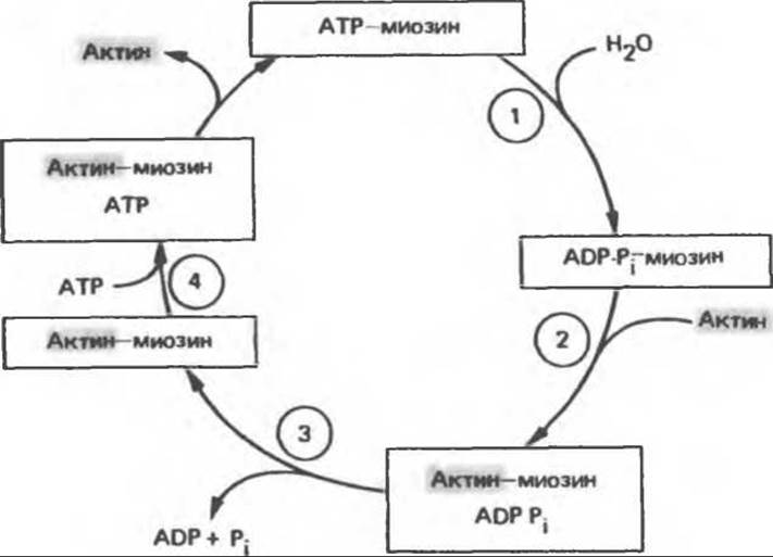
Fig. 56.7. ATP hydrolysis drives the actin-myosin association and dissociation cycle through the five reactions described in the text; Pi — inorganic phosphate. (Modified from Stryer L. Biochemistry, 2nd ed. Freeman, 1981.)
3) This interaction triggers the release of ADP and inorganic phosphate from the actin-myosin complex. Because the Actomyosin bond has its lowest energy at a 45° angle, myosin changes its angle relative to the filament axis from 90° to approximately 45°, pulling the actin filament (by 10–15 nm) toward the center of the sarcomere.
4) A new ATP molecule binds to the myosin-F-actin complex.
5) The myosin-ATP complex exhibits a low affinity for actin, causing the myosin (ATP) head to detach from F-actin. This final stage represents actual relaxation, which thus directly depends on ATP binding to the actin-myosin complex. ATP is subsequently rehydrolyzed by the myosin head without releasing ADP and inorganic phosphate, and the cycle begins anew.
Thus, ATP detaches the myosin head from the thin filament and serves as the driving force for contraction. The efficiency of this contraction is about 50%, whereas the efficiency of an internal combustion engine is less than 20%.
Regulation of Muscle Contraction and Relaxation
The contraction of any muscle proceeds According to the general mechanism described above. Muscle fibers from different organisms, and even from different Tissues of the same Organism, may employ distinct molecular mechanisms to regulate contraction and relaxation. Notably, Ca2+ ions play a pivotal regulatory role in all cases. There are two primary mechanisms of muscle contraction regulation: actin-linked and myosin-linked.
Actin-linked Regulation
Actin-linked regulation is characteristic of vertebrate striated muscles—both skeletal and cardiac. According to the general mechanism discussed above, ATP is the only potentially limiting factor in the muscle contraction cycle. Skeletal muscles are inhibited at rest and disinhibited upon the activation of contraction. In striated muscles, this inhibitory role is performed by the troponin system, which is associated with tropomyosin and F-actin in the thin filaments (Fig. 56.3). In the absence of the tropomyosin-troponin system, The regulation of striated muscle contraction (or ATPase activity as a biochemical indicator of contraction) does not occur. As noted above, tropomyosin is localized within the groove of F-actin, while the three troponin components—TnT, TnI, and TnC—are bound to the F-actin-tropomyosin complex. TnI prevents the attachment of the myosin head to the corresponding binding site on F-actin, either by altering the conformation of F-actin (via tropomyosin molecules) or simply by shifting ("rotating") tropomyosin into a position that blocks the myosin head binding sites on F-actin. In either case, the activation of myosin ATPase mediated by this binding is prevented. Consequently, the TnI system blocks the contraction cycle at step 2 of the scheme shown in Fig. 56.7. This underlies the inhibited state of a relaxed striated muscle.
Muscle contraction is mediated by Ca2+. In the sarcoplasm of a resting muscle, the calcium concentration ranges from 10-7 to 10-8 mol/L. Calcium is sequestered into the sarcoplasmic reticulum via Active Transport involving a Ca-binding protein called calsequestrin. The sarcomere is surrounded by an excitable membrane with transverse tubules extending toward the sarcoplasmic reticulum. Upon excitation of the sarcomere membrane—for example, through the interaction of acetylcholine receptors with acetylcholine—Ca2+ is rapidly released from the sarcoplasmic reticulum into the sarcoplasm, raising its concentration to 10-5 mol/L. The Ca2+-binding sites on TnC within the thin filament rapidly become saturated with Ca2+. The TnC-4Ca2+ complex interacts with TnI and TnT, modulating their interaction with tropomyosin. Depending on this, tropomyosin either simply dissociates or alters the conformation of F-actin, enabling the ADP-Pi-myosin head to interact with F-actin and initiating the contractile cycle.
Relaxation occurs when 1) the Ca2+ concentration in the sarcoplasm drops below 10-7 mol/L due to its uptake by the sarcoplasmic reticulum; 2) the TnC-4Ca2+ complex loses its bound Ca2+; 3) troponin interacts with tropomyosin to inhibit further interaction between the myosin head and F-actin; and 4) in the presence of ATP, the myosin heads detach from F-actin, resulting in relaxation. Thus, Ca2+ regulates muscle contraction via an allosteric mechanism mediated by TnC, TnI, TnT, tropomyosin, and F-actin within the muscle.
In cardiac muscle, extracellular fluid serves as the primary source of Ca2+ ions for excitation. If Ca2+ is absent from the extracellular fluid, cardiac contraction ceases within a minute, whereas skeletal muscle can continue to contract for hours under such conditions.
The depletion of ATP in the sarcoplasm leads to the following consequences: 1) the Ca2+ pump of the sarcoplasmic reticulum can no longer maintain a low Ca2+ concentration in the sarcoplasm, thereby stimulating the interaction between myosin heads and F-actin; 2) the ATP-dependent detachment of myosin heads from F-actin fails to occur, resulting in rigor mortis.
Muscle contraction does not operate on an "all-or-none" basis, as the reader might initially assume. Rather, it represents a delicate dynamic equilibrium between the attachment and detachment processes of myosin heads to and from F-actin. The system is subject to complex regulatory influences from the nervous system.
Myosin Regulation of Contraction
As noted above, actin, myosin, and tropomyosin are present in all types of muscles, whereas the troponin system is found exclusively in vertebrate striated muscles. Consequently, the mechanisms regulating contraction must differ among various contractile systems.
Although the molecular structures of smooth muscles closely resemble those of striated muscles, the arrangement of their sarcomeres does not produce the characteristic striated pattern. Similar to skeletal muscles, smooth muscles contain $\alpha$-actinin and tropomyosin molecules, but they lack the troponin system; furthermore, the light chains of smooth muscle myosin differ from their counterparts in striated muscles. Nevertheless, smooth muscle contraction, much like that of striated muscle, is regulated by Ca2+.
When smooth muscle myosin binds to F-actin in the absence of other muscle proteins, such as tropomyosin, the resulting complex exhibits virtually no detectable ATPase activity. This contrasts sharply with the interaction between striated muscle myosin and F-actin, which displays high ATPase activity. Smooth muscle myosin contains a specific light chain (the p-light chain) that prevents myosin heads from binding to F-actin. For this light chain to permit the activation of myosin ATPase upon interacting with F-actin, it must first undergo phosphorylation. The phosphorylation of the p-light chain triggers the association-dissociation cycles in the smooth muscle contractile system.
The sarcoplasm of smooth muscles contains a calcium-dependent myosin light chain kinase. The activation of this enzyme by calcium requires the binding of its 105,000-dalton subunit to calmodulin·4Ca2+ (Fig. 56.8). Activated by calmodulin·4Ca2+, the light chain kinase phosphorylates the p-light chain, which thereupon ceases to inhibit the interaction between myosin and F-actin. Thus, the contractile cycle is initiated (Fig. 56.8).

Fig. 56.8. Calcium Regulation of smooth muscle contraction. (Adapted from Adelstein R. S., Eisenberg R. Regulation and kinetics of actin myosin ATP interaction. Annu. Rev. Biochem., 1980, 49, 921.)
Smooth muscle relaxation occurs when: 1) the concentration of Ca2+ ions in the sarcoplasm drops below 10-7 mol/L; 2) Ca2+ dissociates from calmodulin, which in turn detaches from the myosin light chain kinase, thereby inactivating it; 3) no further phosphorylation of the p-light chain takes place, and light chain protein phosphatase—which is constitutively active and calcium-independent—cleaves the previously attached phosphates from the p-light chain; 4) the dephosphorylated myosin p-light chain inhibits the binding of myosin heads to F-actin and suppresses ATPase activity; 5) in the presence of ATP, the myosin heads detach from F-actin, while re-attachment is prevented by the presence of the dephosphorylated p-light chain in the system. As a result of these events, muscle relaxation ensues.
Table 56.1 summarizes and compares data regarding the regulation of actin-myosin interaction (activation of myosin ATPase) in striated and smooth muscles.
Myosin light chain kinase is not a direct target of cAMP-mediated activation. However, the standard cAMP-activated protein kinase (see Chapter 44) can phosphorylate this enzyme (rather than the p-light chain directly). Phosphorylated myosin light chain kinase exhibits a significantly lower affinity for calmodulin-Ca2+ and is therefore less sensitive to activation. Accordingly, elevated cAMP levels attenuate the contractile response of smooth muscles to increases in sarcoplasmic Ca2+. This molecular mechanism can account for the relaxing effect of $\beta$-adrenergic stimulation on smooth muscle. Phenothiazines, which are widely used as antipsychotic agents, bind to calmodulin and prevent its interaction with calcium-dependent enzymes, thereby also inducing smooth muscle relaxation.
The striated muscles of Mollusks, such as the sea scallop, feature a myosin-regulated contraction system. Both scallop myosin and F-actin, similarly to these proteins in smooth muscle, lack ATPase activity due to the inhibitory Properties of the scallop's "regulatory" light chain. The inhibition of the actin-myosin interaction in the sea scallop is relieved by the direct binding of Ca2+ to a specific site on the myosin molecule. This regulatory mechanism does not require Covalent Modification of myosin and/or the addition of specific proteins such as calmodulin or TnC.
Table 56.1. Actin-Myosin Interactions in Striated and Smooth Muscles
|
Striated muscles |
Smooth muscles (and non-muscle cells) |
|
|
Muscle filament proteins |
Actin |
Actin |
|
Myosin (hexamer) |
Myosin (hexamer) 1) |
|
|
Tropomyosin |
Tropomyosin |
|
|
Troponin (TnI, TnT, TnC) |
||
|
Spontaneous interaction of F-actin with isolated myosin (spontaneous activation of myosin ATPase by F-actin) |
Present |
Absent |
|
Inhibitor of F-actin-myosin interaction (inhibitor of F-actin-dependent ATPase activation) |
Troponin system (TnI) |
Unphosphorylated regulatory myosin light chain |
|
Contraction is activated by |
Ca2+ |
Ca2+ |
|
Direct action of Ca2+ |
4Ca2+ bind to TnC |
4Ca2+ bind to calmodulin |
|
Action of Protein-bound Ca2+ |
TnC∙4Ca2+ overcomes the inhibitory effect of TnI on the F-actin-myosin interaction (enables ATPase activation by F-actin) |
Calmodulin-4Ca2+ activates myosin light chain kinase, which phosphorylates the regulatory myosin light chain. The phosphorylated regulatory light chain ceases to inhibit the F-actin-myosin interaction (enables ATPase activation by F-actin) |
1) Myosin light chains differ between striated and smooth muscles.
Phosphorylation of Muscle Proteins
Phosphorylation of the smooth muscle myosin light chain relieves its inhibitory effect on the actin-myosin interaction, thereby triggering the contractile cycle. Thus, phosphorylation is required to initiate the actin-myosin interaction in smooth muscles.
One pair of skeletal muscle myosin light chains can also undergo phosphorylation; however, this does not affect actin-activated myosin ATPase (unlike smooth muscle myosin). It is hypothesized that the phosphate groups on the myosin light chains may form a chelate with Ca2+ (bound to the tropomyosin-TnC-actin complex), thereby increasing The rate of cross-bridge formation between myosin heads and actin.
Emerging evidence suggests that phosphorylation of myosin heavy chains is a prerequisite for their assembly into thick filaments in skeletal muscle, smooth muscle, and non-muscle cells (see below).
TnI and the peptide component of the sarcoplasmic reticulum Ca2+ pump in cardiac muscle can be phosphorylated by cAMP-dependent protein kinase. There is a certain correlation between TnI phosphorylation and the enhanced cardiac contraction caused by catecholamines. This mechanism may account for the inotropic effect (increased contractility) of ß-adrenergic compounds on The Heart.
Muscle METABOLISM
ATP, which is required as a constant energy source for the muscle contraction-relaxation cycle, can be generated via Glycolysis, Oxidative Phosphorylation, creatine phosphate, or from two ADP molecules. ATP stores in skeletal muscle are rapidly depleted during contraction, lasting for less than a second of activity. In slow skeletal muscles, which possess significant O2 reserves in Myoglobin, Oxidative phosphorylation is the primary source of ATP regeneration. Fast skeletal muscles regenerate ATP predominantly through glycolysis.
Phosphagens, such as creatine phosphate, prevent the rapid depletion of ATP stores by providing a readily available high-energy phosphate required for the resynthesis of ATP from ADP. Creatine phosphate is formed from ATP and creatine during muscle relaxation when the demand for ATP is low. Creatine phosphorylation is catalyzed by creatine phosphokinase (CPK), a muscle-specific enzyme used in the Diagnosis of acute or chronic muscle disorders.
Skeletal muscle sarcoplasm contains large glycogen stores localized in granules adjacent to the I-band. The release of glucose from glycogen depends on the muscle-specific enzyme Glycogen phosphorylase (see Ch. 19). To ensure The production of glucose-6-phosphate required for glycolysis, glycogen phosphorylase b must be converted into its active form, phosphorylase a. This activation requires the phosphorylation of phosphorylase b by phosphorylase b kinase (see Ch. 19). The activation of phosphorylase b kinase, which also occurs via enzyme phosphorylation, is stimulated by Ca2+. Thus, Ca2+ not only triggers muscle contraction but also enhances the production of ATP, the energy source required for this process. Muscle glycogen phosphorylase b is absent in a specific myopathy known as McArdle's disease, which is a form of glycogen storage disease.
ATP in muscle tissue is also formed during oxidative phosphorylation, a process that requires a continuous supply of oxygen. Muscles characterized by a high oxygen demand due to a prolonged state of contraction (e.g., to maintain a specific posture) have The ability to store oxygen in myoglobin (see Ch. 6). Because Oxygen binds to the heme group in myoglobin, muscles containing myoglobin appear red, in contrast to white skeletal muscles, which lack it. Table 56.2 presents comparative data on some of The properties of fast (or white) and slow (or red) skeletal muscles.
Myoadenylate kinase, an enzyme present in muscles, catalyzes The formation of one molecule of ATP and one molecule of AMP from two molecules of ADP. This reaction, shown in Fig. 56.9, is coupled with the hydrolysis of ATP by myosin ATPase during muscle contraction. The figure also illustrates the relationship between various sources of ATP and its consumption during muscle contraction.
In humans, skeletal muscle protein serves as the primary energy reserve following fat. This accounts for the significant loss of muscle mass (especially in adults) observed during prolonged caloric deficiency.
The Study of in vivo tissue protein degradation is hindered by the fact that Amino Acids released during intracellular protein breakdown can be largely reutilized for Protein Synthesis within The Cell or transported to other Organs to participate in anabolic processes. However, actin and myosin are methylated after their peptide bonds are formed, yielding 3-methylhistidine. During the intracellular breakdown of actin and myosin, 3-methylhistidine is released and excreted in the urine. Administration of a label to rats or humans has demonstrated that the urinary excretion of this methylated amino acid serves as a reliable indicator of the rate of myofibrillar protein degradation in muscles. The fractional rate of muscle protein degradation in elderly individuals differs little from that in young people, but because muscle mass decreases with Aging, THE CONTRIBUTION OF this tissue to the overall age-related increase in whole-body protein degradation is reduced.
Table 56.2. Characteristics of fast and slow skeletal muscles
|
Fast skeletal muscle |
Slow skeletal muscle |
|
|
Myosin ATPase activity |
High |
Low |
|
Energy utilization |
High |
Low |
|
Color |
White |
Red |
|
Myoglobin |
Absent |
Present |
|
Contraction frequency |
High |
Low |
|
Contraction duration |
Short |
Long |
As noted above, skeletal muscles serve as the primary protein reserve in the body. They also exhibit high activity in degrading Certain amino acids and synthesizing others. In mammals, muscles are the main site of branched-chain Amino Acid Catabolism. Muscle tissue oxidizes leucine to CO2 and converts the carbon skeletons of aspartate, asparagine, glutamate, isoleucine, and valine into Tricarboxylic Acid Cycle intermediates. The ability of muscles to degrade branched-chain amino acids increases 3- to 5-fold during starvation and diabetes.
Muscles also synthesize and release large amounts of Alanine and glutamine. The synthesis of these compounds utilizes amino groups generated by The breakdown of branched-chain amino acids, which are then transferred to α-ketoglutarate and Pyruvate via Transamination reactions. The source of nearly all the pyruvate used for alanine synthesis is glycolysis derived from exogenous glucose. These reactions constitute the glucose-alanine cycle, in which muscle-derived alanine is utilized in hepatic Gluconeogenesis while simultaneously delivering amino groups to the Liver, where they are removed in the form of urea.
The carbon Skeleton of amino acids degraded and incorporated into The Tricarboxylic Acid Cycle in muscle tissue is converted mainly into glutamine and pyruvate, which is subsequently oxidized or converted to lactate. Thus, during starvation or in the postprandial period, most of the amino acids generated by muscle protein breakdown leave the muscle; exceptions include isoleucine, valine, glutamate, aspartate, and asparagine, which participate in the formation of glutamine, which is released by muscles and utilized by other tissues.
It has long been known that working muscle releases ammonia. As has now been established, the direct source of ammonia in skeletal muscle is AMP, which is deaminated to IMP by the action of adenylate deaminase. IMP can be reconverted to AMP via reactions utilizing aspartate and catalyzed by adenylosuccinate synthetase and adenylosuccinase (see Ch. 35).

Fig. 56.9. Multiple sources of ATP in muscle.
Last update: 06/08/2026
Editorial and Educational Adaptation: This material has been compiled based on the primary/original source text. The project team performed an editorial review, corrected technical inaccuracies, structured sections, and adapted the content for an educational format.
What was processed:
- elimination of formatting defects (OCR errors, structural breaks, corrupted characters);
- editorial organization of content;
- standardization of terminology in accordance with academic sources;
- verification of factual statements against the original source text.
All mentions of the author, publication year, and origin of the primary text have been preserved in accordance with the source.