Genetics - A. V. Sivolob 2008
Gene Expression
Protein Synthesis
The Ribosome and the Mechanism of Translation
The ribosome is a ribonucleoprotein complex composed of two subunits. The small subunit of a prokaryotic ribosome contains a single 16S rRNA molecule (ribosomal components are conventionally designated by their sedimentation coefficients in Svedberg units, S) and 21 ribosomal Proteins. The large subunit contains two rRNA molecules (23S and 5S) and 36 types of proteins. The eukaryotic ribosome contains a slightly larger 18S rRNA instead of 16S, two rRNAs (28S and 5.8S) instead of 23S, a 5S rRNA, and a greater number of proteins. The Structure of both types of Ribosomes and the principles of their operation are similar.
The synthesis of eukaryotic 18S, 5.8S, and 28S rRNAs takes place in the nucleolus, which is formed on tandem repeats of the corresponding rRNA Gene cluster (this cluster is repeated 100 to 1,000 times in various species). The primary METABOLISM/31.html">Transcription product contains three future rRNA fragments separated by spacers, meaning the cluster is transcribed as a single unit. rRNA Processing—the degradation of spacers and specific chemical modifications—is mediated by approximately 150 types of small nucleolar RNAs (much like Small nuclear RNAs participate in mRNA Processing, as discussed below). The genes for another type of rRNA, 5S, are also tandemly repeated elsewhere in The Genome; the 5S rRNAs are then transported to the nucleolus, where ribosomal proteins also arrive, and ribosomal subunit assembly takes place. The primary transcription product of prokaryotic rRNA genes contains regions corresponding to all three prokaryotic rRNAs as well as several future tRNAs. Partial degradation of the transcript yields mature molecules that interact with ribosomal proteins to form the two ribosomal subunits. The final assembly of the ribosome from its two subunits, in both PROKARYOTES AND EUKARYOTES, occurs during Translation initiation.
Ribosomal RNAs account for about 2/3 of the ribosome's mass and dictate its Structure and function. The polynucleotide chain of rRNA forms a multitude of Double helices that fold into a complex three-dimensional structure. Ribosomal proteins are located On the surface of the rRNA (and consequently on The surface of the ribosome), stabilizing its functionally active spatial Organization.
During ribosomal functioning, its small subunit interacts with mRNA. Working together, the two subunits create binding sites for three tRNA molecules (Fig. 2.4): the A-site, where aminoacyl-tRNA (aa-tRNA) binding occurs; the P-site, where peptidyl-tRNA (the tRNA attached to the growing peptide chain) interacts with the ribosome; and the E-site (from exit), where the deacylated tRNA moves before being released from the ribosome.
Information from the mRNA is read by the ribosome in the 5'- to 3'-direction, and the polypeptide chain is synthesized from the N- to the C-terminus. Translation begins with the initiation stage (Fig. 2.4), during which the ribosome recognizes the start codon that sets the beginning and the reading frame of the genetic message. The initiator aa-tRNA is simultaneously loaded onto this codon and into the P-site of the ribosome. The efficiency of these steps is ensured by a set of specific protein initiation factors.
The start codon is predominantly the Methionine codon AUG; accordingly, the initiator is always Met-tRNAiMet (the subscript "i" indicates that this is specifically the initiator methionine tRNA, meaning its structure differs from that of regular tRNAMet used for incorporating Met within a polypeptide chain). Thus, methionine is always the first amino acid (though it is typically cleaved off post-translationally).
Class="center">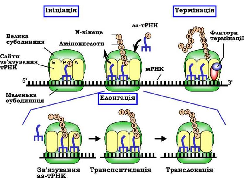
Fig. 2.4. Scheme of the translation process. The bottom part illustrates the events of a single elongation cycle
Subsequently, ribosome operation during Translation elongation involves sequential (triplet-by-triplet) reading of the mRNA information and the corresponding addition of Amino Acids to the polypeptide chain (Fig. 2.4). Each such step consists of three cyclically repeating operations (the elongation cycle). The cycle begins with the binding of aa-tRNA to the A-site. The ribosome ensures high Specificity in codon-anticodon interactions—only the tRNA cognate to the given codon is selected by the system. The positioning of aa-tRNA in the A-site is often accompanied by the dissociation of the deacylated tRNA, left over from the previous cycle, from the E-site.
The consequence of this binding is transpeptidation—The transfer of the peptidyl moiety from peptidyl-tRNA to The amino acid on the aa-tRNA. The catalytically active center responsible for transpeptidation resides on the large ribosomal subunit and is formed exclusively by ribosomal RNA. As a result of transpeptidation, the A-site holds a peptidyl-tRNA with a peptide elongated by one amino acid, while the P-site holds a deacylated tRNA.
The third operation, translocation, involves the movement of the ribosome by one codon along the mRNA (with the tRNA molecules remaining bound to their respective codons), after which the next elongation cycle begins. The efficiency and speed of the elongation cycle depend on two protein elongation factors.
When, following a regular elongation cycle (which turns out to be the last one), one of the three stop codons appears in the A-site, it is recognized by Translation termination factors, as no tRNA contains corresponding anticodons (Fig. 2.4). Termination factors ensure the release of the synthesized amino acid chain and prepare the ribosome for a new round of translation: the dissociation of the ribosomal subunits from each other and from the mRNA.
Last update: 11/08/2026
Editorial and Educational Adaptation: This material has been compiled based on the primary/original source text. The project team performed an editorial review, corrected technical inaccuracies, structured sections, and adapted the content for an educational format.
What was processed:
- elimination of formatting defects (OCR errors, structural breaks, corrupted characters);
- editorial organization of content;
- standardization of terminology in accordance with academic sources;
- verification of factual statements against the original source text.
All mentions of the author, publication year, and origin of the primary text have been preserved in accordance with the source.