Fundamentals of Molecular Biology. Part 2: Molecular Genetic Mechanisms - A. N. Ogurtsov 2011
Protein synthesis on ribosomes
Translation initiation
Much like METABOLISM/31.html">Transcription, the complex process of Translation can be divided into three stages: initiation, elongation, and termination. Let us examine translation in Eukaryotic Cells, keeping in mind that The Mechanism of translation is broadly similar across all organisms.
In most mRNAs, AUG serves as the start codon encoding Methionine. Therefore, the recognition of this codon is critical for establishing the correct reading frame for the entire mRNA.
Both in PROKARYOTES AND EUKARYOTES, There are two distinct methionine tRNAs:
1) tRNAiMet - capable of initiating Protein Synthesis,
2) tRNAMet - capable only of adding methionine to a growing polypeptide chain.
The same aminoacyl-tRNA synthetase (MetSR) aminoacylates both tRNAs with methionine. However, only Met-tRNAiMet (i.e., tRNAiMet with an attached activated methionine) can bind to a specific Active Site—the Y-site—on the small ribosomal subunit to initiate Polypeptide chain synthesis. Ordinary Met-tRNAMet, as well as other aminoacylated tRNAs, binds exclusively to another Active Site of the ribosome, the A-site.
Following the Termination of the preceding translation event, the dissociated 40S and 60S ribosomal subunits bind to the initiation factors eIF3 and eIF6, respectively, rendering them ready for the next round of translation (Figure 41).
In the designation "eIF", the letter "e" stands for eukaryotic factor, "I" denotes the initiation process, and "F" is an abbreviation for "factor".
Class="center">
Figure 41 - Binding of ribosomal subunits to initiation factors
During the first stage of translation, a complex is assembled from the ribosomal subunit, mRNA, and the activated initiator tRNA, which is correctly positioned at the start codon.
The translation preinitiation complex is formed when the (40S subunit-eIF3) complex binds to eIF1A and the ternary complex (Met-tRNAiMet-eIF2-GTP) (Figure 42, step 1) (GTP stands for guanosine triphosphate).
The Cell can regulate protein synthesis via phosphorylation of a Serine residue in the GTP-bound initiation factor eIF2. As a result, phosphorylated eIF2 is unable to exchange the used GDP for GTP and, consequently, cannot bind to Met-tRNAiMet, thereby inhibiting protein synthesis.
During translation initiation, the 5'-cap of the mRNA is bound by the eIF4E subunit of the eIF4 cap-complex.
The (mRNA-eIF4) complex then joins the preinitiation complex through the interaction between the eIF4G subunit and eIF3, forming the initiation complex (Figure 42, step 2).
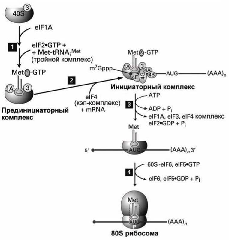
Figure 42 - Stages of translation initiation
Next, the initiation complex moves along—or scans—the mRNA, while the eIF4A subunit, which possesses helicase activity, unwinds the Introduction/11.html">Secondary Structure of the mRNA using the energy of ATP Hydrolysis.
Scanning halts when the anticodon of Met-tRNAiMet recognizes the start codon, which is the first AUG downstream from the 5'-end in most eukaryotic mRNAs (Figure 42, step 3).
Recognition of the start codon triggers the hydrolysis of the GTP bound to eIF2. This irreversible event prevents any further scanning of the mRNA.
The Selection of the initiating AUG codon is facilitated by its specific nucleotide environment, known as the Kozak sequence (named after Marilyn Kozak, who discovered it):

The first A and the last G in this sequence exert the strongest influence on the efficiency of translation initiation.
As soon as the small ribosomal subunit with its attached Met-tRNAiMet is correctly positioned at the start codon, the joining of the large (60S) subunit completes The formation of the 80S ribosome.
This step requires an additional factor, eIF5, and the hydrolysis of its bound GTP (Figure 42, step 4).
The fact that the joining of the large and small subunits is coupled to GTP hydrolysis makes it an irreversible step, meaning that ribosomal subunits do not dissociate until the entire mRNA has been translated and protein synthesis termination has occurred.
During elongation, the growing polypeptide remains attached to the P-site of the ribosome.
In most eukaryotes, the protein synthesis process described above begins approximately 100 NUCLEOTIDES downstream from the capped 5' end of the mRNA. However, some cellular mRNAs contain a so-called internal ribosome entry site, or IRES, located far from the 5' end. In addition, the translation of certain viral mRNAs lacking a 5' cap is initiated at the IRES through mechanisms of virus-host interaction in the infected cell that are not yet fully understood.
Apparently, the same translation initiation factors that promote ribosome scanning from the 5' end in search of an AUG codon are also required for recognizing AUG within an internal ribosome entry site, although the precise details of IRES identification remain unknown.
Preliminary evidence suggests that the IRES folds into a specific tertiary structure that binds to the third active site of the ribosome, the E-site, thereby properly positioning the ribosomal P-site opposite the internal AUG start codon.
Last update: 12/08/2026
Editorial and Educational Adaptation: This material has been compiled based on the primary/original source text. The project team performed an editorial review, corrected technical inaccuracies, structured sections, and adapted the content for an educational format.
What was processed:
- elimination of formatting defects (OCR errors, structural breaks, corrupted characters);
- editorial organization of content;
- standardization of terminology in accordance with academic sources;
- verification of factual statements against the original source text.
All mentions of the author, publication year, and origin of the primary text have been preserved in accordance with the source.