Human Biochemistry, Volume 1 - Murray R. 1993
Structure and Functions of Proteins and Enzymes
Enzymes: Kinetics
Active Site
Protein molecules are significantly larger than low-molecular-weight substrates, which led to the concept that only a restricted region of the enzyme molecule participates in catalysis. We refer to this region as the catalytic center. Initially, it was unclear why enzyme molecules are so large if only a fraction of their Structure is involved in substrate binding and direct catalysis. However, as the analysis of three-dimensional enzyme structures has shown, a much larger portion of the protein molecule interacts with the substrate than previously assumed. When one also accounts for the involvement of allosteric centers of a similar magnitude (see Chapter 10), the sheer bulk of Enzymes is hardly surprising.
Class="center">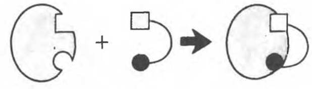
Fig. 8.5. Formation of the Enz S complex according to Fischer's "lock-and-key" model.
The Lock-and-Key Model
The initial model of the catalytic center, proposed by Emil Fischer, treated the interaction between substrate and enzyme by analogy with a "lock and key" system. This model, sometimes referred to as the "rigid template" model (Fig. 8.5), has retained its significance for understanding certain METABOLISM/8.html">Properties of Enzymes—such as their ability to strictly bind two or more substrates in a defined manner (Fig. 8.6)—or for explaining substrate saturation kinetics.
The Induced-Fit Model
A shortcoming of Fischer's model is the inherent rigidity of the catalytic center it implies. The induced-fit model, proposed by Koshland, is of a more general nature. This model is based on highly convincing experimental data. Its essential feature is The flexibility of the catalytic center. In Fischer's model, the catalytic center is considered pre-formed to fit the shape of the substrate molecule. In the induced-fit model, however, the substrate induces Conformational Changes in the enzyme, and only As a result of these changes do The amino acid residues and other groups of the enzyme acquire the spatial orientation required for substrate binding and catalysis. Meanwhile, other amino acid residues may become buried deep within the enzyme molecule.
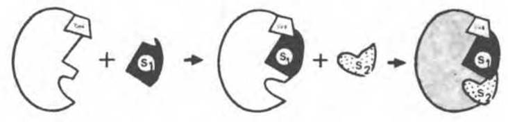
Fig. 8.6. Sequential binding of a coenzyme (CoE) and two substrates (S1 and S2) by an enzyme within the framework of the "rigid template" hypothesis. It is assumed that the coenzyme contains a site capable of binding the first substrate (S1); binding of the first substrate subsequently facilitates the binding of the second substrate S2.
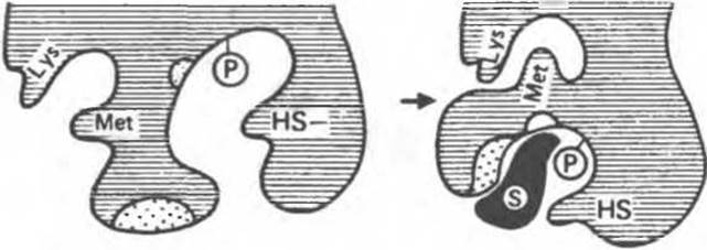
Fig. 8.7. Schematic representation of conformational changes in an enzyme molecule upon substrate binding According to the induced-fit model. Note the arrangement of key residues before and after substrate binding (after Koshland).
In the example shown in Fig. 8.7, hydrophobic and charged groups (regions highlighted by dots) participate in substrate binding. The phosphoserine residue (P) and the Cysteine —SH group take direct part in catalysis. Other residues, not involved in either process, are represented by Lys and Met residues. In the absence of the substrate, the catalytic and substrate-binding groups lie at a distance from each other several times exceeding the bond length. As the substrate approaches the enzyme, it induces conformational Changes in the latter, causing the respective groups to assume the positions necessary for substrate binding and catalysis. Simultaneously, the spatial arrangement of other residues changes—Lys and Met are now brought into close proximity (Fig. 8.7).
Substrate analogues can also trigger conformational changes, but not all of them are "correct" (Fig. 8.8). Upon binding the true substrate (A), all groups (black circles) assume the correct positions. However, when a substrate analogue is bound—whether it is bulkier (Fig. 8.8, B) or, conversely, smaller in size (Fig. 8.8, C)—an incorrect arrangement of these groups is induced. Another structural feature of the enzyme is a small cleft on the right-hand side. Imagine that a regulatory molecule enters this cleft and "prevents" one of the polypeptide segments bearing a catalytic group from moving. In this case, only substrate binding will occur, but not catalysis.

Fig. 8.8. Schematic representation of conformational changes in an enzyme upon binding the true substrate (A) and its analogues (B, C).

Fig. 8.9. Scheme of alternative reaction pathways when substrate-induced conformational changes occur in an enzyme. The enzyme first undergoes a conformational change (A) and then binds the substrate (B). In an alternative pathway, the enzyme first binds the substrate (C) and subsequently undergoes a conformational change (D). Finally, both processes can proceed in a concerted manner (G) to form the final conformation (E).
It remains to clarify the exact sequence of events that constitute substrate-induced conformational changes. Several pathways are possible here, as illustrated in Fig. 8.9.
Let us assume that the complete Introduction/19.html">Primary Structure of an enzyme is known. Even in this case, it is usually difficult to determine precisely which residues form the catalytic center. As the induced-fit model implies, these residues may be located far apart in the primary structure, yet be brought close together in the three-dimensional (tertiary) structure.
Amino acid residues from several polypeptide chain segments take part in forming the catalytic center, as in the case of Hemoglobin (Chapter 6) or Chymotrypsin (Chapter 9).
The Catalytic Center of Lysozyme
Lysozyme is present in tears, nasal mucus, saliva, gastric juice, various Tissues, as well as in milk and egg white. This enzyme catalyzes the Hydrolysis of ß-1,4-glycosidic bonds of N-acetylneuraminic acid (see Chapters 13 and 33), which is a component of Proteoglycans and Glycosaminoglycans. Lysozyme found in tears and nasal mucus destroys The Cell walls of many airborne Gram-positive Bacteria. Lysozyme (molecular weight approximately 15,000) consists of a single polypeptide chain containing 129 residues. Since the molecule contains neither a coenzyme nor Metal Ions, the catalysis, Specificity, and three-dimensional structure of lysozyme are entirely determined by its Amino Acid Sequence. The molecule contains small regions of pleated sheet, several short a-helices, and fairly large segments with an irregular structure. A color photograph of the three-dimensional model of lysozyme complexed with a substrate can be found in J. Biol. Chem. 1968: 243, 1663. A deep cleft runs across the middle of the lysozyme molecule, harboring the residues that form the catalytic center and the substrate-binding region; the latter contains six subsites that interact with different substrates or inhibitors (Fig. 8.10). The residues responsible for bond Cleavage are located between subsites D and E; the most critical role is played by the carboxyl groups of Asp 52 and Glu 35 residues. The latter presumably protonates the glycosidic bond of the substrate, whereas the negatively charged Asp 52 residue, situated on the opposite side, stabilizes the resulting carbonium ion.

Fig. 8.10. Schematic representation of the catalytic center located in the cleft running across the middle of the lysozyme molecule. The glycosyl units of the hexasaccharide are designated by letters from A to F. Certain residues lining the cleft of the active center and their positions in the amino acid sequence of lysozyme are indicated.
The Catalytic Center of Ribonuclease
Unlike the case of lysozyme, information regarding the catalytic site of ribonuclease was largely obtained even before its three-dimensional structure was determined. Conclusions drawn from chemical studies have been fully supported by crystallographic data. The ribonuclease molecule also features a cleft, similar to that in lysozyme, into which two residues, His 12 and His 119, protrude. Previous chemical data had already suggested that these residues are part of the catalytic site. Both of them are located near the binding site for uridylic acid (Fig. 8.11).

Fig. 8.11. Structure of ribonuclease based on X-Ray Diffraction Analysis data. The numbers of specific residues are indicated. (See also Fig. 5.7.)
Amino acid sequences in the region of the catalytic site
The primary structure in the vicinity of the catalytic site appears quite similar across various families of hydrolytic enzymes (Table 8.1). This suggests that the mechanisms of bond hydrolysis in biological systems are relatively limited in number. Therefore, it is hardly surprising that the amino acid sequences around the catalytic site of the same enzyme isolated from different species show even greater similarity.
Last update: 06/08/2026
Editorial and Educational Adaptation: This material has been compiled based on the primary/original source text. The project team performed an editorial review, corrected technical inaccuracies, structured sections, and adapted the content for an educational format.
What was processed:
- elimination of formatting defects (OCR errors, structural breaks, corrupted characters);
- editorial organization of content;
- standardization of terminology in accordance with academic sources;
- verification of factual statements against the original source text.
All mentions of the author, publication year, and origin of the primary text have been preserved in accordance with the source.