BIOCHEMISTRY - L. Stryer - 1984
VOLUME 3
Part IV INFORMATION
CHAPTER 29. EUKARYOTIC CHROMOSOMES AND GENE EXPRESSION IN EUKARYOTES
29.3. The Amino Acid Sequences of Histones H3 and H4 Are Highly Conserved Across Animals and Plants
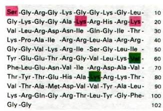
Emil Smith and Robert DeLange demonstrated that The amino acid sequences of histone H4 from pea seedlings and calf Thymus differ at only two out of 102 positions. These substitutions are very conservative: valine replaces isoleucine, and Lysine replaces Arginine. Thus, the Amino Acid Sequence of histone H4 has remained virtually unchanged throughout the 1.2 • 109 years that have elapsed since the divergence of the PLANT AND ANIMAL kingdoms. Histone H3 has also changed very little over this immense evolutionary span. The amino acid sequences of histone H3 from pea seedlings and calf thymus differ by only four residues. It is instructive to compare The rate of evolutionary change in these Histones with that of other Proteins. This is commonly measured by the unit evolutionary period, defined as the time required for a 1% change in amino acid sequence to occur after two evolutionary lineages diverge. For histones H3 and H4, this value is 3 • 108 and 6 • 108 years, respectively, which is significantly greater than for any other proteins investigated to date. For instance, the unit evolutionary period for cytochrome c is 2 • 107 years, for Hemoglobin 6 • 106 years, and for fibrinopeptides 1 • 106 years. The remarkable conservatism of histones H3 and H4 indicates that they perform a critical function that arose at the dawn of eukaryote evolution and has been preserved almost unaltered ever since.
Class="center">Fig. 29.3. Amino acid sequence of histone H4 from calf thymus. Several residues are modified. The α-amino group is acetylated, as is the ε-amino group of Lys-16. The ε-amino group of Lys-20 is methylated or dimethylated. Histone H4 from pea seedlings has the same amino acid sequence except at positions 60 (isoleucine) and 77 (arginine)
29.4. Nucleosomes Are Repeating Subunits of Chromatin
How do histones interact with DNA to form the Chromatin fiber? Drawing on data obtained from various approaches, Roger Kornberg proposed in 1974 that chromatin consists of repeating subunits, each containing 200 Base Pairs of DNA and two molecules each of histones H2A, H2B, H3, and H4. These repeating units are now called nucleosomes. Most of the DNA is wound around a histone core. The remaining DNA, known as linker DNA, connects adjacent nucleosomes and imparts flexibility to the chromatin fiber. Consequently, the chromatin fiber resembles a flexible string of beads.
A wealth of experimental evidence Supports this model of Chromatin Structure.
1. Electron Cell/15.html">Microscopy. Cytology/cytology/93.html">ELECTRON MICROGRAPHS OF chromatin reveal strings of bead-like particles approximately 100 Å in diameter, connected by a thin filament (Fig. 29.4). The degree of extension of the chromatin fiber depends on the Sample preparation method used for microscopy. Some techniques yield electron micrographs with a more compact arrangement of the 100-angstrom beads. Thus, electron microscopy provides direct visual confirmation that chromatin is a chain of nearly spherical particles separated by flexible regions.
Fig. 29.4. Electron micrograph of chromatin. The bead-like particles are approximately 100 Å in diameter

2. X-ray and neutron diffraction. X-Ray Diffraction patterns of chromatin fibers also exhibit a 100 Å repeat. Neutron diffraction studies demonstrate that the DNA is located on the outer surface of the nucleosome.
3. Nuclease Digestion. Free DNA in solution can be cleaved at any phosphodiester bond by pancreatic deoxyribonuclease I (DNase I) or micrococcal nuclease. By contrast, DNA within chromatin, except at exposed linker regions, is largely protected from nuclease Hydrolysis. The pattern of chromatin digestion is strikingly simple: Electrophoresis reveals a distinct ladder of bands (Fig. 29.5). These fragments correspond to DNA lengths that are integral multiples of the fundamental repeat of roughly 200 base pairs. Electron micrographs show that the number of spherical particles in a chromatin fragment equals the number of 200-base-pair repeats (Fig. 29.6). For example, a fragment containing 600 base pairs of DNA consists of three 100-angstrom particles. Therefore, the beads visible in electron micrographs correspond to the nucleosomes generated by nuclease digestion.
4. Reconstitution. When histones are added to adenovirus or SV-40 viral DNA, a chromatin-like fiber can be assembled in vitro. The amount of DNA associated with a nucleosome in such reconstitution systems is approximately 200 base pairs. Furthermore, The formation of nucleosomes requires equimolar amounts of histones H2A, H2B, H3, and H4. If any histone is deficient in the reconstitution mixture, characteristic beads fail to form. However, histone H1 is not required for reconstitution—a fact consistent with the observation that histone H1 is absent from certain nucleosomes in Eukaryotic Cells. X-ray diffraction studies also show that histones H2A, H2B, H3, and H4 are both necessary and sufficient to yield the diffraction pattern characteristic of native chromatin.
Fig. 29.5. Gel electrophoresis of chromatin DNA fragments generated by limited digestion with micrococcal nuclease. Lane A shows unfractionated digest. Sucrose density gradient centrifugation resolved fractions of monomers (B), dimers (C), trimers (D), and tetramers

Fig. 29.6. Electron micrographs of nucleosome monomers (A), dimers (B), trimers (C), and tetramers (D) isolated as described in the legend to Fig. 29.5

29.5. The Minimal Nucleosome (Nucleosome Core Particle) Consists of 140 Base Pairs of DNA Wrapped Around a Histone Octamer
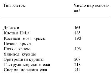
The DNA content of nucleosomes varies from 160 to 240 base pairs across different organisms and cell types (Table 29.2). What is the basis for this variation? Nucleases have again proven invaluable in answering this question. Nucleosomes can be further digested with micrococcal nuclease to yield minimal nucleosome particles (nucleosome core particles) containing 140 base pairs of DNA, regardless of the initial DNA content per nucleosome. This core particle is remarkably uniform across virtually all eukaryotes. It consists of a 140-base-pair DNA fragment bound to a histone octamer containing two molecules each of H2A, H2B, H3, and H4.
Table 29.2. DNA Content of Nucleosomes
Minimal nucleosomes have been obtained in crystalline form and are currently being investigated using electron microscopy (Fig. 29.7) and X-ray diffraction. The crystallization of these particles demonstrates that chromatin nucleosomes are highly homogeneous. Aaron Klug and John Finch established that the minimal nucleosome (nucleosome "core") is a flattened particle measuring 110 x 100 x 55 A and consists of two layers. A DNA fragment
140 base pairs in length is wound externally around the core, forming 13/4 turns of a left-handed superhelix with a pitch of approximately 28 A (Fig. 29.8).
Fig. 29.7. Electron micrograph of a minimal nucleosome crystal. The centers of adjacent nucleosomes in this hexagonal layer are spaced 100 A apart from each other

Fig. 29.8. Schematic representation of a nucleosome. The DNA double helix (red band) is wrapped around a histone octamer (two molecules each of Н2А, Н2В, Н3, and Н4; shown in blue). Histone H1 (yellow) binds to the outer surface of this minimal nucleosome and to the linker DNA

As already mentioned, histone H1 is not always present in the nucleosome. The amino acid sequence of histone H1 is the most variable among all five histones. Furthermore, histone H1 differs from the other histones in its stoichiometry: there is one molecule of histone H1 per nucleosome. In addition, histone H1 is easily dissociated from nucleosomes, which indicates its peripheral Location, meaning it is not part of the histone core. Nucleosomes lose histone H1 when their constituent DNA is shortened from 160 to 140 base pairs. Thus, histone H1 is almost certainly located outside the nucleosome core, closer to the linker DNA. Histone H1 may serve as a bridge between different nucleosomes, thereby contributing to a higher degree of chromatin compaction.
Last update: 06/08/2026
Editorial and Educational Adaptation: This material has been compiled based on the primary/original source text. The project team performed an editorial review, corrected technical inaccuracies, structured sections, and adapted the content for an educational format.
What was processed:
- elimination of formatting defects (OCR errors, structural breaks, corrupted characters);
- editorial organization of content;
- standardization of terminology in accordance with academic sources;
- verification of factual statements against the original source text.
All mentions of the author, publication year, and origin of the primary text have been preserved in accordance with the source.