BIOCHEMISTRY - L. Stryer - 1984
VOLUME 1
PART I. CONFORMATION AND DYNAMICS
CHAPTER 2. INTRODUCTION TO PROTEIN STRUCTURE AND FUNCTION
2.10. The Polypeptide Chain Can Reverse Its Direction by 180° via β Turns
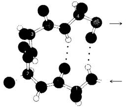
Most Proteins have a compact, globular shape due to extensive folding of their polypeptide chains, which frequently reverse direction by 180°. Studies on the three-dimensional structures of numerous proteins have revealed that this directional reversal is frequently mediated by a common structural motif known as the β turn. This hairpin-like bend is formed through a Hydrogen bond between the CO group of residue n and the NH group of residue (n + 3) in the polypeptide chain (Fig. 2.42), effectively reversing the chain's overall direction.
Class="center">Fig. 2.42. Structure of a β turn. The CO group of residue 1 in the illustrated tetrapeptide forms a hydrogen bond with the NH group of residue 4, creating a hairpin-like bend.
2.11. Structural Levels in Protein Architecture
It is customary to distinguish four LEVELS OF STRUCTURAL Organization in protein molecules. Primary Structure refers simply to the Amino Acid Sequence of a protein and the Location of any disulfide bridges. Thus, a complete description of primary structure encompasses all covalent bonds within the protein. Secondary structure arises from steric interactions between amino acid residues that are close to one another along the linear sequence. Some of these steric interactions occur in a regular pattern, thereby conferring periodicity to the structure. Classical Examples of secondary structure include the α Helix, the pleated β sheet, and the Collagen helix. Tertiary structure is determined by steric interactions between amino acid residues that are far apart in the linear sequence. It should be noted, however, that the distinction between secondary and tertiary structure is somewhat arbitrary. Proteins composed of multiple polypeptide chains exhibit an additional level of organization known as quaternary structure, which describes how these chains are spatially arranged relative to one another. Each individual polypeptide chain in such a protein is termed a subunit. The term domain is also frequently used to designate a compact globular unit of Cell/13.html">Protein Structure. Many proteins consist of multiple domains, typically ranging in molecular mass from 10 to 20 kDa. In very large proteins, individual domains are often connected by relatively flexible segments of the polypeptide chain.
2.12. The Amino Acid Sequence Specifies Three-Dimensional Structure
The relationship between a protein's amino acid sequence and its conformation was first demonstrated by Christian Anfinsen during his studies on Ribonuclease, an RNA-cleaving enzyme. Ribonuclease consists of a single polypeptide chain containing 124 amino acid residues (Fig. 2.43).
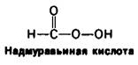
It contains four disulfide bridges, which can be irreversibly oxidized with performic acid to yield cysteic acid residues (see Fig. 2.32). Alternatively, these Disulfide Bonds can be reversibly cleaved by reduction with Reagents such as β-mercaptoethanol, forming mixed disulfides
НО—СН2 —СН2—SН
β-mercaptoethanol
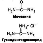
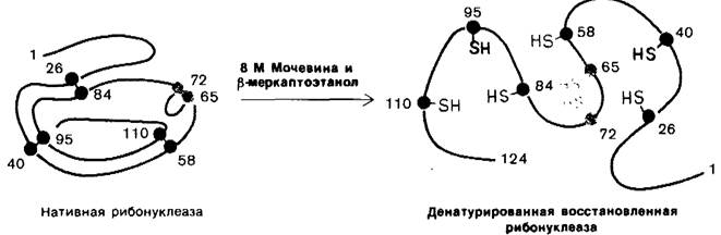
between β-mercaptoethanol and the Cysteine side chains (Fig. 2.44). In the presence of a large excess of β-mercaptoethanol, these mixed disulfides are further reduced, yielding a protein in which all disulfide groups (cystine residues) are fully converted into sulfhydryl groups (cysteine residues). However, it was found that at 37°C and pH 7, the reduction of ribonuclease by β-mercaptoethanol proceeds only if the compact STRUCTURE OF THE protein is first disrupted by partial unfolding with Denaturing Agents such as urea or guanidine hydrochloride. Although the exact MECHANISM OF ACTION of these denaturants is not fully understood, it is clear that they disrupt noncovalent interactions. Deprived of these stabilizing forces, polypeptide chains in 8 M urea or 6 M guanidine hydrochloride collapse into a random conformation—a random coil—as evidenced by physical parameters such as viscosity and optical rotation spectra. When ribonuclease is treated with β-mercaptoethanol in 8 M urea, the reaction product is a fully reduced, randomly coiled polypeptide chain devoid of enzymatic activity. In other words, this Treatment causes the complete Denaturation of ribonuclease (Fig. 2.45).
Fig. 2.43. Amino acid sequence of bovine pancreatic ribonuclease. The four disulfide bonds are highlighted in color.
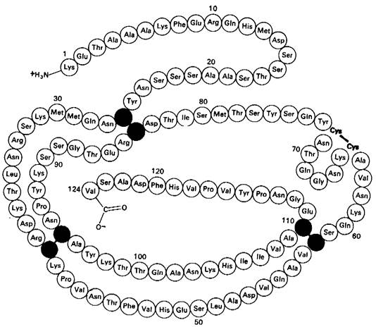
Fig. 2.44. Reduction of protein disulfide bonds by an excess of a sulfhydryl compound such as β-mercaptoethanol.

Fig. 2.45. Reduction and denaturation of ribonuclease.
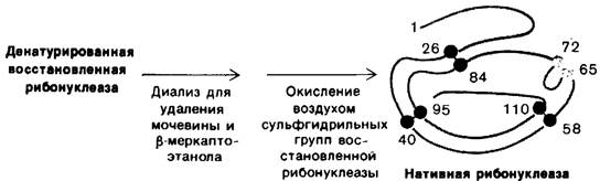
Anfinsen made a crucial observation: when denatured ribonuclease is freed of urea and β-mercaptoethanol via dialysis, the enzymatic activity of the protein gradually recovers. Recognizing the profound significance of this chance observation, Anfinsen proposed that The sulfhydryl groups of the denatured enzyme are reoxidized by atmospheric oxygen, allowing the protein to spontaneously refold into its catalytically active conformation. Subsequent detailed investigations demonstrated that by reoxidizing all sulfhydryl groups under appropriate conditions, virtually 100% of the original enzymatic activity can be restored (Fig. 2.46). All physical and Chemical properties of this renatured enzyme were found to be virtually identical to those of the native protein. These experiments conclusively proved that all the information required to specify the precise three-dimensional structure of ribonuclease is encoded entirely within its amino acid sequence. Subsequent work on other proteins established this principle—that sequence dictates conformation—as a Central dogma of molecular biology.
Fig. 2.46. Renaturation of ribonuclease
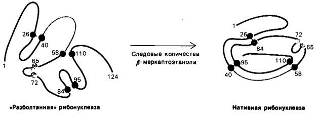
Completely different results are obtained if reduced (denatured) ribonuclease is subjected to oxidation in an 8 M urea solution and urea is removed by dialysis only thereafter. The enzymatic activity of such a ribonuclease preparation is only 1% of that of the native protein. Why is the enzymatic activity restored when denatured ribonuclease is oxidized in the absence of urea, but fails to be restored in its presence? The answer is as follows: when a randomly folded molecule of the reduced protein undergoes oxidation, incorrect disulfide pairs are formed, meaning that disulfide bridges link pairs of Amino Acids different from those in the native protein. There are 105 possible ways to link eight cysteines to form 4 disulfides; only one of these combinations possesses enzymatic activity. The remaining 104 combinations were vividly termed «scrambled» ribonuclease. Anfinsen subsequently discovered that «scrambled» ribonuclease spontaneously converts into the fully active, native form if trace amounts of β-mercaptoethanol are added to the solution of the reoxidizing enzyme protein (Fig. 2.47). The added β-mercaptoethanol catalyzes the rearrangement of disulfide bonds, which ultimately leads to The formation of the native structure, taking about 10 hours. The entire process is driven by a decrease in the Free energy of the system as the scrambled Conformations convert into the stable native conformation of the enzyme. Thus, the native form of ribonuclease appears to be its thermodynamically most stable structure.
Anfinsen (1964) wrote: «It struck me that, in fact, the amino acid sequence in a protein molecule that is to assume a precisely defined geometric shape should be regarded as a melody written in canonical form; by Nature's decree, this melody, when superimposed upon itself, must create such harmonic consonances as correspond to biological function. Extending the analogy further, one might suggest that proteins with “scrambled” disulfide bonds, which I mentioned above, produce dissonant consonances; however, if the possibility of rearrangement arises upon The addition of mercaptoethanol, the pleasant harmony characteristic of the native protein is once again achieved through gradual modulation. Whether on this basis Mozart's music possesses greater thermodynamic stability than Schönberg's is a question I leave to the philosophers in this audience.»
Fig. 2.47. Formation of native ribonuclease from scrambled ribonuclease in the presence of trace amounts of β-mercaptoethanol
Last update: 06/08/2026
Editorial and Educational Adaptation: This material has been compiled based on the primary/original source text. The project team performed an editorial review, corrected technical inaccuracies, structured sections, and adapted the content for an educational format.
What was processed:
- elimination of formatting defects (OCR errors, structural breaks, corrupted characters);
- editorial organization of content;
- standardization of terminology in accordance with academic sources;
- verification of factual statements against the original source text.
All mentions of the author, publication year, and origin of the primary text have been preserved in accordance with the source.