Principles of Protein Structure - H. Schulz 1982
Mechanisms of polypeptide chain folding and association
Secondary structure
Collagen helix
The Collagen Structure is a superhelix formed by three parallel, elongated left-handed helices.
The collagen helix is shown in Fig. 5.6. It is a right-handed helix consisting of three single left-handed helices arranged in parallel. This formation is known as a superhelix. X-Ray Diffraction Analysis of collagen presents significant difficulties [191–194]: the most crucial information for deciphering the collagen X-ray diffraction pattern can be obtained from similar X-ray diffraction patterns of synthetic polymers—poly-Gly-II and poly-L-Pro-II (poly-L-Pro-I contains cis-peptide bonds). Model Building for Pro homopolymers is particularly relevant because their dihedral angle ∅ is fixed near –60°, while The formation of an α-Helix-like structure (ψ = –60°) is impossible for steric reasons; only values in the region (∅, ψ) = (–60°, +140°) (Fig. 2.3) prove acceptable, which adequately describe The structure of an individual collagen helical strand.
* Since the cavity cannot be filled with Water, the binding energy is lowered by pAV (p is pressure, AV is the free volume of the cavity). However, at atmospheric pressure, this reduction is only 0.001 kcal/mol for a 50-residue n-helix, i.e., negligibly small.
Class="center">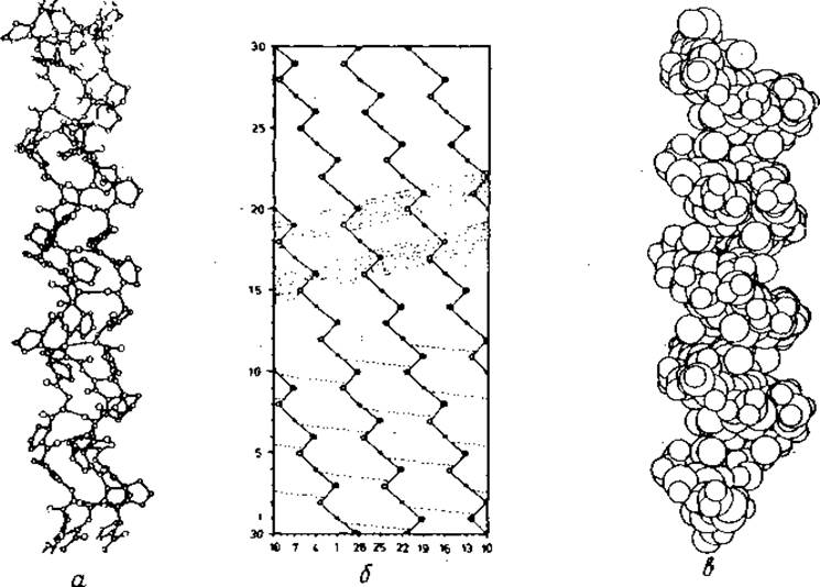
Fig. 5.6. Collagen structure.
a — ORTEP drawing [324] of the collagen triple helix with the (Gly-Pro-Pr●)n sequence and coordinates from [193]. Only those hydrogen atoms participating in bonds are shown (thin, nearly horizontal lines). b — cylindrical diagram of the full identity period of the collagen triple helix [325]. The helical path and Ca atoms are indicated. Three Ca atoms in the (Gly-X-Y)n sequence are marked with dots: (• O ●). Hydrogen Bonds are indicated by dashed lines. c — ORTEP drawing of the collagen triple helix corresponding to a, but with atoms represented by their Van der Waals radii and all hydrogen atoms included. The protruding Pro residues arranged helically with a 9 Å pitch are clearly visible. In Fig. b, they are shown as shaded bands.
The structural unit of the superhelix is a triplet. Knowledge of the Amino Acid Sequence proved decisive for a detailed Analysis of the Spatial Structure of collagen [195–197]. About 96% of the chain can be described by the formula (Gly-X-Y)m. Position X is frequently occupied by Pro, and position Y by Hyp (hydroxyproline = Pro with an OH group at the Cm atom; see Fig. 1.1). The content of each of the Pro and Hyp residues is 11%. Until recently, three models built from triple helices formed by parallel peptide chains were discussed [191, 192]. The structure of so-called collagen-II is consistent with the most frequently occurring triplet (Gly-Pro-Hyp); all other models eventually had to be ruled out [194].
The superhelix is best described using the cylindrical projection shown in Fig. 5.6, b. It is a linear group with a triplet (Gly-X-Y) acting as the group element. There are 10 triplets per polypeptide chain per turn of the superhelix; the superhelix pitch is 86 Å. The three chains are stabilized by van der Waals forces as well as hydrogen bonds (one per triplet). The dipoles forming the hydrogen bonds are arranged linearly and approximately perpendicular to the major axis (Fig. 5.6, b).
An individual left-handed polypeptide chain is insufficiently ordered. The group element of the left-handed single peptide chain helix should be considered the triplet, as it serves as the group element of the superhelix. Not all, but only every third pair of torsion angle values (∅, ψ) are identical. However, the deviations in the (∅, ψ) angles for all residues are small, at about 10°. Therefore, the structure of an individual chain can be approximated by a helix (linear group) with a single residue as the group element, which corresponds to a single conformational point on the (∅, ψ) map (Fig. 2.3). The parameters of the single polypeptide chain helix are given in Table 5.1. With a helical parameter of d = 2.9 Å, the chain is so extended that its geometry is stable only upon aggregation with other chains.
A Gly residue at every third position is essential for stability. Since the parameter n = -3.3 for the left-handed helix of an individual chain is non-integer and the right-handed superhelix contains 10 triplets per turn, every third Ca atom ends up near the major axis. This explains the necessity of the (Gly-X-Y)m sequence, since a compact, hydrogen-bonded structure can only be formed in the absence of side chains for residues near the axis; Gly is the only such residue.
The side chains of collagen residues face the surrounding environment. The overall appearance of the collagen helix is shaped by the side chains of the residues at the X and Y positions. This is particularly noticeable in the molecular model of (Gly-Pro-Hyp)m. As seen in Fig. 5.6, c, the Pro and Hyp side chains form protruding ridges arranged on the helical cylindrical surface with a pitch of 9 Å.
Collagen is the most abundant protein in mammals.
Consisting of highly extended polypeptide chains, collagen can withstand significant mechanical stress along its axis because this direction coincides with the orientation of the covalent bonds in its peptide backbone. Therefore, the structure of collagen is well-suited to its physiological Functions—transmitting tension in tendons and forming protective layers in Skin and other Organs. Collagen is undeniably one of the most widespread Proteins in nature.
The collagen triple helices of most vertebrates consist of two a1 chains and a homologous a2 chain. So far, The amino acid sequence is known only for the a1 chain, which comprises 1052 residues [195–197]. With the exception of 16 N-terminal and 25 C-terminal residues, the (Gly-X-Y)m composition is strictly maintained. Using this formula, collagen-like structures can be readily identified in various Amino acid sequences. Such structures have not yet been discovered in Globular proteins. However, it is very likely that they are present in the Clq component of the human Complement system [198], which recognizes Antibodies bound to Antigens. As established by electron micrographs, this protein contains a bundle of 18 parallel chains organized into six collagen-like fibrils that merge into six globules. It is possible that other hybrid proteins containing collagen-like structures will be identified in the future.
Last update: 06/08/2026
Editorial and Educational Adaptation: This material has been compiled based on the primary/original source text. The project team performed an editorial review, corrected technical inaccuracies, structured sections, and adapted the content for an educational format.
What was processed:
- elimination of formatting defects (OCR errors, structural breaks, corrupted characters);
- editorial organization of content;
- standardization of terminology in accordance with academic sources;
- verification of factual statements against the original source text.
All mentions of the author, publication year, and origin of the primary text have been preserved in accordance with the source.