Principles of Biochemistry, Volume 1 - A. Lehninger 1985
Biomolecules
Water
Chapter Summary
Each of the 20 Amino Acids commonly found as products of Protein Hydrolysis contains an α-carboxyl group, an α-amino group, and an amino acid-specific R group substituting for a hydrogen atom at the α-carbon atom. The α-carbon atom in all amino acids (except Glycine) is asymmetric, and consequently, each of these Amino acids can exist in at least two stereoisomeric forms. Proteins contain exclusively L-stereoisomers, which correspond in configuration to L-glyceraldehyde. The Classification of amino acids is based on differences in the polarity of their R groups. The Class of nonpolar amino acids includes Alanine, leucine, isoleucine, valine, Proline, phenylalanine, Tryptophan, and Methionine. The class of polar neutral amino acids comprises glycine, Serine, Threonine, Cysteine, Tyrosine, asparagine, and glutamine. The class of negatively charged (acidic) amino acids includes aspartic and glutamic acids, while the class of positively charged (basic) amino acids comprises Arginine, Lysine, and Histidine.
At low pH values, monoaminomonocarboxylic amino acids act as dibasic acids (+NH3CHRCOOH). As the pH rises to approximately 6—that is, to the isoelectric point—a proton is abstracted from the carboxyl group, resulting in The formation of zwitterions, which are electrically neutral bipolar ions of the type +NH3CHRCOO-. A further increase in pH leads to the removal of a second proton, yielding ions of the type NH2CHRCOO-. Amino acids with ionizable R groups can also exist in other ionic forms, depending on the pH. The reaction of amino acids with ninhydrin produces colored products. Electrophoresis and Ion-exchange Chromatography are employed to separate complex amino acid mixtures, as well as to identify and quantify the separated amino acids.
Amino acids covalently linked together by peptide bonds form Peptides, which can also be obtained as products of partial polypeptide hydrolysis. The acid-base properties of a peptide are determined by its terminal NH2 and COOH groups, as well as by the ionizable R groups it contains. Complete hydrolysis of peptides yields free amino acids. The reaction of the amino-terminal residue of a peptide with 1-fluoro-2,4-dinitrobenzene yields a derivative with a characteristic yellow color. Certain peptides occur in a free state in Cells and Tissues, performing specific biological Functions. These include numerous Hormones, Antibiotics, and Other Compounds endowed with high biological activity.
Cantor С. R., Schimmel P.R. Biophysical Chemistry, pt. I. The Conformation of Biological Macromolecules, Freeman, San Francisco, 1980. An excellent textbook discussing The properties of biological macromolecules and their constituent building blocks.
Cooper T. G. The Tools of Biochemistry, Wiley, New York, 1977. Theory and practical guidelines for the chromatography and electrophoresis of amino acids.
Corrigan J. Т. D-Amino Acids in Animals, Science, 164, 142-148 (1969).
Dickerson R.E., Geis I. Proteins: Structure, Function and Evolution, 2d ed., Benjamin/Cummings, Menlo Park, Calif., 1983.
Haschemeyer R., Haschemeyer A.H. Proteins: A Guide to Study by Physical and Chemical Methods, Wiley, New York, 1973.
Lehninger A. L. Biochemistry, 2d ed., Worth, New York, 1975. Chapters 4 and 5 provide a more detailed Description of the properties of Amino Acids and peptides.
Meister A.: Biochemistry of the Amino Acids, 2d ed., 2 vols. Academic, New York, 1965. An encyclopedic Treatment of the properties and distribution of amino acids, as well as their Participation in the metabolic processes of living organisms.
Segel I. Н. Biochemical Calculations, 2d ed., Wiley, New York, 1976.
1. Specific optical rotation of an amino acid isolated from watermelon. The amino acid citrulline was first isolated from the watermelon (Citrullus vulgaris), but it is also present in most animal tissues. Although citrulline is not a protein component, it serves as a precursor of arginine and of urea, the excreted end product of Amino Group METABOLISM. The structural formula of citrulline is as follows:
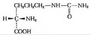
At 25°C, a 20 cm Glass tube filled with a 5% citrulline solution in 0.3 N HCl rotates the plane of polarized light by 1.79° to the right. What is the specific optical rotation of citrulline? Can one determine whether citrulline is a D- or an L-amino acid from its specific rotation?
2. Absolute configuration of citrulline. What is the configuration (D or L) of the citrulline isolated from watermelon (see the formula given in the previous question)? Provide a reasoned answer.
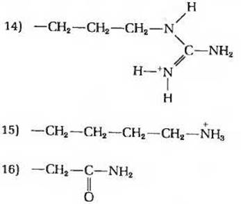
3. Relationship between the structure and Chemical properties of amino acids. Because amino acids serve as the Building Blocks of proteins, a thorough knowledge of their Structure and Chemical properties is essential for understanding how proteins perform their biological functions. Below are the structural formulas of the side chains (R groups) of 16 amino acids (Ala, Arg, Asn, Asp, Cys, Glu, Gly, His, Lys, Met, Phe, Pro, Ser, Trp, Tyr, and Val). Name the amino acids to which the R groups shown here belong. Which of the properties listed below are characteristic of each of these amino acids? Some of these properties can be used to characterize more than one amino acid. Properties of R groups and corresponding amino acids.
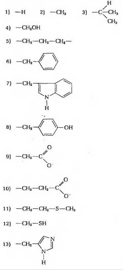
a) A small polar R group containing a hydroxyl group. The corresponding amino acid plays a vital role in the catalytic function of the active sites of certain Enzymes.
b) The R group imposes the least steric constraints.
c) The R group has a pK' ≈ 10.5 and bears a positive charge at physiological pH values.
d) A sulfur-containing R group; neutral at all pH values.
e) An aromatic R group; hydrophobic in nature and neutral at all pH values.
e) An R group consisting of a saturated hydrocarbon residue; makes an important contribution to hydrophobic interactions.
f) The only amino acid containing an ionizable R group with a pK' value close to 7. Plays a vital role in the functioning of active sites in A number of enzymes.
g) The only amino acid containing a substituted α-amino group. Affects protein chain folding by serving as a site of enforced chain bending.
h) The R group has a pK' value of about 4 and carries a negative charge at pH 7.
i) An aromatic R group capable of participating in Hydrogen bond formation; has a pK' value close to 10.
j) Forms disulfide cross-links between polypeptide chains; the pK' value of the functional group is close to 8.
k) An R group with a pK' ≈ 12; carries a positive charge at all physiological pH values. In some proteins, plays an important role in binding negatively charged phosphate groups.
l) If this polar, uncharged R group undergoes hydrolysis, the amino acid containing it is converted into another amino acid with a negatively charged R group at around pH 7.
4. The relationship between The titration curve and the acid-base properties of glycine. A 0.1 M glycine solution (100 mL) with a pH of 1.72 was titrated with a 2 M NaOH solution. pH measurements were recorded during the titration, and the obtained data were plotted on the graph shown in the figure. The most important points on the graph are designated by Roman numerals from I to V. Which of these five points on the titration curve should be indicated when answering the questions below? Explain your choice.
Problem 4

a) Which point corresponds to the pH at which this amino acid exists in the +NH3—CH2—COOH ionic form in a 0.1 M glycine solution?
b) At which point is the average net charge of a glycine molecule equal to 
c) At which point are the amino groups ionized in half of the glycine molecules?
d) At which point is the pH value equal to the pK' of the carboxyl group ionization of glycine?
e) At which point is the pH value equal to the pK' of the protonated amino group (—NH+3) ionization of glycine?
f) At which point does glycine exhibit maximum buffer capacity?
g) At which point is the average net charge of glycine equal to zero?
h) At which point is the carboxyl group of glycine completely titrated (first equivalence point)?
i) At which point are half of the carboxyl groups ionized?
j) At which point is glycine completely titrated (second equivalence point)?
k) At which point does glycine exist predominantly in the form of H3N+—СН2—СОО- ions?
l) At which point are 50% of the glycine molecules converted into H3N+—СН2—СОО- ions and 50% into H2N—СН2—СОО- ions?
m) At which point is the average net charge of a glycine molecule equal to —1?
n) At which point are 50% of the glycine molecules converted into H3N+—СН2—СООН, and the remaining 50% into H3N+—СН2—СОО- ions?
п) Which point corresponds to the isoelectric point of glycine?
р) At which point is the average net charge of glycine equal to 
с) Which point corresponds to the end point of titration?
т) To use glycine as an effective buffer, it is necessary to know at what pH values a glycine solution exhibits minimal buffer capacity. Indicate the corresponding points on the glycine titration curve.
у) At which point during the titration do H2N-CH2-COO- ions become the predominant form of glycine?
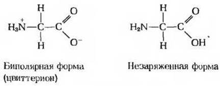
5. What fraction do the fully uncharged forms of glycine represent? At the pH corresponding to the isoelectric point, the net charge of glycine is zero. Although two forms of glycine (zwitterionic and uncharged) have a zero net charge, the zwitterionic form is its predominant form at the isoelectric point.
а) Explain why glycine exists predominantly as the zwitterionic form rather than the fully uncharged form at its isoelectric point.
б) Determine the fraction of glycine molecules that are in the fully uncharged form at the isoelectric point. Support your estimate with reasoning.
Problem 5
б. Ionization state of amino acids. Each ionizable group of an amino acid can exist in one of two states: charged or neutral. The electrical charge on a functional group is determined by the relationship between the pK' value of that group and the pH of the solution. This relationship is described by the Henderson-Hasselbalch equation.
а) Histidine has three ionizable functional groups. Write the equations for the three corresponding ionization processes of histidine and indicate the approximate values of the equilibrium constants (pK') characterizing each of these processes. Draw The structure of histidine in all three ionization states. What is the net charge of the histidine molecule in each ionization state?
б) Draw the ionic structures of histidine that predominate at pH 1, 4, 8, and 12; keep in mind that the ionization state can be determined by treating each ionizable group independently of the others.
в) What is the net charge of the histidine molecule at pH 1, 4, 8, and 12? In which direction will histidine move during electrophoresis at each of these pH values—toward the anode (+) or the cathode (—)?
7 Preparation of a glycine buffer. A glycine solution is frequently used as a buffer. To prepare a 0.1 M glycine buffer, 0.1 M solutions of glycine hydrochloride (+NH3—СН2—СООН-Сl-) and glycine (+NH3—СН2—СООН-)—the two commercially available forms of glycine—are used. What volumes of these two solutions should be mixed to prepare 1 L of a 0.1 M glycine buffer with a pH of 3.2?
8. Paper electrophoresis of amino acids. A drop of a solution containing a mixture of glycine, alanine, glutamic acid, lysine, arginine, and histidine was applied to the center of a paper strip and allowed to dry. The paper was then moistened with a buffer at pH 6.0, and an electrical voltage was applied to the ends of the strip.
а) Which amino acid(s) will migrate toward the anode?
б) Which amino acid(s) will migrate toward the cathode?
в) Which amino acid(s) will remain at or near the origin?
9. Separation of amino acids by ion-exchange chromatography. The analysis of an amino acid mixture begins with the separation of its components by ion-exchange chromatography. A small amount of the mixture is applied to the top of a Column packed with polystyrene particles containing sulfonic acid residues (see Fig. 5-14). A buffer solution is then passed through the column. Amino acids pass through the column at different rates because their migration is retarded by two factors: (1) electrostatic attraction between the negatively charged sulfonic acid residues and the positively charged Functional groups of the amino acids, and (2) hydrophobic interaction between the amino acid side chains and the strongly hydrophobic backbone of the polystyrene resin. For each of the pairs of amino acids listed below, determine which amino acid of the pair will elute from the column first (i.e., experience the least retardation) when a buffer at pH 7.0 is passed through the column.
а) Asp and Lys
б) Arg and Met
в) Glu and Val
г) Gly and Leu
д) Ser and Ala
10 Tripeptide Set. Suppose you wish to synthesize tripeptides using glycine, alanine, and serine as building blocks.
a) How many different tripeptides can be prepared assuming that any of these Three amino acids can occupy any of the three possible positions, and each amino acid can be used more than once?
b) How many different tripeptides can be prepared if each amino acid is used only once?
11. Designation of optical isomers of isoleucine. The structural formula of isoleucine is shown below
Problem 11
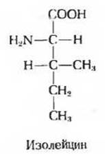
a) How many chiral centers does the isoleucine molecule have?
b) How many optical isomers can isoleucine have?
c) Draw perspective formulas for all optical isomers of isoleucine.
d) How would you designate each of these isomers within the RS system? (Hint: In terms of priority, the CH3CH2 group occupies an intermediate position between the C6H-5 and CH3 groups.)
12. Comparison of pK' values of free amino acids and peptides. The titration curve of the amino acid alanine reflects the ionization processes of two functional groups with pK' values of 2.34 and 9.69, which correspond to The ionization of the carboxylic acid and the protonated amine, respectively. Titration of alanine di-, tri-, and oligopeptides containing more than four residues of this amino acid indicates the ionization of only two functional groups, although the experimentally determined pK' values differ.
|
Amino acid or peptide |
pK'1 |
рK'2 |
|
Ala |
2,34 |
9,69 |
|
Ala-Ala |
3,12 |
8,30 |
|
Ala-Ala-Ala |
3,39 |
8,03 |
|
Аlа-(Аlа)n-Аlа, n > 4 |
3,42 |
7,94 |
a) Draw the structural formula of the Ala-Ala-Ala peptide. Indicate the functional groups corresponding to pK'1 and pK'2.
b) When moving from Ala to oligopeptides composed of Ala residues, the pK'1 value increases. Explain why this happens.
c) When moving from Ala to oligopeptides composed of Ala residues, the pK'2 value decreases. Explain the reason for this.
Last update: 06/08/2026
Editorial and Educational Adaptation: This material has been compiled based on the primary/original source text. The project team performed an editorial review, corrected technical inaccuracies, structured sections, and adapted the content for an educational format.
What was processed:
- elimination of formatting defects (OCR errors, structural breaks, corrupted characters);
- editorial organization of content;
- standardization of terminology in accordance with academic sources;
- verification of factual statements against the original source text.
All mentions of the author, publication year, and origin of the primary text have been preserved in accordance with the source.