Chemistry and Biology of Proteins - F. Haurowitz 1953
Internal structure of globular proteins
Hydrogen bond
In this formula, the shared electron pair of the electron octet is represented by standard dashes, while the unshared pair is shown as two dots. It is clear from the given formula that bonding should occur through the positively charged proton (H+) and the unshared electron pair of oxygen, as illustrated on the right side of the equation. Initially, it was assumed that Hydrogen Bonds were symmetrically arranged and that the proton was attracted with equal strength to both the nitrogen and oxygen atoms. This, however, is not the case, as symmetric Hydrogen bonds are energetically impossible [98]. Currently, it is believed that the hydrogen Nucleus forming the hydrogen bond lies closer to the nitrogen atom than to the oxygen atom. In addition to hydrogen bonds between nitrogen and oxygen in Proteins, There are also hydrogen bonds between two nitrogen atoms or two oxygen atoms. The presence of hydrogen bonds between the hydroxyl group of the Tyrosine ring and free carboxyl groups has been proved by determining the shift of the specific absorption maximum of tyrosine in the ultraviolet region of the spectrum [99].
Class="center">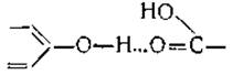
The energy of various hydrogen bonds ranges from 2 to 9 cal [100]. Since a Water molecule is a typical dipole, one might expect that hydrogen bonds between NH and CO groups would be disrupted by water molecules, allowing water to penetrate the clefts between the folded peptide chains [101]. However, X-Ray Diffraction patterns of crystalline proteins have shown that the peptide chains remain in the same folded state even in the Presence of water [2].
Hydrogen bonds can form not only between atoms involved in a peptide bond, but also between other groups of the protein molecule. One such group donates a hydrogen ion, while another, containing nitrogen or oxygen, provides an unshared electron pair whose negative charge attracts the positively charged hydrogen ion [102]. Nevertheless, the majority of hydrogen bonds occur between atoms belonging to peptide groups; consequently, these hydrogen bonds form cross-bridges between parallel peptide chains [103, 104] and stabilize the internal specific Structure OF THE protein molecule [105]. As already mentioned in Chapter IV, upon exposure to urea, protein molecules undergo disaggregation and break down into smaller structural units. This effect of urea is also likely associated with the disruption of hydrogen bonds. So far, however, it remains unknown what types of bonds hold these smaller structural units together within the protein macromolecule. It is possible that we are dealing here with a phenomenon governed by surface geometry. The surfaces of individual smaller structural units may fit together so closely that short-range attractive forces between polar groups become effective, ensuring the tight binding of these structural units (see Chapter XIV).
Last update: 06/08/2026
Editorial and Educational Adaptation: This material has been compiled based on the primary/original source text. The project team performed an editorial review, corrected technical inaccuracies, structured sections, and adapted the content for an educational format.
What was processed:
- elimination of formatting defects (OCR errors, structural breaks, corrupted characters);
- editorial organization of content;
- standardization of terminology in accordance with academic sources;
- verification of factual statements against the original source text.
All mentions of the author, publication year, and origin of the primary text have been preserved in accordance with the source.