Fundamentals of Molecular Biology. Part 2: Molecular Genetic Mechanisms - A. N. Ogurtsov 2011
Viruses
Structure of the Viral Capsid
The virion's nucleic acid is enclosed within a protein shell called a capsid, which is composed of identical Proteins or multiple repeats of a small number of different proteins encoded by a single viral Gene.
This specific Organization allows the virus to encode the information needed to build a relatively large capsid using a very small number of genes.
Such efficiency in utilizing Genetic information is critical, as the highly limited volume of the capsid can only accommodate a very limited set of genes.
The capsid, together with the nucleic acid (RNA or DNA) enclosed within it, is referred to as the nucleocapsid.
There are two primary modes of nucleocapsid organization.
The first mode involves identical capsid proteins forming a helical Structure that surrounds and protects the viral RNA (or DNA), which lies along the helical groove inside the protein tube (Figure 63).
Viruses with such a helical structure (such as the tobacco mosaic virus) have a rod-like shape.
The second primary mode of capsid formation is icosahedral.
An icosahedron is a sphere-like surface constructed from 20 identical equilateral triangles. The number and arrangement of proteins on the faces of icosahedral or quasi-spherical viral capsids vary depending on the size of the virion.
Class="center">
Figure 63 - Structure and self-assembly of the tobacco mosaic virus nucleocapsid from protein subunits and RNA molecules
In small quasi-spherical viruses, each of the 20 triangular faces is built from three identical copies of capsid proteins. The entire capsid is thus constructed from 60 proteins, with each capsid protein occupying an equivalent position and having an equivalent environment (Figure 64). Each vertex of the icosahedron possesses a fivefold axis of Symmetry.
In large quasi-spherical viruses, each face of the icosahedron is formed by more than three protein subunits.
As a result, the number of neighbors for each protein subunit depends on whether it is located at an icosahedral vertex or not. The positions of the proteins within the capsid are no longer equivalent, but rather quasi-equivalent.
For example, each face of an adenovirus icosahedron is formed by four subunits (Figure 65). Consequently, the vertices of the icosahedron exhibit fivefold symmetry, while the junctions between the faces exhibit threefold symmetry.

Figure 64 - Structure of small icosahedral viruses: poliovirus (a human RNA virus), cowpea mosaic virus (CPMV; an RNA virus; Vigna is a genus of annual and perennial plants of the legume family), and simian virus 40 (SV40, a monkey DNA virus)
In small viruses, such as poliovirus, the depression surrounding each vertex of the icosahedron interacts with receptors On the surface of the host Cell.

Figure 65 - Structure of a large icosahedral adenovirus
In large viruses (such as adenovirus), long Fibrillar Proteins protruding above the nucleocapsid surface interact with the surface receptors of the host cell.
In many DNA Bacteriophages, the viral DNA is located within an icosahedral "HEAD" connected to a tubular channel or "tail" (Figure 62(d)). During infection, viral proteins at the tip of the tail bind to receptors on the host cell surface, after which the viral DNA is transferred from the head through the tail's tubular channel into the Cytoplasm of the infected cell.
In some viruses, the symmetrical nucleocapsid is surrounded by an outer envelope, which is a lipoprotein membrane consisting primarily of a phospholipid bilayer and one or two TYPES OF VIRAL proteins (Figure 66).

Figure 66 - Electron micrograph of an Influenza virus
The Phospholipids of the viral lipoprotein envelope are identical to those of The Plasma Membrane of the host cell. In fact, the viral envelope itself is derived from the host cell's plasma membrane during the virus's Replication cycle, while also incorporating A large number of viral proteins.
Figure 66 shows an electron micrograph of the influenza virus, clearly displaying the surface proteins protruding from the viral envelope: neuraminidase (a tetrameric protein) or hemagglutinin.
Study of viruses. The number of viruses in a sample can be determined using the plaque assay. A diluted virus-containing solution is applied to The surface of a Petri dish containing susceptible Cells. Following infection, The Cell layer is covered with an Agar overlay for immobilization and then incubated. Next, the number of lesions—referred to as foci, plaques, or negative colonies—that develop after a specific period of time is counted (Figure 67).
A plaque develops in the Petri dish at the site where a single virion initially infected a single cell. The virus multiplies within this cell and subsequently lyses it, releasing numerous progeny virions that infect neighboring cells in the dish. After several infection cycles, a sufficient number of cells are destroyed to form a visible spot (plaque) amidst the surrounding uninfected cell monolayer.
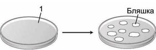
Figure 67 - Determination of the number of infectious particles by the plaque assay; 1 - cell seeding
Since the progeny virions within a plaque originate from a single parental virus, they form a viral clone. This technique is standard for studying bacterial and animal viruses (Figure 68). Plant viruses are investigated similarly by counting the number of local lesions on virus-infected plant leaves. Indeed, the analysis of viral mutants isolated from plaques has made a major contribution to our current understanding of molecular cellular processes. Furthermore, the plaque assay is a vital component in isolating clones of bacteriophage λ, which is widely used to transport segments of cellular DNA in Recombinant DNA technology.

Figure 68 - Viral clones: a - plaques formed by bacteriophage λ in E. coli cells; b - plaques formed by poliovirus in HeLa cells
Last update: 12/08/2026
Editorial and Educational Adaptation: This material has been compiled based on the primary/original source text. The project team performed an editorial review, corrected technical inaccuracies, structured sections, and adapted the content for an educational format.
What was processed:
- elimination of formatting defects (OCR errors, structural breaks, corrupted characters);
- editorial organization of content;
- standardization of terminology in accordance with academic sources;
- verification of factual statements against the original source text.
All mentions of the author, publication year, and origin of the primary text have been preserved in accordance with the source.