BIOCHEMISTRY - L. Stryer - 1984
VOLUME 1
PART I. CONFORMATION AND DYNAMICS
CHAPTER 5. MOLECULAR DISEASES: SICKLE-CELL ANEMIA
5.6. The Surface of Sickle-Cell Hemoglobin Contains "Sticky" Patches
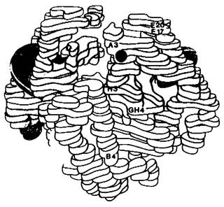
Valine has a nonpolar side chain, whereas glutamate is highly polar. The substitution of glutamate by valine at the 6th position of the β chain results in a nonpolar residue On the surface of Hemoglobin S (Fig. 5.8). As a consequence, the solubility of deoxygenated hemoglobin S decreases significantly, whereas the solubility of oxygenated hemoglobin S remains virtually unchanged. This fact underlies the entire clinical picture of Sickle-Cell Anemia, as well as the features characteristic of the sickle-cell trait.
Class="center">Fig. 5.8. Low-resolution model of deoxyhemoglobin A. α Chains are shown in yellow, β chains in blue. The region where The amino acid substitution occurred in hemoglobin S is marked in red. Note that this region is located on the surface of the molecule
The molecular mechanism behind the appearance of sickle-shaped erythrocytes can be envisioned as follows.
1. The replacement of glutamic acid by valine leads to the appearance of a "sticky patch" on the outer surface of each β chain of hemoglobin S (Fig. 5.9). Such a "sticky patch" is present in both oxy- and deoxyhemoglobin S, but not in hemoglobin A.
Fig. 5.9. Red triangles indicate the "sticky patches" present in both oxy- and deoxyhemoglobin S, but absent in hemoglobin A. The complementary site is depicted as a notch matching the triangle. This complementary site is present in deoxyhemoglobin S and possibly also in deoxyhemoglobin A
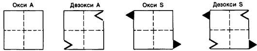
2. Deoxyhemoglobin S possesses a site complementary to the "sticky patch" (Fig. 5.9). The localization of this complementary site has not yet been established. The complementary site of one deoxyhemoglobin S molecule interacts with the "sticky patch" of another deoxyhemoglobin S molecule, leading to The formation of long aggregates that deform the erythrocyte.
3. In oxyhemoglobin S, the complementary site is masked. The "sticky patch" is present, however, the unavailability of the complementary site prevents hemoglobin S molecules from binding to one another.
4. Thus, sickle-shaped erythrocytes appear under conditions where the deoxygenated form of hemoglobin S reaches a high concentration (Fig. 5.10).
Fig. 5.10. The interaction of the "sticky patch" in deoxyhemoglobin S with the complementary site of another deoxyhemoglobin S molecule leads to the formation of long aggregates. One of the helical fiber strands formed in this manner is shown here

Based on these facts, A number of Clinical symptoms of sickle-cell anemia can be explained. For instance, when sickle-shaped erythrocytes appear in the tiniest Blood Vessels, a vicious circle arises. The sickle Cells block the blood vessel, which creates a local oxygen deficiency. As a result, more hemoglobin in this region transitions into the deoxy form, leading to the formation of even more deformed, sickle-shaped erythrocytes. In carriers of the sickle-cell trait, disease symptoms are usually not observed because the concentration of hemoglobin S in this case does not exceed half of the total hemoglobin. At a normal oxygen concentration, this level of hemoglobin S is not high enough to cause erythrocyte deformation. However, upon a significant drop in the partial pressure of oxygen (at high altitudes, for example), sickle-shaped erythrocytes may also appear in carriers of the sickle-cell trait.
5.7. Deoxyhemoglobin S Forms Long Helical Fibers
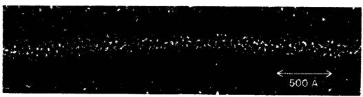
As noted above, deoxyhemoglobin S forms a fibrous precipitate that deforms erythrocytes, giving them a sickle shape (Fig. 5.2). Electron Microscopy reveals fibers of two types: 170 Å in diameter (Fig. 5.3) and, more commonly, 215 Å in diameter (Fig. 5.11). The predominant structures appear to be fibers consisting of a 14-stranded helix, in which 10 hemoglobin S molecules are located on the outside and 4 molecules on the inside (Fig. 5.12). An essential feature of this stranded helix is that each hemoglobin S molecule contacts at least eight others. Clearly, the helix is stabilized by numerous bonds. The bond involving valine-6 in the β chain shifts the thermodynamic equilibrium of the deoxy form of hemoglobin toward fiber formation, though this is not the only factor stabilizing the helix.
Fig. 5.11. Electron micrograph of a negatively stained deoxyhemoglobin S fiber
5.8. The Rate of Fiber Formation Strongly Depends on the Concentration of Deoxyhemoglobin S
The kinetics of deoxyhemoglobin S fiber formation is of great importance, as it determines whether an erythrocyte has time to assume a sickle shape during its passage through the capillaries—that is, in approximately 1 s. The most crucial factor in this process is the concentration of deoxyhemoglobin S; this was demonstrated in in vitro experiments that exploited the fact that the solubility of deoxyhemoglobin S is significantly higher at low temperatures than at high temperatures. The polymerization of deoxyhemoglobin S was triggered by rapidly raising the Temperature of the solution from 4 to 37 °C. Fiber formation was monitored by Changes in the Physical Properties of the solution, specifically light scattering. At a sufficiently low hemoglobin S concentration, fiber formation exhibits a delay of many minutes (Fig. 5.13). The delay time τ depends on the concentration c (more precisely, on the thermodynamic activity) of deoxyhemoglobin S in accordance with the equation
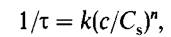
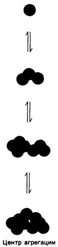
where C8 is the solubility of hemoglobin S at equilibrium, k is a constant depending on environmental conditions, and n is the exponent. Determined experimentally, the value of n turned out to be strikingly large—around 10. In other words, The rate of fiber formation is proportional to the 10th power of hemoglobin S activity. Consequently, fiber formation is a highly cooperative process. Kinetic studies of this process have demonstrated that the rate-limiting phase of fiber formation is the generation of an aggregation center (nucleation). Once a critical-size aggregate forms (consisting of approximately 10 deoxyhemoglobin S molecules), subsequent fiber growth proceeds very rapidly (Fig. 5.14). This aggregate size apparently corresponds to the major part of a single turn of the 14-stranded helix (Fig. 5.12). These findings are of great clinical significance, demonstrating that both kinetic and thermodynamic factors play a crucial role in sickle Cell Formation. Even at high deoxyhemoglobin S concentrations, erythrocytes will not sickle if their transit time from the tissue capillaries to the lung alveoli, where oxygenation takes place, is shorter than the lag phase of fiber formation. The high degree of concentration dependence of the polymerization rate on deoxyhemoglobin S also explains why the sickle cell trait is typically asymptomatic. Specifically, the concentration of deoxyhemoglobin S in heterozygote erythrocytes is roughly half that of corresponding homozygotes; consequently, the rate of fiber formation must be approximately 1000 times slower (210 = 1024).
Fig. 5.12. Model of a deoxyhemoglobin S fiber composed of 14 helical strands. A — longitudinal section, B — cross section. Each circle represents a hemoglobin S tetramer.
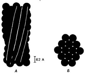
Fig. 5.13. Kinetics of in vitro deoxyhemoglobin S fiber formation based on light-scattering measurements. Polymerization was triggered by a rapid temperature jump from 4 to 37°C. The prolonged lag phase corresponds to the aggregation (complex formation) stage.
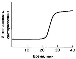
Fig. 5.14. Aggregation phase during the formation of deoxyhemoglobin A fibers. The assembly of aggregation centers proceeds more slowly than the subsequent growth of the aggregates.
Last update: 06/08/2026
Editorial and Educational Adaptation: This material has been compiled based on the primary/original source text. The project team performed an editorial review, corrected technical inaccuracies, structured sections, and adapted the content for an educational format.
What was processed:
- elimination of formatting defects (OCR errors, structural breaks, corrupted characters);
- editorial organization of content;
- standardization of terminology in accordance with academic sources;
- verification of factual statements against the original source text.
All mentions of the author, publication year, and origin of the primary text have been preserved in accordance with the source.