BIOCHEMISTRY - L. Stryer - 1984
VOLUME 1
PART I. CONFORMATION AND DYNAMICS
CHAPTER 5. MOLECULAR DISEASES: SICKLE-CELL ANEMIA
5.4. Peptide Mapping: Identification of the Amino Acid Substitution in Sickle-Cell Hemoglobin
Thus, electrophoretic analysis revealed that Hemoglobin S carries 2 to 4 more positive charges than hemoglobin A. There are several possible explanations for such a difference in molecular charge:
Class="center">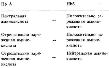
The exact Nature of the substitution in hemoglobin S was elucidated in 1954, when Vernon Ingram developed a novel method for identifying amino acid replacements in Proteins. To carry out the analysis, the hemoglobin molecule was cleaved into smaller fragments, since it is unquestionably easier to detect an amino acid substitution in small Peptides containing about 20 Amino Acids than in an intact protein molecule roughly 10 times larger. Hemoglobin was subjected to specific Trypsin Cleavage at peptide bonds formed by the carboxyl groups of Lysine and Arginine. Because an αβ-half of hemoglobin contains a total of 27 lysine and arginine residues, tryptic Hydrolysis yields 28 distinct peptides. The next step was to separate the resulting peptides, for which a two-dimensional Separation technique was employed (Fig. 5.6). The peptide mixture was applied as a small spot near the corner of a large sheet of filter paper. Electrophoresis was then performed in one direction, thereby separating the peptides according to their net charge. However, this did not achieve complete separation of the mixture, as many peptides overlapped. The separation process was therefore continued using paper Chromatography, carried out in a direction perpendicular to the electrophoretic run.
Fig. 5.6. Peptide mixtures are separated by horizontal electrophoresis followed by vertical chromatography

Chromatography — a term introduced by Mikhail Tsvet in 1906 with reference to the separation of a mixture of plant leaf pigments on a calcium carbonate Column. Tsvet compared this process to "resolving light into a spectrum."
Derived from the Greek words chroma (color) and graphein (to draw, to write).
The Procedure was conducted as follows: the edge of the paper closest to the applied peptides was placed in a mixture of organic Solvents and Water poured at the bottom of a tightly sealed Glass jar. The solvent then ascended the paper by capillary action. Under these conditions, each peptide could either migrate with the solvent (nonpolar environment) or remain bound to the hydrated paper Cellulose (highly polar environment). This separation technique is known as partition chromatography. Peptides with pronounced nonpolar properties dissolve in the solvent and consequently travel up the paper with the solvent front, whereas the most polar peptides remain near the bottom of the paper. Paper chromatography and electrophoresis Complement each other because they separate peptides based on independent properties: the former based on differences in polarity, and the latter based on differences in net charge. The entire analytical procedure—Selective Cleavage of a protein into small peptides followed by two-dimensional separation—is termed peptide mapping (or fingerprinting).
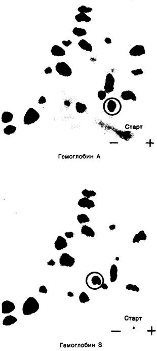
The resulting peptide maps provide exceptionally clear and informative results. Following ninhydrin staining, the peptide spots become clearly visible. A comparison of the maps of hemoglobin A and hemoglobin S showed that all their peptide spots were identical except for one. This spot was eluted from each peptide map and determined in both cases to correspond to an 8-amino-acid peptide. Subsequent Amino acid analysis revealed that the hemoglobin S peptide differs from the hemoglobin A peptide by a single amino acid.
5.5. A Single Amino Acid Substitution Occurs in the β-Chain
The α- and β-chains were separated by Ion-exchange chromatography, after which their peptide maps were obtained. It turned out that hemoglobin S differs from hemoglobin A in its β-chain, specifically in the N-terminal tryptic peptide of the β-chain. By determining the Amino Acid Sequence of this peptide, Ingram demonstrated that valine replaces glutamate at position 6 in the β-chain of hemoglobin S:

Fig. 5.7. Comparison of ninhydrin-stained peptide maps of hemoglobin A and hemoglobin S. The red circle encloses the peptide that differs between the two Hemoglobins
Last update: 06/08/2026
Editorial and Educational Adaptation: This material has been compiled based on the primary/original source text. The project team performed an editorial review, corrected technical inaccuracies, structured sections, and adapted the content for an educational format.
What was processed:
- elimination of formatting defects (OCR errors, structural breaks, corrupted characters);
- editorial organization of content;
- standardization of terminology in accordance with academic sources;
- verification of factual statements against the original source text.
All mentions of the author, publication year, and origin of the primary text have been preserved in accordance with the source.