Molecular Biology: Protein Structure and Functions - Stepanov V.M. 2005
Globins
Hemoglobin
All mammalian Hemoglobins have a Quaternary Structure and consist of four subunits. Almost always, these are two pairs of identical subunits, so hemoglobins can stoichiometrically be represented by formulas such as a2ß2, a2у2, a2ß2, etc. The peptide chains corresponding to the hemoglobin subunits exhibit Primary Structure similarities, meaning they are homologous. For instance, when comparing the primary structures of the a- and ß-chains of human hemoglobin A, which consist of 141 and 147 Amino Acids respectively, about half of the residues match. Homology is also revealed when comparing the sequences of hemoglobins and myoglobins, although it is less pronounced.
At the same time, the tertiary structure and spatial folding of The polypeptide chains in Myoglobin and hemoglobin subunits are very close, reflecting their evolutionary relationship and descent from a common ancestor. Thus, hemoglobin subunits are structured almost identically to the aforementioned myoglobin molecule: the a-subunit is formed by seven, and the ß-subunit by eight a-helices. The binding of an oxygen molecule to hemoglobin subunits, each of which contains a heme group, proceeds in a first approximation similarly to the saturation of myoglobin.
11 mmHg = 1.3 hPa.
The main difference in the Functional Properties of myoglobin and hemoglobin is that The quaternary structure grants the hemoglobin molecule The ability to regulate oxygen binding and release, imparting cooperative properties that myoglobin lacks. Given this, further Discussion of hemoglobin properties will focus on analyzing the Quaternary Structure of this protein and its functional role.
8.2.1. Quaternary Structure of Hemoglobin
In the quaternary structure of hemoglobin, the subunits form a tetrahedron stabilized by intersubunit contacts—a system of non-covalent bonds. These contacts, which account for a total of roughly 1/5 of the entire surface of the four subunits, vary considerably in the number of non-covalent interactions and, consequently, in strength (Fig. 8.4). The most developed and stable contact is between the a-subunit and one of the ß-subunits—the so-called a1—ß1 contact. Its formation involves 34 amino acid residues, with 110 atoms brought within a distance of less than 4 Å of each other. If the total area of intersubunit contacts is taken as 100%, the a1—ß1 contact and the identical a2—ß2 contact account for 60%, which corresponds to a highly complex network of predominantly hydrophobic interactions and several Hydrogen Bonds. The contact of the same a1-subunit with the other ß-subunit, the so-called a1—ß2 contact, looks different because it is formed by other areas of the globule surfaces. It involves 19 amino acid residues forming hydrophobic contacts and hydrogen bonds. It accounts for 33% of the total intersubunit contact surface (along with the identical a2—ß1 contact). As a consequence, it is significantly weaker than the a1—ß1 contact.
Class="center">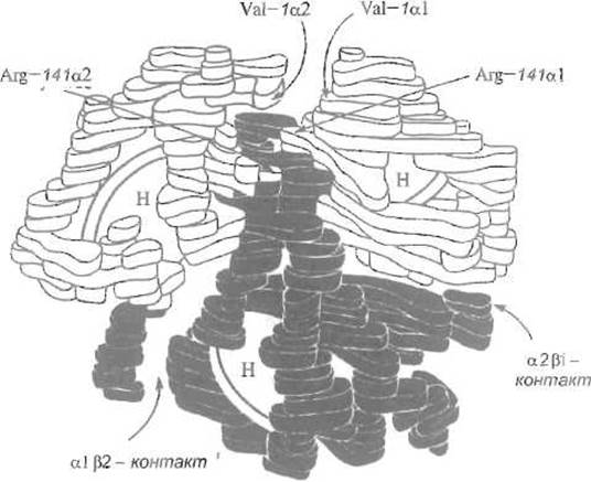
Fig. 8.4. Model of the quaternary structure of hemoglobin.
Discs labeled with the letter H are Hemes. The a1ß2 and a2ß1 contacts are visible. It is shown that the weak a1a2 contact depends on the interaction of the N-terminal valine and C-terminal Arginine residues
Even weaker are the contacts between identical subunits, a1 - a2 and ß1 - ß2, which are represented by a few, predominantly ionic, interactions. They account for about 7% of the total contact surface stabilizing the quaternary structure. Thus, two contacts, i.e., a1 - ß1 and a2 - ß2, dominate The structure of the hemoglobin molecule. Under certain conditions, such as in 2 M NaCl, hemoglobin dissociates into a-ß dimers, which remain intact thanks to the strong a1-ß1 contact. Practically speaking, the quaternary structure of hemoglobin is formed as if by two relatively rigidly linked a-ß dimers held together by weaker interactions (a dimer of dimers).
Under certain conditions, the relative arrangement of the dimers can change due to alterations in The Nature of the weaker a1-ß2 and, especially, a1-a2 and ß1-ß2 contacts. It should not be assumed that weak subunit contacts, which are not overly significant for maintaining molecular integrity, are functionally unimportant. On the contrary, it is precisely the combination of weak and strong interactions that underlies the cooperativity of the hemoglobin quaternary structure, a feature that also appears characteristic of many other Proteins possessing this level of Organization.
As X-Ray Diffraction Analysis has shown, the quaternary STRUCTURE OF THE hemoglobin molecule can exist in one of two states, differing by the Rotation of the a-ß dimers relative to each other (Fig. 8.5). One state is stable when the subunits are saturated with oxygen and the molecule contains four oxygen molecules—oxyhemoglobin. The other is stable when the subunits contain no oxygen—deoxyhemoglobin. Intermediate forms are unstable, and their concentration at any given moment is low. Hemoglobin owes its cooperativity in oxygen binding and release, as well as A number of other functionally important properties, to the existence of these two interconverting quaternary structure forms.
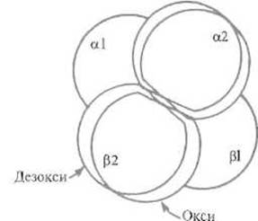
Fig. 8.5. Rotation and displacement of aß-dimers in the quaternary structure of hemoglobin relative to each other during the transition of the protein from the deoxy- to the oxy-form.
The rotation is approximately 15°, and the displacement is 0.8 Å. The dimers are fixed in the new position due to Changes in the a1ß2 contact (see Fig. 8.9)
8.2.2. Oxygen Binding to Hemoglobin and Associated Tertiary Structure Changes
The following rather simplified Description of the sequence of events accompanying the binding of an oxygen molecule to any of the hemoglobin subunits is generally accepted.
An oxygen molecule penetrates inside the subunit and lodges between the heme iron atom and the imidazole group of the distal Histidine, which presumably forms a Hydrogen bond with one of the oxygen atoms. Oxygen binding alters the electronic state of the iron atom, which was previously located somewhat "above" the heme plane, but now shifts by 0.4–0.8 Å, approaching the oxygen and being drawn into the plane of the porphyrin ring (Fig. 8.6). This movement leads to a corresponding Displacement of the imidazole of the proximal histidine, which forms a nearly covalent bond with the iron. The displacement of the imidazole further causes a shift of the entire His F8 histidine residue and the entire F a-helix in which this residue is located. Naturally, the structural change initiated by a small displacement of the iron atom encompasses a considerable number of protein atoms.
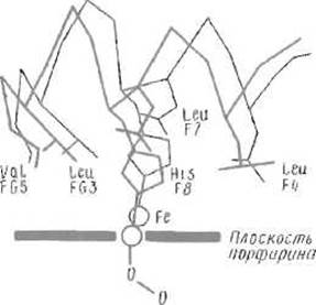
Fig. 8.6. Conformational changes near the heme accompanying oxygen binding to the iron atom and the transition of the hemoglobin subunit from the deoxy- to the oxy-form.
The iron atom is drawn into the plane of the porphyrin ring, causing the displacement of the attached histidine F8 residue, followed by the movement of the entire F helix and the rotation of FG
An important consequence of such a structural transition is that the lateral p-hydroxybenzyl group of the Tyr HC2 Tyrosine residue, which in deoxyhemoglobin was located between the F and H helices, is forced—due to their coming closer together—to leave this position in the structure and emerge onto The surface of the molecule (Fig. 8.7). Note that the Tyr HC2 residue precedes the C-terminal Arg HC3.
A drastic shift in THE POSITION OF the neighboring tyrosine entails A change in the Location of this arginine residue as well, and hence of two charged groups simultaneously: the a-carboxylate ion and the guanidinium group that it possesses. Prior to oxygen binding, the charged Functional groups of the C-terminal Arg HC3 residue of one of the chains (the a-chain) participated in The formation of two salt bridges. The carboxylate ion interacted with the protonated a-amino group of the N-terminal residue of the other a-chain (a2), contributing to the a1-a2 contact. The cationic guanidino group formed a salt bridge with the Asp-126 carboxylate ion in the same a1 chain. Following oxygen binding, the C-terminal arginine loses both of these bonds and acquires the ability to rotate almost freely. The same happens with the C-terminal arginine of the a2 chain, so that in oxyhemoglobin the contact between identical a1-a2 subunits, already weak to begin with, becomes negligible.
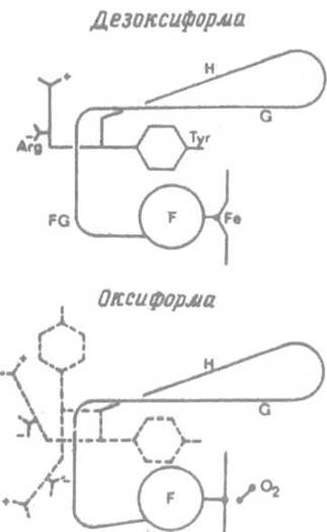
Fig. 8.7. Transmission of the F-helix displacement effect to the C-terminal arginine. The side chain of the Tyr HC2 tyrosine is "squeezed out" of the space between the F and G helices due to their approximation, which sets the C-terminal arginine residue in motion (shown by the dashed line). This entails the rupture of salt bridges involving the terminal amino acid residues
Similarly, the binding of an oxygen molecule to the heme of the ß-subunit (ß2) triggers a conformational rearrangement that begins with the approach of the distal histidine toward the heme iron. The ultimate result is the rupture of the salt bridges formed by the C-terminal His-146 residue. One of these bridges is located between the carboxylate ion of the C-terminal histidine and the protonated amino group of the Lys-40 residue in the a1 chain, while the other forms between the imidazolium cation of His-146 and the Asp-94 carboxylate in the same ß-subunit chain. This entails a weakening of the a1-ß2 contact (Fig. 8.8).
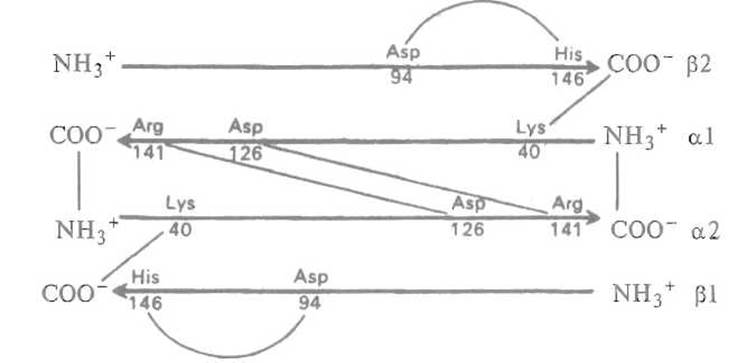
Fig. 8.8. Schematic representation of Electrostatic Interactions (indicated by thin lines) in the hemoglobin molecule.
Subunits are conventionally depicted by arrows pointing toward the C-termini. These bonds are lost upon oxygen binding (oxygenation).
8.2.3. Functional Role of the Quaternary Structure of Hemoglobin
Overall, the transition from deoxyhemoglobin to a fully oxygenated, oxygen-saturated molecule entails the loss of eight ionic bonds, both within and between subunits, specifically affecting the relatively weak a1-a2 and ß1-ß2 contacts. The weakening of the latter leads to the relaxation of the hemoglobin molecule, allowing its halves—the a-ß dimers—to alter their relative orientation. This displacement reaches as much as 7 Å (see Fig. 8.6), whereas the most robust a1-ß1 contact remains virtually unaffected.
The displacement of the a-ß dimers occurs in an abrupt, stepwise manner due to the existence of two alternative modes of non-covalent interactions at the a1-ß2 contact interface. Its surface can be roughly envisioned as a “swallowtail” joint (Fig. 8.9), where in one form (the deoxy- or T-form), the relative arrangement of the subunits is stabilized, notably by a hydrogen bond between the TyrC7 residue of the a1-subunit and the AspG1 carboxyl group of the ß2-subunit. Upon transition to the oxy- (R) form, this bond is lost; however, the carboxyl group of AspG1 in the a1-subunit, having moved into a sterically favorable position following the relative displacement of the dimers, forms a hydrogen bond with AsnG4 of the ß2-subunit. Thus, the a1-ß2 contact Functions as a molecular switch capable of adopting one of two distinct states depending on whether oxygen has bound to the respective subunits.
In summary, the binding of an oxygen molecule to a hemoglobin subunit initiates a cascade of structural changes that propagate from the heme group outward to the periphery of the globule, shifting ionized groups on its surface. This phenomenon—long-range allosteric communication—is characteristic of the Tertiary Structure of proteins. If such an effect were confined solely to the tertiary structure, its contribution would be relatively minor. In hemoglobin, however, it alters ionic interactions within the weak intersubunit contact, modifying it and driving the cooperative transition of the quaternary structure into an alternative state.
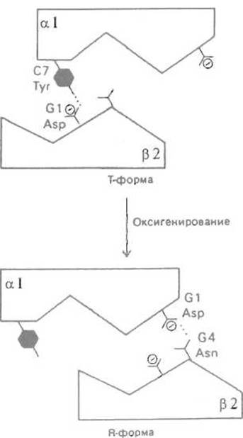
Fig. 8.9. Transition of the a1ß2 contact during hemoglobin oxygenation. The swallowtail-shaped contact can also exist in one of two states upon oxygenation and the transition of the molecule from the T- to the R-state. The loss of the Tyr C7–Asp G1 hydrogen bond between the a1- and ß2-subunits is compensated by the formation of a bond between Asp G1 and Asn G4.
Consequently, the changes occurring in one subunit upon oxygen binding can be transmitted to the remaining subunits, making it energetically favorable for them to alter their conformation so that their tertiary structure conforms to the reorganized quaternary structure, thereby facilitating the binding of further oxygen molecules. Naturally, such a rearrangement of the quaternary structure—its transition from the T-form characteristic of deoxyhemoglobin (from “tight”) to the relaxed R-form typical of oxyhemoglobin (from “relaxed”)—is unlikely (though not entirely impossible) if an oxygen molecule has bound to only one of the four subunits. Furthermore, the energetic barrier imposed by these requisite tertiary structural changes, which conflict with the native deoxy conformation, hinders the binding of the first oxygen molecule while favoring its release.
Consequently, the binding of the first, and to a lesser extent the second, oxygen molecule proceeds with difficulty. It requires a markedly higher partial pressure of oxygen than is necessary for the saturation of myoglobin, which exhibits no such kinetic delays. Indeed, this initial phase of hemoglobin oxygenation takes place in the pulmonary alveoli, where the partial pressure of oxygen is exceptionally high.
Crucially, after two oxygen molecules have bound to hemoglobin, the quaternary structure is equally likely to adopt either the T- or the R-form. The latter promotes the binding of the third oxygen molecule, after which the probability that the entire quaternary structure—including the fourth subunit, not yet loaded with oxygen—will switch into the conformation that facilitates oxygen binding becomes very high. This leads to the rapid completion of saturation for the entire tetrameric molecule. Thus, hemoglobin oxygenation, as a cooperative process, follows a principle akin to “to those who have, more shall be given,” thereby ensuring the complete loading of oxygen carrier molecules in the Lungs.
In peripheral Tissues, the first oxygen molecule is released with a certain degree of difficulty because this process requires alterations in subunit contacts that run counter to the quaternary structure characteristic of oxyhemoglobin. Nevertheless, these energetic hurdles are overcome owing to the low partial pressure of oxygen, which renders the reverse reaction highly improbable. The dissociation of subsequent oxygen molecules is facilitated at each step, resulting in the complete unloading of the transport molecule. Thus, cooperativity ensures the efficient delivery of oxygen from the lungs to the tissues. As physiologists note, in the absence of cooperative effects—for instance, if erythrocytes contained myoglobin instead of hemoglobin—animals would succumb to tissue Hypoxia.
8.2.4. 2,3-Bisphosphoglycerate: An Effector Regulating Hemoglobin Function
Evidently, the Cooperativity of oxygen binding and release by hemoglobin depends on how easily or, conversely, how difficultly the coordinated transition of the quaternary structure from the T- to the R-state occurs. It has been established that 2,3-bisphosphoglycerate (BPG), present in erythrocytes, is capable of interacting selectively with deoxyhemoglobin, thereby stabilizing its quaternary structure (the T-form). 2,3-Bisphosphoglycerate bears five negative charges distributed across eight oxygen atoms. Its molecule binds within the central cavity formed by both ß-subunits, where it simultaneously engages seven cationic groups of these polypeptide chains. These include the a-amino groups of the N-terminal Val-1 residues, the imidazole rings of His-2 and His-143 in both ß-subunits, and the amino group of the Lys-82 residue from one of the chains (Fig. 8.10).
Stabilization of the deoxy conformation diminishes the oxygen affinity of hemoglobin complexed with 2,3-bisphosphoglycerate. In the absence of this effector, adult human hemoglobin A is 50% saturated with oxygen at an oxygen partial pressure of 12 mmHg, whereas in the presence of bisphosphoglycerate, a partial pressure of 50 mmHg is required to achieve the same 50% saturation. 2,3-Bisphosphoglycerate is a compound structurally unrelated to oxygen and binds to a site on the hemoglobin molecule remote from the oxygen-binding pocket. Nevertheless, it exerts a profound influence on oxygen-hemoglobin binding. Compounds possessing this property, which play a major role in regulating protein function within the Organism, are designated as allosteric (structurally distant) effectors. Notably, the very capacity of such an effector to influence a functionally distinct center located far away relies on the inherent flexibility of the quaternary structure, enabling it to respond to relatively subtle effectors.
Alterations within the 2,3-bisphosphoglycerate binding pocket can significantly modulate hemoglobin's oxygen affinity. For instance, in the ß-chains of the South American llama, which inhabits high altitudes and requires adaptation to reduced oxygen partial pressures, the His-2 residues are replaced by asparagine residues. These lack a cationic group and are unable to participate in 2,3-bisphosphoglycerate binding. Consequently, the deoxy conformation of llama hemoglobin is less stabilized, resulting in a higher oxygen affinity. Fifty percent saturation of llama hemoglobin is achieved at an oxygen partial pressure of just 24 mmHg. The functional rationale for this amino acid substitution is entirely transparent, especially given that the dromedary camel (an evolutionary ancestor of the llama) inhabiting lowland plains retains the canonical 2,3-bisphosphoglycerate binding site comprising seven cationic groups, including the His-2 residues of the ß-chains.
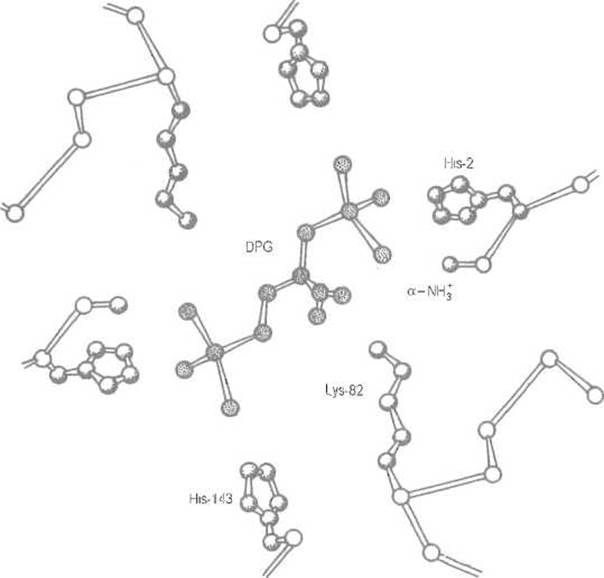
Fig. 8.10. Binding of 2,3-bisphosphoglycerate (BPG) to the cationic groups of hemoglobin ß-subunits.
Electrostatic interactions between the anionic groups of 2,3-bisphosphoglycerate and the cationic groups of deoxyhemoglobin (shaded) substantially reinforce subunit contacts, particularly the very weak ß1ß2 contact. Stabilization of the deoxy form is equivalent to a decrease in oxygen affinity. Affinity can be enhanced in hemoglobins adapted for oxygen binding at low partial pressures through the attenuation of effector interactions. Thus, in llama hemoglobin, binding is weakened by the substitution of His-2 with Asn-2, whereas in fetal hemoglobin (hemoglobin F), it is reduced by replacing His-143 with Ser-143, which diminishes the number of protein cationic groups interacting with the anionic moieties of the effector.
Human fetal hemoglobin (hemoglobin F) is synthesized exclusively during gestation and must ensure the efficient transfer of oxygen from the Placenta, where the oxygen partial pressure is only about 12 mmHg, to fetal tissues (expressed colloquially by physiologists as “the fetus breathes at the altitude of Everest”). Clearly, hemoglobin F must exhibit a higher oxygen affinity than adult hemoglobin. This adaptation is likewise achieved by substituting the His-143 residue in the ß-chain of hemoglobin F with Serine, which weakens 2,3-bisphosphoglycerate binding and reduces the stabilization of the deoxyhemoglobin conformation.
8.2.5. Participation of Hemoglobin in CO2 and Hydrogen Ion Transport
The cooperative effects of the quaternary structure endow hemoglobin with the capacity to perform, alongside Oxygen transport, a series of other physiological functions vital to the Circulatory system.
Hemoglobin transports hydrogen ions and CO2 from the tissues toward the lungs. The transport of hydrogen ions is governed by the Bohr effect. As previously mentioned, the saturation of hemoglobin with oxygen leads to the disruption of ionic bonds between individual functional groups, whereas their release promotes the reformation of these bonds. The rupture or formation of ionic bonds directly influences the propensity of these functional groups to bind protons. This is particularly characteristic of relatively weak bases whose pKa values lie close to the physiological pH. Under such conditions, even minor shifts in pKa substantially alter a group's ability to bind and, consequently, transport a proton. Such groups include the a-amino group and the imidazole ring of histidine. For instance, in oxyhemoglobin, the imidazole ring of the C-terminal His-143 residue in the ß-chains is unliganded and presumably exhibits a normal histidine pKa of approximately 6. Following oxygen release in the tissues, this imidazole ring re-establishes an ionic bond with the carboxylate anion of the Asp-94 residue in the same chain, placing it within a localized negative electrostatic field. Naturally, a proton finds it considerably more difficult to dissociate from the imidazolium cation when the latter's positive charge is compensated by a nearby negatively charged group.
Thus, the ability of the imidazole ring of the C-terminal histidine to bind a proton is regulated by the position of this ring within the Spatial Structure and its participation (or lack thereof) in an ensemble with a carboxyl group. As a consequence, deoxyhemoglobin—in which the basicity of the His-143 imidazole group increases—binds a proton and transports it from the tissues to the lungs, where, upon hemoglobin oxygenation and the transition of its quaternary structure to the relaxed R-form, the hydrogen ion is released.
CO2 transport by hemoglobin is mediated by its ability to bind to the alpha-amino groups of the protein, forming a carbamic acid derivative known as a carbamate (a carbonic acid half-amide):

This reaction takes place in tissues containing the amino groups of deoxyhemoglobin. The resulting negatively charged carbamate residues engage in ionic interactions with the cationic groups of the protein, further stabilizing the deoxy form. In the lungs, upon hemoglobin saturation with oxygen, the reverse reaction occurs, leading to the release of CO2.
8.2.6. Abnormal Hemoglobins
Normal hemoglobin A predominates in the human population; however, abnormal hemoglobins—mutant proteins characterized by a single amino acid substitution—occur with a frequency of approximately 1/1000. Such substitutions can occur at many positions: about 200 abnormal hemoglobins are known, whereas the alpha and beta chains contain a total of 288 amino acid residues. The functional consequences of these substitutions vary significantly. Many of them, particularly those occurring On the surface that do not involve a radical change in the nature of The amino acid residue, have no effect on functional properties, remaining neutral and clinically silent. Nevertheless, a considerable number of abnormal hemoglobins are known whose carriers suffer to a greater or lesser extent from impaired hemoglobin function. The severity of such molecular diseases varies and is typically mitigated in heterozygotes by the presence of normal hemoglobin A.
Abnormal hemoglobins are conventionally named after the geographical location where the given mutation was first discovered. In many cases, structural data on hemoglobin make it possible to elucidate the molecular mechanisms underlying particular disruptions of its Structural and functional properties.
In hemoglobin Vienna, the Tyr-130 residue of the beta chain is replaced by aspartic acid. Its carboxyl group is pulled into the Hydrophobic core, occupying the position of the tyrosine phenyl ring, which destabilizes the molecule. Carriers of this mutation suffer from hemolytic anemia.
In hemoglobin Hammersmith, the replacement of Phe-42 in the beta chain by serine leads to the loss of a crucial hydrophobic contact with the heme. Furthermore, a hydrophilic group—followed by Water molecules—invades the hydrophobic environment of the heme. As a result of the destabilization of the hydrophobic core, the beta chain becomes unstable, readily loses the heme, and undergoes undesirable iron oxidation.
In hemoglobin Boston, the distal His E7 of the alpha chain is replaced by tyrosine, whose phenolic group forms an ionic bond with the heme iron ion, leaving no room for oxygen binding. An analogous substitution of the distal histidine by tyrosine in the beta chain results in the formation of abnormal hemoglobin Saskatoon.
Amino Acid Substitutions in the subunit contact interfaces lead to very serious consequences. For instance, the replacement of Tyr-35 in the beta chain by phenylalanine disorganizes the alpha1-beta1 contact, causing the protein to dissociate into monomers.
As already noted, Mutations affecting amino acid residues on the molecular surface are frequently neutral. However, in hemoglobin S (Sickle-Cell Anemia), the replacement of the surface Glu-6 residue in the beta chain by valine causes the deoxy form to assemble into supramolecular aggregates—extended fibers that subsequently form crystalline structures. Apparently, the appearance of a hydrophobic amino acid at this position is detrimental because its side chain finds a complementary contact site elsewhere on the hemoglobin S molecule, thereby triggering uncontrolled aggregation. It should be borne in mind that the hemoglobin concentration in erythrocytes is extremely high, approaching 35%, which makes the risk of aggregate and even precipitate formation very real. Hemoglobin S aggregates distort erythrocyte Morphology, impairing microcirculation in the capillaries.
There is reason to believe that the mutation responsible for The Emergence of hemoglobin S instead of hemoglobin A occurred in the Middle Ages on the Arabian Peninsula, from where it spread along migration routes. In certain tropical malaria-endemic regions, this mutation became established because the parasite adapts poorly to survival within erythrocytes altered by the presence of abnormal hemoglobin aggregates. This phenomenon serves as a prime model of mutant protein retention in a population under Selection pressure.
Of course, abnormal variants are not restricted to hemoglobins and can affect any other proteins as well. In particular, a number of abnormal human serum albumins have been described.
Last update: 13/08/2026
Editorial and Educational Adaptation: This material has been compiled based on the primary/original source text. The project team performed an editorial review, corrected technical inaccuracies, structured sections, and adapted the content for an educational format.
What was processed:
- elimination of formatting defects (OCR errors, structural breaks, corrupted characters);
- editorial organization of content;
- standardization of terminology in accordance with academic sources;
- verification of factual statements against the original source text.
All mentions of the author, publication year, and origin of the primary text have been preserved in accordance with the source.