Human Anatomy (with the Fundamentals of Dynamic and Sports Morphology) - Ivanitsky, M. F. 2008
General Dynamic Morphology
Displacement of the heart, diaphragm, and internal organs in various body positions
Physical education teachers and coaches must understand the morphological and Functional Characteristics of Internal Organs that result from changes in an athlete's body position during various physical exercises.
The displacement of internal organs during body position changes is studied using contrast radiography. For this method, special contrast agents are injected directly into the organ (such as The Stomach or Urinary Tract). Scans are first taken in the standard upright position and then while performing an exercise. The resulting radiographs are used to evaluate Changes in the borders of a given organ by utilizing bone landmarks.
Heart. Changes in the shape and size of The Heart are accompanied by simultaneous alterations in systemic Blood Circulation. This occurs during exercises such as a back-hollow hang, handstand, and bridge (i.e., when the direction of blood flow relative to the heart changes), as well as exercises that cause an increase in intrathoracic pressure, such as a cross-support on rings, front support (push-up position), and L-sit support.
In a front support, the heart shifts toward the HEAD by approximately 1.5–2 cm; in a hang on rings, it shifts by 6.1 cm in novices and 3.4 cm in highly skilled gymnasts. Performing this exercise very often decreases the transverse diameter of the heart while increasing its longitudinal diameter. This is apparently due to the tension of the Pericardium (heart sac), which exerts lateral pressure on the heart. The displacement of the lower border of the heart can be explained by the tensioning of the subclavian vessels when the upper Regions of the chest are fixed.
During a handstand, back-hollow hang, and L-sit support, the heart takes on a more horizontal position in some athletes, and the cardiac silhouette "waist" becomes less pronounced. The greatest displacement of the heart toward the head is observed during a handstand (Fig. 166): by 8.2 cm in novices and by 6.1 cm in 1st-category gymnasts. In this position, the heart not only shifts toward the head but also assumes a more horizontal orientation. In a back-hollow hang, the displacement of the heart reaches 3.6 cm in 1st-category gymnasts and 3.5 cm in master-level athletes. As with the handstand, the heart occupies a more horizontal position, becomes elongated, and its apex shifts to the left. In a bent-knee hang and an inverted hang, the displacement of the heart's borders is somewhat smaller than in a handstand or headstand.
Changes in the heart borders associated with the performance of specific exercises are more pronounced during expiration than inspiration. They are accompanied by changes in the anterior surface area of the heart (visible on radiographs) and the total heart volume. In a handstand, the area of the anterior surface of the heart generally decreases compared to the initial position, whereas in a headstand and a bent-knee hang, it increases. In novice athletes, changes in heart surface area relative to the initial position are more pronounced than in well-trained individuals.
Roentgenokymography has shown that the heart Functions more intensively when the body is in upside-down positions, which indicates The Need for strict dosage and regulation of such exercises.
Diaphragm. As is well known, THE POSITION OF the diaphragm depends largely, on the one hand, on the mobility of the internal organs, but on the other hand, it can itself exert a significant influence on the position of the organs in the thoracic and abdominal cavities.
The most mobile part of the diaphragm is its muscular portion. The central tendon typically shifts toward the head and rarely downward (such as in a cross-support on rings or an L-sit support). Cranial displacement of the diaphragm occurs most frequently during gymnastic exercises and is explained by increased pressure exerted upon it by the abdominal organs. With appropriate training, a person can voluntarily regulate tension in the diaphragm and thereby provide the necessary resistance to the forces acting upon it. When performing a handstand, novices among gymnasts and wrestlers show a relatively greater diaphragmatic displacement compared to the heart than highly qualified athletes do. Evidently, The process of training in gymnastics and wrestling creates favorable conditions for The Development of this Muscle, As a result of which its tension reduces the displaceability of the heart. Upon exhalation, the displaceability of the diaphragm, like that of the heart, is greater than upon inhalation. Significant displacements of the diaphragm when the body is upside down impede its movement during inhalation due to high pressure from the abdominal organs. This impacts not only external Respiration but also blood circulation.
The displaceability of the right dome of the diaphragm is greater than that of the left, which is associated with the pressure of the Liver against the right dome; in highly skilled gymnasts, the displaceability of the diaphragmatic domes is significantly smaller than in novice athletes.
Class="center">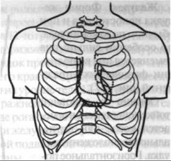
Fig. 166. Projection of heart borders in the standing position and during gymnastic exercises: solid line — standing position; dashed line — hang on rings; dots — handstand (observations by M.L. Jafarov)
Stomach. The shape of the stomach is variable even in a normal body position, but it changes particularly drastically during Physical Exercise (Fig. 167).
Typically, athletes show an oblique or vertical stomach layout in the basic standing position. It generally assumes a horizontal position during a handstand, back-hollow hang, and bridge position. In a hang on rings, the lower part of the stomach is usually pulled upward (by up to 7.2 cm). When performing a cross-support on rings, L-sit support, or front support—situations that create increased intra-abdominal pressure—the stomach shadow generally decreases, and its position and shape change accordingly. The greatest deviations from the starting position are observed during a handstand: the stomach border shifts cranially by 18–19 cm. While in the standing position the shape of the stomach resembles an elongated hook, the cardiac orifice (stomach entrance) is projected to the left of the body's midline at the level of the 6th thoracic vertebra, the fundus is above the upper edge of the 11th rib, the lower border of the greater curvature reaches the 4th lumbar vertebra, and the pyloric part is located at the level of the 2nd lumbar vertebra; conversely, in a hang on rings, the stomach shape resembles a fishing hook, the entrance is projected at the 10th rib level, the fundus rises slightly above the 10th rib, the lowest point of the greater curvature is located at the level of the 2nd lumbar vertebra, and the pyloric part is at the level of the 12th thoracic to 1st lumbar vertebra.
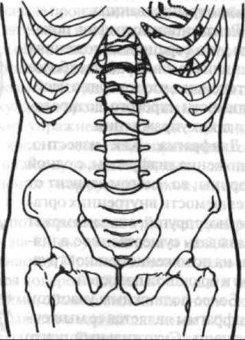
Fig. 167. Position and shape of the stomach during the performance of certain gymnastic exercises (observations by M.A. Jafarov):
solid line — stomach border in the standing position; dashed line — in a hang on rings; dotted line — in a back-hollow hang; double line — in a handstand
In a handstand, the stomach entrance is located on the left between the 9th and 10th Ribs, the fundus is strongly displaced to the left and projected between the 10th and 11th ribs, and the pyloric part is to the right of the 12th thoracic vertebra; that is, the stomach is positioned higher, lies almost horizontally, and has a horn-like shape.
In a back-hollow hang, the stomach entrance is projected between the 8th and 9th ribs, the fundus is at the level of the lower edge of the 8th rib, and the pyloric part is shifted to the right and located at the level of the 12th thoracic vertebra body. During this exercise, the stomach position is at its highest, and it lies almost horizontally in the shape of a horn. After completing the exercises, the stomach walls straighten out, and the organ returns to its initial position. The most mobile part of the stomach is the region of its greater curvature.
Large Intestine. The most mobile section of the large intestine is the transverse colon, which can shift in a cranial direction by up to 20 cm (in the median plane). Its right flexure (up to 14 cm) and left flexure (up to 11.6 cm) shift to a lesser degree; the cecum shifts by 11.2 cm (Fig. 168).
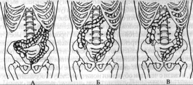
Fig. 168. Displacement of the large intestine in the standing position and during certain gymnastic exercises (observations by M.A. Jafarov):
A — standing position; B — handstand; C — back-hollow hang
The ascending colon tends to change its length and width more frequently than the descending colon. During the execution of almost all exercises, the angle of the right colic flexure is larger and located lower than the angle of the left colic flexure. When performing exercises such as a rest lying crosswise over a bar (front rest across a bar), the ascending colon and the beginning of the right half of the transverse colon, as well as the left half of the transverse colon together with the descending colon, arrange themselves into a so-called double-barreled configuration (sometimes simultaneously on the right and left sides). The appearance of these sharp bends is associated with individual morphological characteristics, increased intra-abdominal pressure, and the point of contact with the bar. If the transverse colon is positioned above this point of contact, it is significantly displaced upward (even more so than in a handstand); if it is below, part of it drops into the pelvic region.
Given the high mobility of the transverse colon, its shape also changes frequently. The appearance and smoothing of characteristic haustra (pouches) of the large intestine (especially along the transverse colon) are accompanied by its expansion and shortening or elongation and narrowing. During the performance of exercises such as a handstand, back-hollow hang, or bridge, the transverse colon may exhibit a convexity facing toward the head.
From all the above, it can be concluded that There is a certain correlation between the shape and position of the heart, diaphragm, stomach, and large intestine, on the one hand, and the position of the body in space, on the other. Almost all of these organs shift upward when performing a hang on rings. During exercises that cause an increase in intra-abdominal pressure (such as a cross on rings or an L-sit), these organs more frequently descend. The greatest changes in their shape and position are observed during exercises such as a handstand, a back arched hang (bridge hang), or a bridge—i.e., when the gravitational force of the internal organs is directed toward the head. The stomach and large intestine undergo more changes in shape and position than other organs. Their position during physical exercise is heavily influenced by the condition of the Abdominal Muscles and the diaphragm. Therefore, when training beginners, special attention should be paid to developing these muscles before moving on to learning complex exercises. Liver and Gallbladder. Despite being fairly securely fixed in the Abdominal cavity, the liver can shift during physical exercise, with particularly significant displacement occurring during a handstand. The gallbladder shifts along with the liver. On an X-ray in the upright position, its shadow typically has a pear shape in most athletes (66.6%). The lower edge of the shadow contour is generally located at the level of the 3rd–4th lumbar vertebrae. In the prone position, it shifts to the level of the 2nd–3rd lumbar vertebrae, and in the upside-down body position (for example, during a back arched hang on rings)—to the level of the 1st–2nd lumbar vertebrae. When transitioning to the prone position, which serves as the starting position for A number of physical exercises, not only does the position of the gallbladder change, but also its shape (becoming bean-shaped in 34.7% of cases), which affects its evacuation function.
Judging by the changes in the position of The Liver and gallbladder during exercise, these organs are subject to translational, rotational, and combined translatory-rotational movements. Specifically, the following patterns of gallbladder displacement have been established: 1) movement of the entire gallbladder upward and laterally; 2) movement of the entire gallbladder upward and medially; 3) movement of the neck of the gallbladder upward and medially, and the fundus upward and laterally; 4) movement of the neck upward and laterally, and the fundus upward and medially.
In addition to body position (standing, lying down, upside down), the gallbladder is significantly affected by The Nature of the efforts exerted by the abdominal muscles and the diaphragm. Thus, it has been established that after flexing the legs in a supine position, the volume of the gallbladder decreases; after running, it can either decrease or increase; and after jumping on both feet or performing exercises such as a bridge or front support, it increases. These findings indicate that during brief increases or decreases in abdominal muscle tension, the tone of the gallbladder increases slightly, whereas with prolonged reduction, it decreases and the gallbladder fills more with Bile.
In women, when standing, the gallbladder is located more medially and higher, its shadow is shorter and narrower, and its volume is smaller than in men. After leg flexion and running, the volume and dimensions of the gallbladder in men decrease, whereas after performing a front support, they increase. In women, after running, performing a bridge exercise, or jumping, the volume and dimensions of the gallbladder generally increase due to a certain degree of bile stagnation.
Kidneys, renal pelves, Ureters. In the standing position, the kidneys are typically located at the level of the 12th thoracic to the 3rd lumbar vertebrae.
In a back arched hang, the shadow of the renal calyces and pelvis appears higher than its initial position. The ureters straighten during this exercise and shift slightly laterally. As a rule, the upper end of the Ureter shifts more than its middle and lower thirds. The right ureter deviates less than the left.
After jumping, the shadow of the renal calyces and pelvis is located below the initial position. On the right side, it drops by 3–10 mm and shifts laterally by 2–4 mm, while on the left side, it drops by 4–14 mm and shifts by 2–7 mm, respectively. During a barbell bench press, the shadow of the renal pelvis and calyces shifts medially and downward.
The upward displacement of the kidneys is often accompanied by a decrease in their angle of inclination in the frontal plane, whereas downward displacement is accompanied by an increase. Such rotations occur more frequently on the right side and are more pronounced than on the left. Among the external and internal forces determining the magnitude and direction of the resultant pressure force on the Kidney, the force of the abdominal muscles and the diaphragm should be considered the most active.
Uterus and fallopian tubes. These organs also undergo some positional changes during physical exercise. The longitudinal axis of the uterus relative to the body's midline is usually located asymmetrically both before and after exercise. The greater part of the uterine cavity shadow lies to the left or right of this line. If the uterus deviates to the side during exercise, it is generally in the direction of its greater natural tilt. Typically, parts adjacent toalei the uterus (the ISTHMUS OF THE fallopian tubes) shift along with the uterus itself. The magnitude of displacement of this part of the fallopian tubes is greater than that of the ampullary part.
When transitioning from a supine to a sitting position—i.e., when intra-abdominal pressure increases due to the contraction of the abdominal wall muscles—the uterus and fallopian tubes undergo relatively minor topographical changes, despite even the additional load. The left and right corners of the uterine cavity do not always shift uniformly.
When performing parallel bar exercises with support against the bar using the anterior abdominal wall, a slight displacement of the uterine fundus is observed. As the point of pressure approaches the line connecting the anterior superior iliac spines, the mechanical impact on the uterus and fallopian tubes increases, which may be facilitated by a certain shift of the intestines. Based on this, excessive pressure of sports apparatuses against the anterior abdominal wall should be avoided, especially impacts of a Shock nature.
Jumping almost always has a uniform effect on the position of the uterus and fallopian tubes, causing them to shift downward. However, jumping can also cause the uterus to deviate either forward or backward—i.e., its natural tilt may decrease. In the first case, the uterine fundus descends more than the cervix; In the second case, the cervix descends more than the fundus. Characteristically, the second variant of uterine displacement is typically observed when landing with pronated feet. Therefore, it is necessary to land on both feet (in vaulting over the buck, playing volleyball, etc.) with slight supination. Incidentally, this is advisable both for biomechanical reasons (better shock absorption conditions) and technical sports reasons (better balance maintenance).
The nature of uterine displacement during physical exercise may depend on The Structure of body movements, individual technique characteristics, and anatomical and functional features (physical development, pelvic tilt angle, etc.). In this regard, ongoing gynecological monitoring of female athletes is essential, especially those specializing in athletic jumps, discus throwing, shot put, backstroke swimming, etc.
GENERAL PATTERNS OF internal organ displacement during human body movements.
Studies have shown that human body movements produce an inertial force effect on internal organs. For instance, when the body moves downward, the Organs of the abdominal cavity initially lag behind the walls of the cavity they inhabit due to inertia, temporarily ending up higher than their initial level. Upon landing, conversely, the internal organs experience impact overloads because, during a sudden halt of the body, inertial forces continue to act upon them.
Thus, during downward body movement (initiation of movement, falling, landing), the pressure exerted by the abdominal organs on the cavity walls changes twice: first it decreases, and then it increases compared to the baseline value. Moreover, the increase in pressure prevails over its decrease.
The Effect of inertial forces on internal organs is more pronounced in the lower sections of the abdominal cavity. Given the presence of poorly protected areas in its anterior wall, as well as the relatively smaller mass and smaller physiological cross-section of the muscles in the lower half of the abdomen, attention must be paid to developing the strength of these muscles, particularly in sports specializations such as pole vaulting, long jump, high jump, running, etc. Constant dynamic and static overloads on abdominal organs supported by weak abdominal muscles can become one of the causes of visceral ptosis (dropping of internal organs).
The degree to which the inertial force of internal organs manifests depends not so much on body mass as on the mass of the organs themselves. The Structural and functional Features of the pelvis also matter. The same intra-abdominal pressure in men and women, combined with differing abdominal press muscle strength and varying minor pelvis dimensions, places the female body at a comparative disadvantage, which obliges coaches to be especially careful in selecting and dosing physical exercises for women.
Last update: 08/08/2026
Editorial and Educational Adaptation: This material has been compiled based on the primary/original source text. The project team performed an editorial review, corrected technical inaccuracies, structured sections, and adapted the content for an educational format.
What was processed:
- elimination of formatting defects (OCR errors, structural breaks, corrupted characters);
- editorial organization of content;
- standardization of terminology in accordance with academic sources;
- verification of factual statements against the original source text.
All mentions of the author, publication year, and origin of the primary text have been preserved in accordance with the source.