Anatomy and Physiology of Children and Adolescents - M. R. Sapin 2007
Urogenital System
Urinary System
Kidneys
The Kidney is a paired, bean-shaped organ with a firm consistency. The kidneys are located on the posterior abdominal wall, on either side of THE Vertebral Column, extending from the level of the XII thoracic to the I–II lumbar vertebrae. The right kidney lies slightly lower than the left. A suprarenal (adrenal) gland sits superiorly on top of each kidney. Anteriorly, the kidneys are related to the Peritoneum; the right kidney is in contact (through the peritoneum) with the Liver, the right colic flexure, and the descending part of the duodenum. The anterior surface of the left kidney is adjacent to The Stomach, Pancreas, left colic flexure, and loops of the Small Intestine.
Class="center">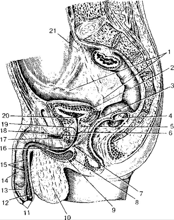
Fig. 61. Topography of the male URINARY AND REPRODUCTIVE Organs in the lesser pelvis cavity:
1 — Ductus deferens (vas deferens); 2 — Urinary Bladder; 3 — Seminal Vesicle; 4 — rectum; 5 — Prostate Gland; 6 — ejaculatory duct; 7 — urethral sphincter; 8 — bulbospongiosus Muscle; 9 — bulb of Penis; 10 — Scrotum; 11 — prepuce of penis; 12 — external urethral orifice; 13 — glans penis; 14 — corpus cavernosum of penis; 15 — spongy part of male Urethra (penile urethra); 16 — membranous part of male urethra; 17 — deep transverse perineal muscle; 18 — seminal colliculus (verumontanum); 19 — prostatic part of urethra; 20 — internal urethral orifice; 21 — Ureter

Fig. 62. Topography of the female urinary and reproductive organs in the lesser pelvis cavity:
1 — Uterine tube (fallopian tube); 2 — Ovary; 3 — Uterus; 4 — rectum; 5 — rectouterine pouch (pouch of Douglas); 6 — posterior part of vaginal fornix; 7 — external orifice of uterus (uterine os); 8 — Vagina; 9 — vaginal orifice; 10 — labium majus; 11 — labium minus; 12 — glans clitoridis; 13 — body of Clitoris; 14 — female urethra; 15 — urinary bladder; 16 — vesicouterine pouch; 17 — round ligament of uterus
The kidney features superior and inferior poles, anterior and posterior surfaces, and two borders: a convex lateral border and a concave medial border. A depression is located on the medial border — the renal hilum, which leads into the renal sinus. Entering the kidney through the hilum are the renal artery and nerves, while the renal vein and Lymphatic vessels exit the organ. The renal sinus contains the Major and minor calyces, the renal pelvis, and surrounding adipose tissue. The kidney is enveloped by a tough fibrous capsule, which in turn is surrounded by a layer of fat (the adipose capsule).
On a coronal (frontal) section of the kidney (Fig. 63), two distinct layers are visible: an outer, lighter-colored renal cortex and an inner, darker renal medulla. The cortex contains renal corpuscles, as well as the proximal and distal parts of nephron tubules (renal tubules). The medulla consists of 7–10 renal pyramids. The base of each pyramid faces the cortex, while its tapering apex — the renal papilla — projects into a minor calyces. Extensions of cortical tissue, known as renal columns, project between the pyramids and contain the interlobar Blood Vessels of the kidney.
The Structural and functional unit of the kidney is the nephron. A nephron consists of a glomerular capsule and a system of nephron tubules. The combined length of the tubules in a single nephron is 50–55 mm, while the total length of all nephrons in both kidneys reaches about 100 km. The diameter of the tubules varies from 15 to 60 µm across different segments of the nephron. Each kidney contains over 1 million nephrons. The beginning of each nephron is a double-walled glomerular capsule (Bowman's capsule), which encloses a glomerulus of blood capillaries. The capsule together with the vascular glomerulus forms a renal corpuscle with a diameter of about 200 µm. The nephron comprises the glomerular capsule, the proximal convoluted tubule, the nephron loop (Loop of Henle) consisting of descending and ascending limbs, and the Distal convoluted tubule (Fig. 64).
The glomeruli of all nephrons are located in the renal cortex, whereas their loops extend into the medulla. The distal tubules open into cortical collecting ducts, which originate in the cortex. These collecting ducts then descend through the medullary pyramids and empty into short papillary ducts, which finally open into the minor renal calyces.
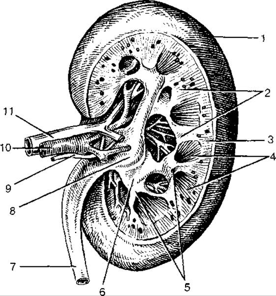
Fig. 63. Right kidney. Frontal (longitudinal) section.
Posterior view:
1 — renal capsule; 2 — renal columns; 3 — renal cortex; 4 — renal medulla (pyramids); 5 — minor renal calyces (opened); 6 — major renal calyces; 7 — ureter; 8 — renal pelvis; 9 — nerve; 10 — renal artery; 11 — renal vein
In its Structure, the glomerular capsule resembles a double-walled goblet. A space (the capsular space) lies between the inner and outer walls of the nephron capsule. The inner wall is tightly adherent to the glomerular capillaries, the number of which can reach 50 in a single corpuscle. Thus, two fused walls — the capillary endothelium and the glomerular visceral layer (making up the renal filtration barrier) — lie between the blood in the capillaries and the lumen of the glomerular capsule. Fluid is filtered across these walls from the blood into the lumen of the nephron capsule, forming primary urine. Over the course of a day, about 180 liters of primary urine are filtered into the capsular spaces of both kidneys.
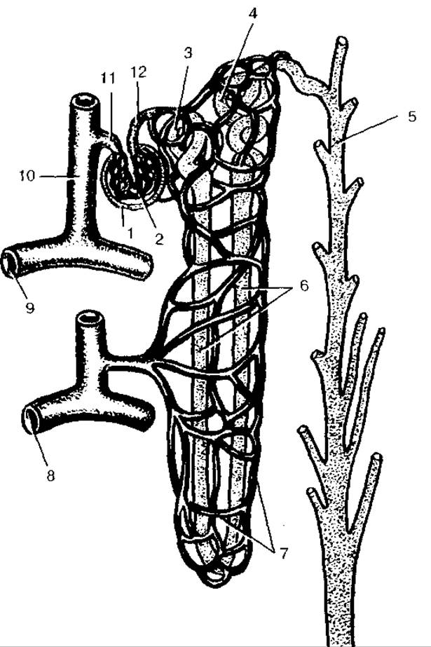
Fig. 64. STRUCTURE OF THE nephron and its vascular supply:
1 — glomerular capsule; 2 — glomerulus (vascular tuft) of renal corpuscle; 3 — proximal convoluted tubule; 4 — distal convoluted tubule; 5 — collecting duct; 6 — nephron loop (loop of Henle); 7 — peritubular capillary network; 8 — arcuate vein; 9 — arcuate artery; 10 — interlobular artery; 11 — afferent glomerular arteriole; 12 — efferent glomerular arteriole
The STRUCTURE AND Functions of nephrons are closely tied to the Organization OF THE renal vascular bed. Upon entering the renal hilum, the renal artery branches out. Its interlobar Arteries course through the renal columns between the medullary pyramids. At the level of the pyramid bases, these vessels arch to form arcuate arteries, situated along the corticomedullary junction. Numerous interlobular arteries branch off the arcuate arteries into the cortex. Intralobular arteries diverge from each interlobular artery, giving rise to afferent glomerular arterioles. Within the renal corpuscle, these arterioles break up into the glomerular capillary network, which is enclosed by the glomerular capsule. These capillaries converge to form the efferent glomerular arteriole, which exits the renal corpuscle and subsequently branches a second time into peritubular capillaries that entwine the nephron tubules. Blood from this secondary capillary network drains into venules, which merge into interlobular Veins, then into arcuate veins, and further into interlobar veins. The latter converge and enlarge to form the renal vein. Thus, the kidneys possess two capillary systems. One of these is the arterial vascular glomerulus, interposed between two arterioles (the so-called "admirable net" or *rete mirabile*). The other is a typical capillary network situated along the pathway between efferent arterioles and venules.
Last update: 10/08/2026
Editorial and Educational Adaptation: This material has been compiled based on the primary/original source text. The project team performed an editorial review, corrected technical inaccuracies, structured sections, and adapted the content for an educational format.
What was processed:
- elimination of formatting defects (OCR errors, structural breaks, corrupted characters);
- editorial organization of content;
- standardization of terminology in accordance with academic sources;
- verification of factual statements against the original source text.
All mentions of the author, publication year, and origin of the primary text have been preserved in accordance with the source.