HUMAN MEDICAL BIOLOGY, ANATOMY, PHYSIOLOGY AND PATHOLOGY - Ya.I. Fedoniuk 2010
ANATOMY, PHYSIOLOGY, PATHOLOGY
SECTION 3. ANATOMIC AND PHYSIOLOGICAL ASPECTS OF BODY FUNCTION AUTOREGULATION
ENDOCRINE GLANDS AND THEIR PATHOLOGY
PITUITARY GLAND
1. STRUCTURE AND FUNCTIONS OF THE PITUITARY GLAND
The Pituitary Gland (hypophysis) lies in the hypophyseal fossa on the floor of the sella turcica of the Sphenoid bone and is separated from the cranial cavity by a fold of the dura mater that forms the diaphragma sellae. Through an opening in this Diaphragm, the pituitary is connected to the infundibulum of the Hypothalamus of the Diencephalon. Externally, the pituitary gland is covered by a capsule, and its mass does not exceed 0.6 g. It consists of anterior and posterior lobes (Fig. 3.77). The anterior lobe comprises the pars distalis, which occupies the anterior part of the hypophyseal fossa; the pars intermedia, located at the boundary with the posterior lobe; and the pars tuberalis, which connects to the infundibulum of the hypothalamus. The parenchyma of the anterior lobe is composed of glandular Cells, whereas the posterior lobe consists of neuroglial cells (pituicytes), nerve fibers extending from neurosecretory nuclei of the hypothalamus into the neurohypophysis, and neurosecretory bodies. The anterior lobe—the adenohypophysis—produces tropic Hormones that regulate The activity of Endocrine glands. The posterior lobe is known as the neurohypophysis.
Class="center">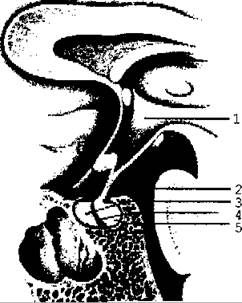
Fig. 3.77. Pituitary gland (diagram)
The pituitary gland occupies a special place among the endocrine glands due to the function of specific tropic hormones that regulate the activity of other endocrine glands.
The adenohypophysis produces two groups of hormones. The first group includes hormones that influence metabolic processes and regulate body GROWTH AND DEVELOPMENT. These are somatotropin (Growth Hormone), Lipoproteins, and prolactin.
The second group comprises tropic hormones, whose primary role is to regulate the function of peripheral glands. These include adrenocorticotropin, thyrotropin, and gonadotropic (luteinizing and follicle-stimulating) hormones.
Hormone effects. Somatotropic hormone, or growth hormone, promotes the synthesis of Proteins and Nucleic Acids within cells, driving linear growth, overall body size, body mass, and the size of individual Organs.
Prolactin stimulates mammary gland growth and milk secretion.
Following childbirth, pituitary secretion of prolactin increases, leading to Lactation—the production and release of milk. Prolactin is also present in males, in whom it stimulates the growth and Development of the Prostate Gland and Seminal Vesicles.
Lipoproteins stimulate the mobilization of fat from adipose depots.
Melanocyte-stimulating hormone regulates pigment METABOLISM and promotes the redistribution of melanin pigment in the Skin, retina, Hair, and iris.
Adrenocorticotropic hormone (ACTH) promotes the Synthesis and Secretion of corticosteroids, the Hormones of the Adrenal Glands.
Thyrotropic hormone stimulates the synthesis and release of THYROID HORMONES. Extirpation of the pituitary gland in animals leads to thyroid atrophy, whereas the administration of thyrotropic hormone results in the proliferation of thyroid tissue.
Gonadotropic hormones include the follicle-stimulating and luteinizing hormones. These hormones are present in both males and females. In males, the follicle-stimulating hormone influences The formation of Male Germ Cells. In females, this hormone promotes the growth of vesicular follicles and the secretion of follicular fluid. Luteotropic hormone stimulates the synthesis of Female Sex Hormones (estrogens) and Male Sex Hormones (androgens). Additionally, in females, this hormone triggers the release of the ovum from the ovarian follicle and the formation of a Cytology/practical/130.html">Corpus luteum at the site of the ruptured follicle.
Hypofunction of the anterior pituitary lobe halts The Development of the Reproductive System. Conversely, hyperfunction leads to precocious Puberty.
Thus, the activity of the anterior pituitary lobe is closely linked to the function of other endocrine glands and is, in turn, regulated by the hypothalamus via releasing hormones.
Secretory Cells of the hypothalamic nuclei produce antidiuretic hormone and oxytocin. These hormones are transported along axons forming the hypothalamo-hypophyseal tract to the posterior pituitary lobe (neurohypophysis), from which they enter the bloodstream.
Antidiuretic hormone promotes enhanced Water reabsorption in the distal renal tubules, thereby reducing urine output and increasing its concentration.
When its concentration in the Blood increases, antidiuretic hormone exerts a pressor effect by constricting arterioles, which leads to an elevation in blood pressure.
Oxytocin enhances uterine contractions, ensuring a normal labor process, and promotes milk ejection after childbirth.
Last update: 08/08/2026
Editorial and Educational Adaptation: This material has been compiled based on the primary/original source text. The project team performed an editorial review, corrected technical inaccuracies, structured sections, and adapted the content for an educational format.
What was processed:
- elimination of formatting defects (OCR errors, structural breaks, corrupted characters);
- editorial organization of content;
- standardization of terminology in accordance with academic sources;
- verification of factual statements against the original source text.
All mentions of the author, publication year, and origin of the primary text have been preserved in accordance with the source.