Human Anatomy - Kotsan I. Y. 2009
Endocrine glands
Endocrine portion of the pancreas
The Pancreas is classified as a gland of mixed secretion, consisting of both exocrine and endocrine components. The exocrine part is described in the chapter on the Digestive System. The Endocrine Part (pars endocrina pancreatis) is represented by the pancreatic islets (insulae pancreaticae), commonly known as the islets of Langerhans.
The pancreatic islets develop from the same epithelial primordium of the primitive gut as the exocrine portion of the pancreas. They appear as oval or spherical structures measuring up to 0.3 mm, with some reaching 1 mm in diameter. Their total number ranges from 1 to 2 million. Although distributed throughout all PARTS OF THE pancreas, they are most abundant in the tail region. The islets lack excretory ducts. They are formed by clusters of epithelial Cells surrounded by Connective Tissue and a dense network of capillaries penetrating their interior. The pancreatic islets comprise three main Cell types: alpha, beta, and delta cells (Fig. 229). Beta cells are the most abundant (60–80%) and produce the hormone Insulin, which exerts diverse effects on the body, most notably lowering Blood glucose levels. Insulin promotes The conversion of glucose into Glycogen, stimulates lipid synthesis and storage in subcutaneous adipose tissue, and enhances Carbohydrate METABOLISM in Muscles. Insufficient insulin secretion leads to Diabetes Mellitus, a condition characterized by elevated blood sugar (hyperglycemia) and the presence of glucose in the urine (glucosuria). Alpha cells (accounting for 10–30%) secrete Glucagon, a hormone with antagonistic effects to insulin, as it stimulates Glycogenolysis and lipolysis. Delta cells (approximately 10%) synthesize Somatostatin, which inhibits the pituitary release of Growth Hormone, suppresses the secretion of both insulin and glucagon, and reduces The production of pancreatic digestive Enzymes.
Class="center">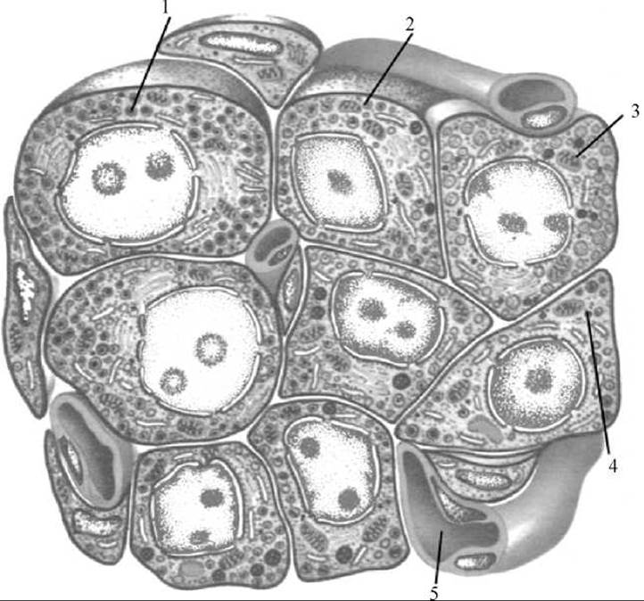
Fig. 229. Diagram of The Structure of a pancreatic islet
1 — alpha cell; 2 — beta cell; 3 — delta cell; 4 — PP cell; 5 — blood capillary
The relative mass of the islet tissue of the pancreas varies across different age groups, being highest in infants under two months of age. By the age of one year, the mass of the islet tissue accounts for 1–0.8% of the total glandular mass, a ratio that remains relatively stable until 40–50 years of age. In infants under six months, insulin production is twice as high as in adults. After the age of 40, The activity of the endocrine apparatus declines: insulin secretion decreases, while glucagon levels rise. This hormonal imbalance can predispose individuals to The Development of diabetes mellitus.
Last update: 08/08/2026
Editorial and Educational Adaptation: This material has been compiled based on the primary/original source text. The project team performed an editorial review, corrected technical inaccuracies, structured sections, and adapted the content for an educational format.
What was processed:
- elimination of formatting defects (OCR errors, structural breaks, corrupted characters);
- editorial organization of content;
- standardization of terminology in accordance with academic sources;
- verification of factual statements against the original source text.
All mentions of the author, publication year, and origin of the primary text have been preserved in accordance with the source.