Review of Medical Physiology - William F. Ganong 2002
Endocrine System, Metabolism, and Reproduction
Energy Balance, Metabolism, and Nutrition
Carbohydrate Metabolism
Dietary CARBOHYDRATES are primarily hexose polymers, the most important of which are glucose, galactose, and fructose (Fig. 17-8). Most Monosaccharides in the body occur as D-isomers. Glucose is the primary product absorbed in the digestive tract and circulating in the Blood. Normally, the plasma glucose level in peripheral venous blood is 70-110 mg/dL (3.9-6.1 mmol/L), while in arterial Blood Plasma it is 15-30 mg/dL higher than in venous blood.
Class="center">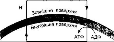
Fig. 17-7. Simplified diagram of proton Transport Across the inner and outer surfaces of The inner mitochondrial membrane via The electron transport system (flavo-cytochrome system), followed by the reverse movement of protons down the proton gradient, resulting in ATP synthesis.
Upon cellular uptake, glucose is predominantly phosphorylated to glucose-6-phosphate. The enzyme catalyzing this reaction is called hexokinase. The Liver also contains glucokinase, which has a higher affinity for glucose; its activity, like that of hexokinase, is enhanced by Insulin and suppressed during fasting and in diabetes. Glucose-6-phosphate is either utilized for Glycogen synthesis or degraded (Catabolism). These pathways are illustrated in Fig. 17-9. The process of glycogen synthesis is termed Glycogenesis, whereas its breakdown is Glycogenolysis. Glycogen, as a storage form of glucose, is found in many body Tissues, but is most abundant in The Liver and skeletal Muscles. The breakdown of glucose to Pyruvate or lactate (or both) is called Glycolysis. Carbohydrate Catabolism proceeds either via the Formation of fructose and its Cleavage into trioses, or through oxidation and decarboxylation yielding pentoses. The pathway leading to pyruvate via triose intermediates is known as the Embden-Meyerhof pathway, whereas the pathway leading to 6-phosphogluconate and pentoses is the oxidative Pentose Phosphate Pathway (pentose monophosphate shunt) (see Fig. 17-9). Pyruvate is converted into acetyl-CoA. During the Interconversion of Carbohydrates, fats, and Proteins, the glycerol moiety of fats is converted into dihydroxyacetone phosphate, and A number of Amino Acids—whose carbon skeletons resemble Intermediates of the Embden-Meyerhof pathway and The Citric Acid Cycle—are converted into these intermediates following deamination. In this manner, as well as through The conversion of lactate to glucose, non-glucose molecules can replenish glucose levels (Gluconeogenesis).

Fig. 17-6. Simplified diagram of Oxidative Phosphorylation. During the METABOLISM of fats, glucose, and amino acids, protons (H+) are generated and pass across the inner mitochondrial membrane (dashed line). The diffusion of these protons down the established concentration gradient drives ATP synthase to convert ADP into ATP. Key Enzymes for this conversion are shown in Fig. 1-7.
Glucose can be converted into fats via acetyl-CoA; however, because the conversion of pyruvate to acetyl-CoA is irreversible (unlike other glycolytic reactions, Fig. 17-10), fats cannot be converted back into glucose through this pathway. The conversion of fats to glucose in the body is minimal (with the exception of a quantitatively minor formation of glucose from glycerol), given the lack of metabolic pathways for this interconversion.
The Citric Acid Cycle
The citric acid cycle (Krebs cycle, Tricarboxylic Acid Cycle) is a sequence of reactions in which acetyl-coenzyme A is metabolized to CO2 and hydrogen atoms. Acetyl-coenzyme A first condenses with the dicarboxylic acid anion oxaloacetate to yield citrate and HS-CoA. Through a series of seven consecutive reactions, two molecules of CO2 are released, and oxaloacetate is regenerated (see Fig. 17-10). Four pairs of hydrogen atoms are transferred to the flavoprotein-cytochrome chain, generating 12 molecules of ATP and 4 molecules of H2O, of which two H2O molecules are consumed within the cycle. The citric acid cycle serves as the common pathway for The oxidation of carbohydrates, fats, and Certain amino acids to CO2 and H2O.
The primary route leading into the citric acid cycle is via acetyl-CoA, although certain Amino acids can be converted into citric acid cycle intermediates via deamination. The citric acid cycle requires O2 and does not operate under anaerobic conditions.

Fig. 17-8. Structure OF THE principal dietary hexoses. Formulas of the major naturally occurring isomers are shown.
Energy Generation
The net yield of high-energy phosphate compounds generated during the metabolism of glucose and glycogen to pyruvate depends on whether metabolism proceeds via the Embden-Meyerhof pathway or The pentose phosphate pathway. In substrate-level phosphorylation, 1 mole of ATP is produced per mole of phosphoglyceraldehyde, and the conversion of 1 mole of phosphoenolpyruvate to pyruvate yields another mole of ATP. Because the conversion of 1 mole of glucose-6-phosphate via the Embden-Meyerhof pathway produces 2 moles of phosphoglyceraldehyde, the metabolism of 1 mole of glucose to pyruvate yields 4 moles of ATP. All these reactions occur without O2 and consequently represent The pathway of anaerobic energy production. However, 1 mole of ATP is consumed in forming fructose-1,6-diphosphate from fructose-6-phosphate, and another mole is used for glucose phosphorylation. Thus, The formation of pyruvate from glycogen under anaerobic conditions yields 3 moles of ATP per mole of glucose-6-phosphate; if pyruvate is formed from 1 mole of glucose, this net yield is only 2 moles of ATP.
The conversion of phosphoglyceraldehyde to phosphoglycerate requires NAD+. One might expect that under anaerobic conditions (anaerobic glycolysis), a metabolic block would occur at the phosphoglyceraldehyde conversion step as the available NAD+ is depleted to NADH. However, this does not happen because NADH transfers its hydrogen to pyruvate acting as a hydrogen acceptor, thereby regenerating NAD+ and forming lactate:

Through this pathway, glucose Metabolism and Energy production can proceed in the absence of O2. The accumulated lactate can be converted back to pyruvate, and in the presence of O2, NADH passes its hydrogen to the flavoprotein-cytochrome chain.
During aerobic glycolysis, the net ATP yield is 19 times greater than under anaerobic conditions. Four ATP molecules are produced via the oxidation in the flavoprotein-cytochrome chain of two NADH molecules generated during the conversion of 2 moles of phosphoglyceraldehyde to phosphoglycerate (see Fig. 17-9); another six ATP molecules are produced from two NADH molecules formed during the conversion of pyruvate to acetyl-CoA; and 24 ATP molecules are generated during two successive turns of the citric acid cycle: of these, 18 result from the oxidation of six NADH molecules, four from the oxidation of two FADH2 molecules, and two from substrate-level phosphorylation when succinyl-CoA is converted to succinate. Although GTP is actually produced in this reaction, it is energetically equivalent to ATP. Consequently, the net yield of ATP per mole of glucose metabolized via the Embden-Meyerhof pathway and the citric acid cycle is 2 + [2 x 3] + [2 x 3] + [2 x 12] = 38.
When glucose is oxidized via the pentose phosphate pathway, significant amounts of NADPH are produced. This reduced coenzyme is essential for numerous metabolic processes. The resulting pentoses serve as building blocks for nucleotide synthesis (see below). The amount of ATP generated depends on the proportion of NADPH that is trans-hydrogenated to NADH for subsequent oxidation.
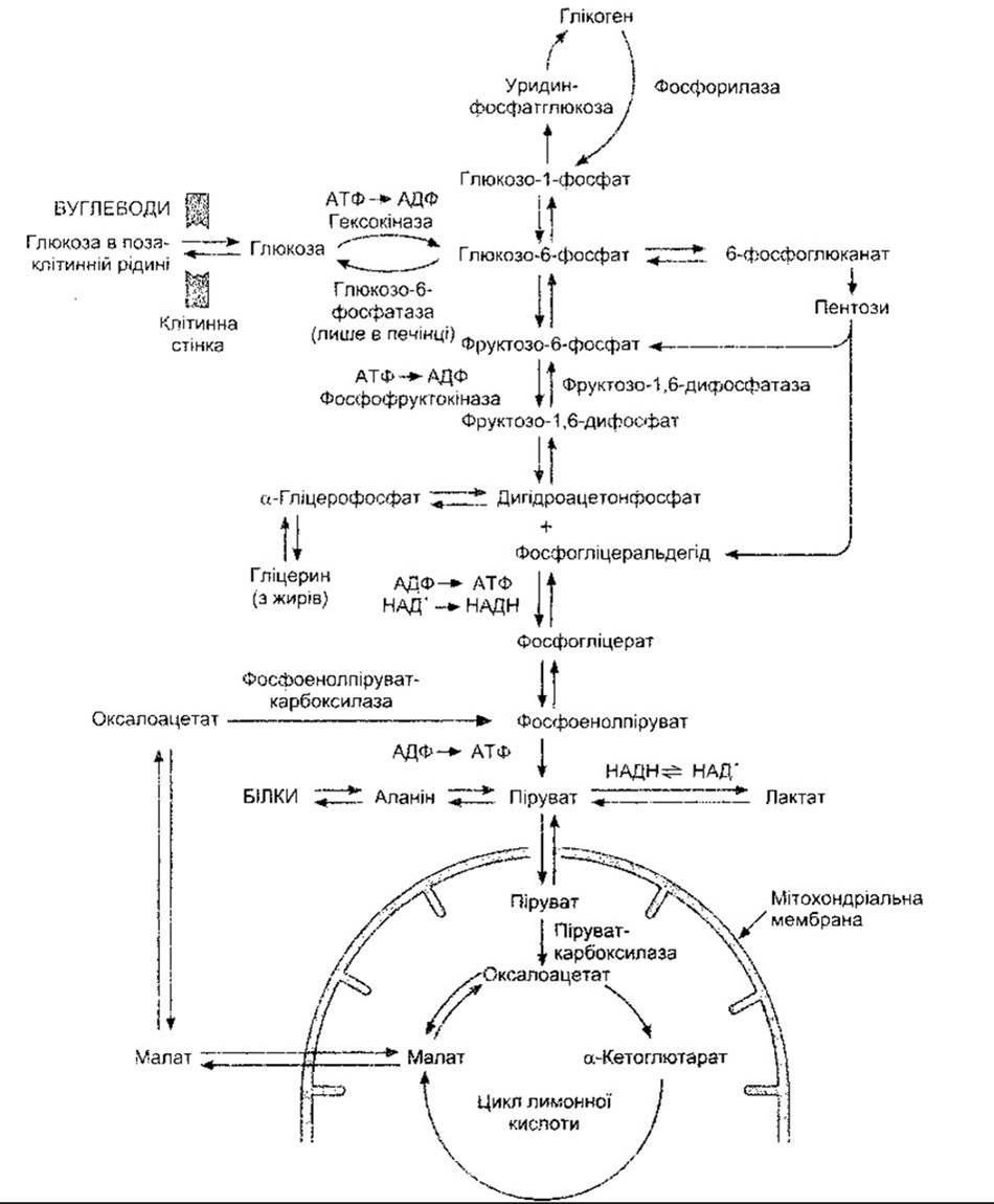
Fig. 17-9. Scheme of cellular Carbohydrate metabolism. The Key Enzymes of carbohydrate conversion are indicated.
"Directional Valves" (One-Way Gates)
Metabolism is regulated by various Hormones and other factors. Controlling certain metabolic processes requires directing Chemical Reactions along a single pathway.
Most Intermediary Metabolism reactions are reversible, but among them are several "directional valves"—that is, reactions driven in one direction by a specific single enzyme or transport mechanism, whereas their reverse path requires a different enzyme. Figure 17-11 illustrates five such reactions in intermediary carbohydrate metabolism. Other Examples include the distinct pathways for fatty acid Synthesis and catabolism (see below). Regulatory factors influence metabolism by acting directly or indirectly on these types of "directional valves."
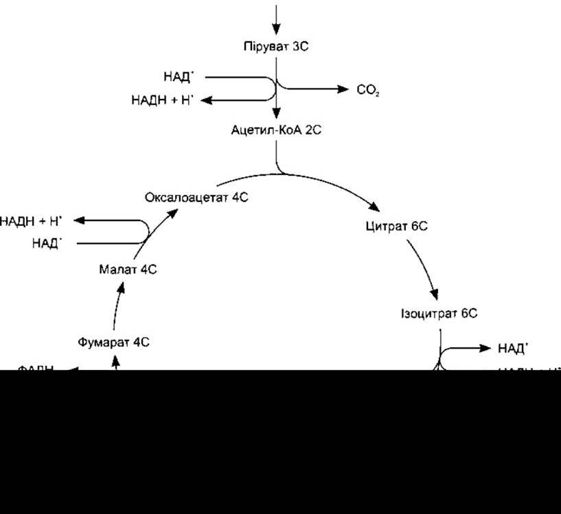
Fig. 17-10. The citric acid cycle. The numbers (6C, 5C, etc.) indicate the number of carbon atoms in the intermediates. The conversion of pyruvate to acetyl-CoA and each turn of the cycle result in the formation of four molecules of NADH and one molecule of FADH2, which are subsequently oxidized in the flavoprotein-cytochrome chain, as well as one molecule of GTP, which is readily converted to ATP (modified and reproduced by permission from Alberts BM et al: Molecular Biology of The Cell, 2nd ed. Garland, 1989).
Synthesis and degradation of Glycogen
Glycogen is a branched polymer of glucose formed by Two Types of glycosidic bonds: 1:4α and 1:6α (Fig. 17-12). It is synthesized on glycogenin—a protein primer—starting from glucose-1-phosphate via uridine diphosphate glucose (UDPG). The enzyme glycogen synthase completes the synthesis. The presence of glycogenin is one of the Factors Determining the extent of glycogen formation. The Cleavage of the 1:4α-linkage is catalyzed by phosphorylase, whereas the cleavage of the 1:6α-linkage is catalyzed by another enzyme.
Phosphorylase is activated in part by epinephrine, which acts on hepatic β2-adrenergic receptors. This, in turn, initiates a cascade of reactions that serves as a classic example of hormonal action mediated by cAMP (Fig. 17-13). Protein kinase A is activated by cAMP and catalyzes The transfer of a phosphate group to phosphorylase kinase, converting it to its active form. Phosphorylase kinase catalyzes the phosphorylation and consequent activation of phosphorylase. Inactive phosphorylase is known as phosphorylase $b$ (dephosphophosphorylase), and the activated form as phosphorylase $a$ (phosphophosphorylase).
The cAMP-mediated activation of protein kinase A not only enhances glycogen breakdown but also inhibits its synthesis. Glycogen synthase (see Fig. 17-12) is active in its dephosphorylated form and inactive when phosphorylated. It is phosphorylated by phosphorylase kinase upon the activation of protein kinase A.
Glycogen is also broken down in response to catecholamines acting on hepatic α1-adrenergic receptors. This degradation is mediated by intracellular Ca2+ and triggered by the Activation of a cAMP-independent phosphorylase kinase. Large doses of vasopressin and angiotensin II can also induce glycogenolysis via this pathway, though this is less physiologically significant, as these hormones play a positive role in glucose Homeostasis. Because the liver contains the enzyme glucose-6-phosphatase, most of the glucose-6-phosphate produced in this organ can be converted to glucose and released into the bloodstream, raising plasma glucose levels. Renal function also contributes to maintaining this level. Other tissues lack this enzyme; therefore, in these tissues, the overwhelming majority of glucose-6-phosphate is catabolized via the Embden-Meyerhof pathway and the hexose monophosphate shunt. Increased Glucose Catabolism in Skeletal Muscle leads to elevated blood lactate levels (see Chapter 4).

Fig. 17-11. Five examples of carbohydrate metabolism "pace-maker valves," i.e., reactions that proceed in one direction via one mechanism and in the reverse direction via another. The double line in Example 5 denotes the mitochondrial membrane. Pyruvate is converted to malate within the Mitochondria, and malate diffuses out of the mitochondria into the Cytosol, where it is converted to phosphoenolpyruvate.
Epinephrine stimulates adenylate cyclase, leading to the activation of phosphorylase in the liver and skeletal muscles. The consequences are an increase in plasma glucose levels and a rise in blood lactate. Glucagon has a similar effect, but it acts exclusively on hepatic phosphorylase. Consequently, glucagon causes an elevation in plasma glucose levels without altering blood lactate levels.
McArdle's Disease
In the condition known as McArdle's disease, or myophosphorylase deficiency glycogenosis, glycogen accumulates in skeletal muscles due to a deficiency of muscle phosphorylase. Affected individuals experience muscle pain and rigidity during physical exertion, along with a marked decrease in exercise tolerance. Skeletal muscle lacks the glycogen breakdown required to supply energy for Muscle contraction (see Chapter 3), and the amount of glucose delivered to the muscles from the bloodstream is sufficient only for very limited movement. Administration of glucagon or epinephrine results in a normal plasma glucose response, indicating that these patients have no deficiency in hepatic phosphorylase.
The "Hepatic Glucostat"
When plasma glucose levels are high, glucose is taken up by the liver; when they are low, glucose is released from the liver into the blood. Thus, the liver acts as a "glucostat" that maintains a constant blood glucose concentration. This function does not occur automatically. The processes of hepatic glucose uptake and release are regulated by numerous hormones. The endocrine regulation of plasma glucose levels and carbohydrate metabolism is discussed in Chapter 19.
Renal Glucose Regulation
Glucose is readily filtered by the Kidneys; however, at normal plasma glucose levels, all but a very small fraction of it is reabsorbed in the proximal tubules (see Chapter 38). As the amount of filtered glucose increases, its reabsorption increases as well, up to a certain limit. That is, once the maximum tubular reabsorptive capacity (TmG) is exceeded, measurable amounts of glucose appear in the urine (glucosuria). The renal threshold for glucose—the arterial blood concentration at which glucosuria occurs—is approximately 180 mg/dL. However, it may be higher if the Glomerular Filtration rate is low.
Glucosuria
Glucosuria occurs when plasma glucose levels rise due to insulin deficiency (Diabetes Mellitus) or excessive glycogenolysis following physical or emotional stress. Some individuals have a congenital defect in renal tubular glucose transport mechanisms, in which case glucosuria occurs even at normal plasma glucose levels. This condition is known as renal glucosuria. Alimentary glucosuria—glucosuria following the ingestion of a carbohydrate-rich meal—necessarily occurs in practically healthy individuals, although a significant number of such persons actually have a mild form of diabetes mellitus. The maximum rate of intestinal Glucose Absorption is 120 g/h.
Factors Determining Plasma Glucose Concentration
Plasma glucose levels depend on the balance between glucose entering the bloodstream and glucose being cleared. The primary factors influencing this balance are dietary glucose intake, The rate of glucose uptake by muscle Cells, adipose tissue, and other tissues, and the glucostatic activity of the liver (Fig. 17-14). Five percent of the glucose absorbed in the intestine is immediately converted into glycogen in the liver, and 30% to 40% into fats. The remainder is metabolized in muscles and other tissues. During fasting, liver glycogen is broken down, and glucose is released from the liver into the bloodstream. During prolonged fasting, glycogen stores become depleted, and the liver engages in gluconeogenesis from Amino Acids and glycerol. In otherwise healthy individuals, prolonged fasting can lower plasma glucose levels to approximately 60 mg/dL; however, symptoms of hypoglycemia do not develop because gluconeogenesis prevents any further drop in glucose levels.

Fig. 17-12. Synthesis and Breakdown of glycogen. The activation process of phosphorylase is illustrated in Fig. 17-13.

Fig. 17-13. The reaction cascade by which epinephrine activates phosphorylase. Glucagon exerts a similar effect in the liver, but not in skeletal muscle.
Carbohydrate Homeostasis During Exercise
In a 70-kg human, carbohydrate reserves amount to approximately 2,500 kcal, stored as 400 g of muscle glycogen, 100 g of liver glycogen, and 20 g of glucose dissolved in the extracellular fluid. For comparison, 112,000 kcal (roughly 80% of the body's energy reserves) are stored as fats, with the remainder in protein compounds. Resting muscles, as well as muscles recovering from physical exertion, rely on Fatty acids for their metabolic needs. During fasting at rest, the human Brain utilizes 70% to 60% of glucose, with red Blood Cells consuming most of the remainder.
During physical work, the muscles' caloric demand is initially met through glycogenolysis and increased glucose uptake. Plasma glucose levels initially rise due to enhanced hepatic glycogenolysis, though they may drop significantly during prolonged, intense exercise. An increase in gluconeogenesis is observed (Fig. 17-15). Plasma insulin levels decrease, while glucagon and adrenaline levels increase. Following exercise, hepatic glycogen stores are replenished via additional gluconeogenesis, while hepatic glucose output declines.
Metabolism of Other Hexoses
In addition to glucose, the gastrointestinal tract absorbs galactose, which is produced during the breakdown of lactose and converted into glucose within the body, as well as fructose, derived partly from dietary sources and partly from the Hydrolysis of sucrose. Following phosphorylation, galactose reacts with UDPG to form uridine diphosphate galactose, which is reconverted to UDPG and participates in glycogen synthesis (see Fig. 17-12). This reaction is reversible; the conversion of UDPG to uridine diphosphate galactose serves to supply galactose for the synthesis of Glycolipids and mucoproteins should dietary galactose intake be insufficient. The utilization of galactose, like that of glucose, is insulin-dependent (see Chapter 19). Inborn errors of metabolism known as galactosemia involve a deficiency of galactose-1-phosphate uridylyltransferase—the enzyme that catalyzes the reaction between galactose-1-phosphate and UDPG—resulting in the accumulation of absorbed galactose in the blood. This condition leads to severe growth and developmental impairments. Adopting a galactose-free diet significantly improves the patient's condition; it prevents galactose deficiency because uridine diphosphate galactose is synthesized from UDPG. Fructose is partially converted into fructose-6-phosphate and further metabolized via fructose-1,6-diphosphate (see Fig. 17-9). The enzyme catalyzing the formation of fructose-6-phosphate is hexokinase, which also catalyzes the conversion of glucose to glucose-6-phosphate. However, a much larger proportion of fructose is converted into fructose-1-phosphate in a reaction catalyzed by fructokinase. Most of the fructose-1-phosphate is cleaved into dihydroxyacetone phosphate and glyceraldehyde, which is phosphorylated and enters glucose metabolism along with dihydroxyacetone phosphate. Because reactions initiated by the phosphorylation of fructose at the first position can proceed at normal rates independently of insulin, fructose is recommended for diabetic patients to replenish their carbohydrate stores. Nevertheless, since the bulk of fructose is metabolized in the intestine and liver, its capacity to replenish carbohydrates in other Organs is limited.

Fig. 17-14. Plasma glucose homeostasis. Note the glucostatic function of the liver, as well as The excretion of glucose in the urine when its renal threshold is exceeded (dashed arrow).
Fructose-6-phosphate can also be phosphorylated at the second position to yield fructose-2,6-diphosphate, a key regulator of hepatic gluconeogenesis. High concentrations of fructose-2,6-diphosphate enhance the conversion of fructose-6-phosphate to fructose-1,6-diphosphate, thereby stimulating glycolysis down to pyruvate. Conversely, a decrease in fructose-2,6-diphosphate levels favors the reverse reaction and thus promotes gluconeogenesis. Protein kinase, under The Influence of glucagon, causes a reduction in hepatic fructose-2,6-diphosphate levels (see Chapter 19).
Last update: 10/08/2026
Editorial and Educational Adaptation: This material has been compiled based on the primary/original source text. The project team performed an editorial review, corrected technical inaccuracies, structured sections, and adapted the content for an educational format.
What was processed:
- elimination of formatting defects (OCR errors, structural breaks, corrupted characters);
- editorial organization of content;
- standardization of terminology in accordance with academic sources;
- verification of factual statements against the original source text.
All mentions of the author, publication year, and origin of the primary text have been preserved in accordance with the source.