Review of Medical Physiology - William F. Ganong 2002
Endocrine System, Metabolism, and Reproduction
Energy Balance, Metabolism, and Nutrition
Protein Metabolism
Proteins consist of A large number of Amino Acids (Fig. 17-16) linked into chains by peptide bonds, which connect the amino group of one acid to the carboxyl group of another. In addition, Some proteins contain CARBOHYDRATES (Glycoproteins) and Lipids (Lipoproteins). Shorter chains of Amino acids are called Peptides or Polypeptides. The boundaries between peptides, polypeptides, and proteins are not sharply defined. In this book, chains 2-10 amino acid residues in length are referred to as peptides, chains containing more than 10 but fewer than 100 residues as polypeptides, and chains containing 100 or more amino acid residues as proteins. The term oligopeptide, sometimes used to denote small peptides, is not used here. The sequence of amino acids in a peptide chain is called the Introduction/19.html">Primary Structure of a protein. Chains that are folded and twisted in a complex manner, and the term Secondary structure of a protein, refer to its spatial Organization formed by such folding and twisting. A typical secondary structure is a regular helix with 3.6 amino acid residues per turn (α-Helix). Another typical secondary structure is the β-pleated sheet. An antiparallel β-pleated sheet is formed when an extended polypeptide chain folds back on itself, establishing Hydrogen Bonds between the peptide linkages of adjacent chains. In addition, parallel β-pleated sheets occur between polypeptide chains. In the so-called ribbon model of a protein (see Fig. 27-8), α-helices are depicted as coils and β-pleated sheets as parallel arrows.
The Tertiary Structure of a protein is the packing of folded chains into layers, crystals, or fibers. The term quaternary structure is used to denote the arrangement of subunits that make up many protein molecules (e.g., Hemoglobin; see Chapter 27).
Class="center">
Fig. 17-15. Output from the Liver of glucose formed by Glycogenolysis (unshaded portion of the bar) and glucose formed presumably by Gluconeogenesis (portion of the bar bounded by the brace). The contributions of various glucose precursors to gluconeogenesis are shown (reproduced with permission from Felig P, Wahren J: Fuel Homeostasis in exercise. N Engl J Med 1975;293:1078).
Amino Acids
The amino acids found in proteins are listed in Table 17-2. They are designated by three- or single-letter Abbreviations. Many other important amino acids, such as Ornithine, 5-hydroxytryptophan, L-DOPA, taurine, and thyroxine (T4), exist in the body but are not found in proteins. In higher animals, the natural forms of amino acids are L-isomers. In particular, L-isomers of Hormones, such as thyroxine, are much more active than D-isomers. Amino acids have a basic, neutral, or acidic reaction depending on The ratio of free acidic (-COOH) and basic (-NH2) groups in the molecule.
Some amino acids are Essential Amino Acids, meaning they must be obtained from the diet, whereas others can be synthesized in vivo in amounts sufficient to meet metabolic needs (see below).
Amino Acid Pool
Although a small amount of proteins and some peptides are absorbed in the gastrointestinal tract, the vast majority of ingested proteins are digested and their constituent amino acids absorbed. The body's own proteins are continuously broken down into Amino Acids and resynthesized. Endogenous protein turnover averages 80-100 g/day, being highest in the intestinal mucosa and near zero in Collagen. Amino acids formed during endogenous protein breakdown are identical to those of dietary proteins. Together, they form The amino acid pool that meets the body's requirements (Fig. 17-17). Most filtered amino acids are reabsorbed in the Kidneys. During growth, the equilibrium between amino acids and body proteins shifts toward proteins, so that synthesis exceeds degradation. At any age, a fraction of protein is lost in the form of Hair; in women, small amounts of protein are lost during menstruation. Some small proteins are excreted in the urine, and unreasorbed proteins of digestive secretions are present in the feces. Losses are replenished by synthesis from the amino acid pool.
Table 17-2. Amino acids found in proteins
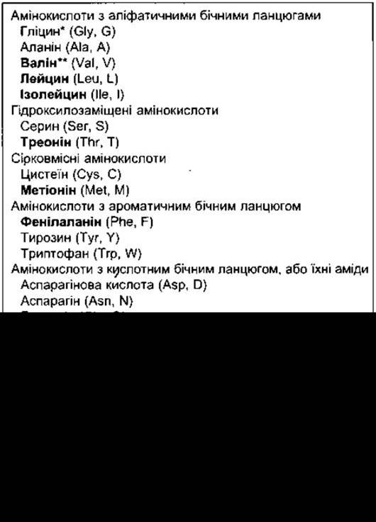
* Standard three- and single-letter abbreviations for amino acids are given in parentheses.
** Essential amino acids are shown in bold.
1 There are no tRNAs for these four amino acids; they are formed by posttranslational Modification of the corresponding unmodified amino acids within the peptide linkage. There are tRNAs for selenocysteine and the other 20 amino acids, and they are incorporated into proteins under direct Genetic control.
2 Arginine and Histidine are sometimes called semi-essential—they are not required for the maintenance of nitrogen balance, but are necessary for normal growth.
3 Selenocysteine is a rare amino acid in which the sulfur of Cysteine is replaced by selenium. The UGA codon is normally a stop codon, but in some instances it encodes selenocysteine.

Fig. 17-16. Amino acid structure and formation of peptide bonds. The dashed line represents The formation of peptide bonds with the release of H2O; R represents the amino acid residue; for example, in Glycine R = H, in glutamate R = -(CH2)2-COO-.
Specific Metabolic Functions of Amino Acids
THYROID HORMONES, catecholamines, histamine, serotonin, melatonin, and intermediates in The Urea Cycle are derived from Certain amino acids. Methionine and cysteine supply the sulfur contained in proteins, CoA, taurine, and other biologically important compounds. Methionine is converted into S-adenosylmethionine, an active methylating agent in the synthesis of compounds such as adrenaline. It is the major donor of biologically labile methyl groups, although methyl groups can also be supplied by formic acid derivatives attached to Folic acid derivatives, provided that sufficient amounts of Folic Acid and cyanocobalamin are present in the diet.
Urinary Sulfates
The oxidation of cysteine is the principal source of urinary sulfates. Most urinary excretion is in the form of sulfates (SO42-), accompanied by equivalent amounts of cations (Na+, K+, NH4+, or H+). Ethereal sulfates in the urine are organic esters of sulfonic acids (R-O-SO3H) formed in the liver from endogenous and exogenous phenols, including estrogens and Other Steroids, indoles, and drugs.
Deamination, Amination, and Transamination
The interconversions between amino acids and the catabolic products of carbohydrates and fats at the level of the general metabolic pool and The Citric Acid Cycle involve the transfer, Cleavage, and formation of amino groups.

Fig. 17-17. Amino acid METABOLISM.
Transamination reactions involve The conversion of one amino acid into the corresponding keto acid, with the simultaneous conversion of another keto acid into an amino acid:

These reactions occur in many Tissues. Transaminases involved in these processes are also present in the Circulation. When extensive damage to active Cells occurs As a result of a pathological process, the level of serum transaminases rises. An example is the elevation of plasma aspartate aminotransferase (AST) levels in myocardial infarction.
Oxidative Deamination of amino acids takes place in the liver. An imino acid is formed via dehydrogenation, after which a Hydrolysis reaction yields the corresponding keto acid with the release of NH4+:

The NH4+ cation exists in equilibrium with NH3. Amino acids can also take up NH4+ to form the corresponding amide. An example of such a reaction is The addition of NH4+ to glutamate, which is observed in the Brain (Fig. 17-18). The reverse reaction occurs in the kidneys, yielding NH4+ for excretion in the urine (see Chapter 38).
The interconversions between the amino acid pool and the general metabolic pool are illustrated in Fig. 17-19. Leucine, isoleucine, phenylalanine, and Tyrosine are termed ketogenic because they are converted into acetoacetate (Ketone Bodies, see below). Alanine and many Other Amino Acids are glucogenic because they yield compounds that can be readily converted into glucose.
Biosynthesis of Urea
Most of the NH4+ generated by AMINO ACID DEAMINATION in the liver is converted into urea. The NH4+ cation forms carbamoyl phosphate, which is transferred to ornithine in the Cell/35.html">Mitochondria to yield citrulline. The enzyme catalyzing this process is ornithine transcarbamylase. Citrulline is then converted into arginine, from which urea is cleaved, regenerating ornithine (the urea cycle; Fig. 17-20). The liver is the primary site of urea synthesis; consequently, in severe liver disease, Blood urea nitrogen (BUN) levels decrease while blood NH3 levels rise. An inherited deficiency of ornithine transcarbamylase can also lead to NH3 toxicity, even in heterozygous individuals.
Creatine is synthesized in the liver from methionine, glycine, and arginine. In Skeletal Muscle, it is phosphorylated to form phosphocreatine (Fig. 17-21), which serves as an important energy reserve for ATP synthesis (see Chapter 3). ATP generated during Glycolysis and Oxidative Phosphorylation reacts with creatine to produce ADP and phosphocreatine. During Physical Exercise, this reaction reverses, ensuring a supply of ATP, which is the immediate energy source for Muscle contraction. Some athletes use creatine as a dietary Supplement, claiming it improves performance in sprinting and Other forms of short-duration high-intensity exercise.

Fig. 17-18. Release and uptake of NH4+ during the interconversion of glutamine and glutamate. The NH4+ cation is in equilibrium with NH3. The reaction equilibrium shifts to the right in the kidneys, and NH3 is secreted into the urine. The reaction equilibrium shifts to the left in the brain, removing neurotoxic NH3.
Urinary creatinine is derived from phosphocreatine. Creatine is not converted directly into creatinine. The rate of creatinine excretion remains relatively constant over long periods. In metabolic studies, urinary creatinine is frequently measured to verify collection accuracy; the average daily creatinine excretion is determined, and the daily output of Other Compounds is referenced to a specific level of excreted creatinine.

Fig. 17-19. Participation of The Citric Acid cycle in transamination and gluconeogenesis. Highlighted arrows indicate the main pathway of gluconeogenesis (reproduced with permission from Murray RK et al: Harper’s Biochemistry, 25th ed. McGraw-Hill, 2000).

Fig. 17-20. The urea cycle.
Creatinuria is occasionally observed in children, in women during and after Pregnancy, and sometimes in nonpregnant women. Normally, adult male urine contains little to no creatine, but its level increases markedly under any condition associated with intensive muscle breakdown. Creatinuria occurs in starvation, thyrotoxicosis, uncompensated Diabetes Mellitus, and various Primary and secondary muscle diseases (myopathies).
Purines and Pyrimidines
Physiologically important purines and pyrimidines are shown in Fig. 17-22. Nucleosides—purines or pyrimidines linked to ribose—are components not only of various Coenzymes and related compounds (NAD+, NADP+, ATP, UDP-glucose, etc.) but also of RNA and DNA (Table 17-3). The STRUCTURE AND FUNCTIONS of DNA and RNA, as well as their role in Protein Synthesis, are discussed in Chapter 1.

Fig. 17-21. Creatine, phosphocreatine, and creatinine.
Dietary Nucleic Acids are broken down (digested) and their constituent purines and pyrimidines are absorbed; however, the majority of purines and pyrimidines are synthesized from amino acids, primarily in the liver, and are subsequently used to form NUCLEOTIDES, RNA, and DNA. RNA exists in dynamic equilibrium with the amino acid pool, whereas DNA, once formed, remains metabolically stable throughout life.
Purines and pyrimidines released from nucleotide breakdown can be either reused or catabolized, with only a small fraction excreted unchanged in the urine. Pyrimidines are catabolized to CO2 and NH3, whereas purines are converted into uric acid.
Protein Degradation
Like protein synthesis, protein degradation is a meticulously regulated and complex process. The conjugation of proteins with ubiquitin, a 74-amino-acid polypeptide, serves as a tag for proteins destined for degradation. This polypeptide is highly conserved and present in all cells, ranging from Bacteria to humans. Ubiquitination of cytosolic proteins—including integral proteins of The Endoplasmic reticulum—targets them for degradation by multi-subunit proteolytic complexes known as 26S proteasomes, whereas ubiquitination of Membrane Proteins, such as growth Hormone Receptors, targets them for degradation in Lysosomes. A strict balance is maintained between the rates of protein Synthesis and degradation; consequently, ubiquitin tagging plays a crucial role in cellular biology, such as in Cell Cycle regulation (see Chapter 1). The metabolic turnover rates of individual proteins vary, and the body therefore possesses mechanisms to recognize and degrade abnormal proteins more rapidly than normal cellular constituents. For instance, abnormal Hemoglobins are rapidly catabolized in individuals with inherited hemoglobinopathies (see Chapter 27). Protein degradation decreases in hypertrophied, conditioned skeletal Muscles and increases during atrophy in denervated or otherwise disused skeletal muscles. Furthermore, it is a key determinant of organ size (e.g., the rate of hepatic protein degradation drops markedly during compensatory hypertrophy following partial hepatectomy).
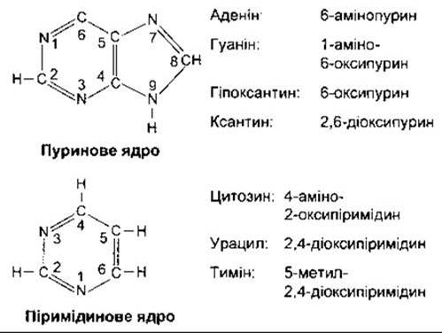
Fig. 17-22. Major physiologically important purines and pyrimidines. Oxypurines and oxypyrimidines can form enolic derivatives (hydroxypurines and hydroxypyrimidines) by shifting a hydrogen atom to an oxygen-containing substituent.
Table 17-3. Purine- and pyrimidine-containing compounds
Compound type |
Components |
Nucleoside |
Purine or pyrimidine plus ribose or 2-deoxyribose |
Nucleotide (mononucleotide) |
Nucleoside plus a phosphoric acid residue |
Nucleic acid |
Multiple nucleotides forming double-stranded structures or two polynucleotide chains |
Nucleoprotein |
Nucleic acid plus one or more simple basic proteins |
Ribose-containing |
Ribonucleic acid |
2-Deoxyribose-containing |
Deoxyribonucleic acid |
Uric Acid
Uric acid is formed via The breakdown of purines and through direct synthesis from 5-phosphoribosyl pyrophosphate (PRPP) and glutamine (Fig. 17-23). In humans, it is excreted in the urine, whereas in certain animals it is oxidized to allantoin prior to excretion. The normal blood uric acid concentration in humans is approximately 4 mg/dL (0.24 mmol/L). The kidneys filter, reabsorb, and secrete uric acid. Typically, 98% of the filtered uric acid is reabsorbed, and the remaining 2% accounts for about 20% of the total uric acid excreted, with the other 80% supplied by tubular secretion. Uric acid excretion on a purine-free diet is about 0.5 g/day, rising to approximately 1 g/day on a normal diet.
Primary and Secondary Gout
Gout is a disorder characterized by recurrent attacks of acute Arthritis, urate deposits in the joints, kidneys, and other tissues, and elevated levels of uric acid in the blood and urine. The first metatarsophalangeal joint of the big toe is typically affected initially. There are two forms of primary gout: in one, uric acid production is increased due to various enzymatic abnormalities; in the other, renal tubular transport of uric acid is selectively impaired. In secondary gout, uric acid levels in Body Fluids rise as a result of decreased excretion or enhanced production secondary to an underlying disease. For example, excretion is reduced in patients taking thiazide Diuretics (see Chapter 38) and in individuals with renal disease. Uric acid production is increased in leukemia and Pneumonia due to the accelerated destruction of leukocytes.

Fig. 17-23. Synthesis and degradation of uric acid. Adenosine is converted to hypoxanthine, which is then transformed into xanthine, and xanthine into uric acid. The final two reactions are catalyzed by xanthine oxidase. Guanosine is converted directly to xanthine. Xanthine oxidase is inhibited by allopurinol, a medication used in the Treatment of gout.
Treatment of gout involves alleviating the symptoms of acute arthritis using medications such as colchicine or nonsteroidal anti-inflammatory drugs, and lowering blood uric acid levels. Colchicine does not affect uric acid metabolism; rather, it appears to reduce gouty attacks by inhibiting the phagocytosis of uric acid crystals by leukocytes, thereby suppressing joint inflammation. Phenylbutazone and probenecid inhibit the reabsorption of uric acid in the renal tubules. Allopurinol, which inhibits xanthine oxidase (see Fig. 17-23), is one of the primary agents used to decrease uric acid production.
Nitrogen Balance
A daily intake of protein is required to replace lost PROTEINS AND AMINO acids. This requirement pertains not to intact proteins per se, but to their constituent amino acids, which can also be supplied in pure form. The loss of proteins and their derivatives in feces is normally very small. Consequently, urinary nitrogen excretion serves as a reliable indicator of the irreversible Breakdown of Proteins and amino acids. When urinary nitrogen equals the nitrogen content of dietary protein, an individual is said to be in nitrogen balance. As protein intake increases, a greater amount of amino acids undergoes deamination, urea excretion rises, and nitrogen balance is re-established. Conversely, when the secretion of catabolic Adrenal Cortical Hormones is elevated or Insulin secretion is diminished—as well as during fasting or enforced immobilization—nitrogen losses exceed intake, resulting in negative nitrogen balance. During growth, convalescence from illness, or the administration of anabolic steroids such as testosterone, nitrogen intake exceeds excretion, yielding a positive nitrogen balance.
If even a single essential amino acid required for Protein synthesis is lacking, protein synthesis fails. The remaining amino acids that should have been incorporated into the protein are deaminated, just like other excess amino acids, and their nitrogen is excreted as urea. This is why removing a single essential amino acid from the diet causes nitrogen balance to become negative.
Response to Starvation
When an individual consumes a diet low in protein but adequate in calories, urea and sulfate ester excretion decline; uric acid excretion drops by 50%, whereas creatine excretion remains unchanged. Consequently, creatine and roughly half of the urinary uric acid are generated solely by "wear-and-tear" processes that are independent of Dietary Protein Intake. Total nitrogen excretion falls below 3.6 g/day during protein starvation, even when caloric intake is sufficient, because nitrogen balance remains negative due to the essential amino acid deficiency.
Under conditions of caloric deprivation, urinary urea nitrogen excretion averages 10 g/day as body proteins are broken down to supply energy. Small amounts of glucose counteract this Catabolism to a marked degree (the protein-sparing effect of glucose). This effect occurs primarily because glucose stimulates insulin secretion, which in turn suppresses muscle protein degradation, and is also observed following the administration of relatively small quantities of amino acids.
Fats also promote nitrogen conservation. During prolonged fasting, ketoacids derived from fats (see below) are utilized by the brain and other tissues. These substances act as metabolic Cofactors in muscles alongside the three branched-chain amino acids: leucine, isoleucine, and valine. As fat-derived ketoacids are utilized, these amino acids apparently accumulate. The administration of nitrogen-free analogues of these amino acids promotes protein-sparing and reduces The production of urea and ammonia in patients with renal and hepatic impairment.
The majority of proteins broken down during total fasting originate from the liver, Spleen, and muscles, with relatively little contribution from the brain and Heart. Once hepatic Glycogen stores are depleted, blood glucose levels drop slightly (see above), yet gluconeogenesis maintains them well above the threshold that triggers hypoglycemic symptoms. Ketoremia is observed, and neutral fats are rapidly catabolized. When fat stores are exhausted, Protein Catabolism accelerates further, leading rapidly to death. An average 70-kg human has 0.1 kg of glycogen in the liver, 0.4 kg of glycogen in muscles, and 12 kg of fat. These glycogen reserves are sufficient for approximately one day of fasting. Hospitalized obese patients given only Water and Vitamins exhibit a weight loss of about 1 kg/day During the first 10 days, after which the rate of weight loss declines and stabilizes at approximately 0.3 kg/day. For a period, patients feel relatively well, though complications such as postural hypotension and gouty arthritis occasionally develop. In humans, the average time from the onset of total starvation to death is 60 days.
Last update: 10/08/2026
Editorial and Educational Adaptation: This material has been compiled based on the primary/original source text. The project team performed an editorial review, corrected technical inaccuracies, structured sections, and adapted the content for an educational format.
What was processed:
- elimination of formatting defects (OCR errors, structural breaks, corrupted characters);
- editorial organization of content;
- standardization of terminology in accordance with academic sources;
- verification of factual statements against the original source text.
All mentions of the author, publication year, and origin of the primary text have been preserved in accordance with the source.