Molecular Biology: Protein Structure and Functions - Stepanov V.M. 2005
Fibrous proteins
Collagen
Fibrillar Proteins predominantly perform structural Functions; however, their role is no less significant than that of Globular proteins. The number of known fibrillar proteins is vast and has grown especially rapidly with the discovery of cytoskeletal and Extracellular matrix proteins. Below, we will examine only a few of the most thoroughly studied fibrillar proteins.
Collagen ("glue-producing") is arguably the most abundant protein in Multicellular Organisms. In mammals, for instance, it accounts for approximately 1/4 of the total body protein mass. Collagen is a vital component of Connective Tissue, forming an integral part of The Structure of Skin, bones, tendons, Blood Vessels, Cartilage, and Teeth. Its primary function is to form highly durable, insoluble fibrils.
There are at least ten structural genes that encode various molecular forms of collagen, which differ in their Primary Structure and, occasionally, in their spatial conformation. The collagens they encode are distributed differently across various Organs and Tissues. Type I collagen—the most prevalent form in the body, found in the skin, tendons, bones, and cornea—is composed of two polypeptide chains: the a1 chain and a distinct primary structure a2 chain. Collagens of other types are formed by three identical polypeptide chains characteristic of that specific type. Each collagen polypeptide chain consists of approximately 1,000 amino acid residues. Collagen undergoes extensive post-translational modification, the extent of which depends on the specific organ or tissue and changes as the animal ages.
The Introduction/19.html">Primary structure of The polypeptide chains forming type I–IV collagen fibrils features a clearly defined repeating motif throughout the entire chain: Gly—Хаа—Yaa (where Хаа and Yaa represent various Amino Acids), with the Gly—Pro—Hyp sequence being particularly frequent. This corresponds to a high content of Glycine, Proline, and hydroxyproline (Hyp).
Hydroxyproline, found exclusively in Collagen and Elastin, is formed As a result of the co-translational modification of proline, which occurs even before the Synthesis of the polypeptide chain is complete. Proline incorporated into the peptide chain is hydroxylated by prolyl hydroxylase, an enzyme containing divalent iron.
The enzyme catalyzes the reaction of molecular oxygen with proline, resulting in the hydroxylation of the 4th carbon atom of proline. Simultaneously, a second molecule of oxygen reacts with a-ketoglutarate, converting it into succinate and СО2. The divalent state of iron is maintained under these conditions thanks to the reducing action of ascorbic acid:
Class="center">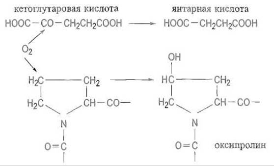
Hydroxylation at C-4 occurs exclusively on proline residues that precede glycine residues in the Pro—Gly—Хаа—Yaa sequence. Similarly (co-translationally), lysyl hydroxylase hydroxylates Lysine residues at their d-carbon atoms when they precede glycine. The hydroxyl groups of hydroxylysine serve as attachment sites for glycosylation.
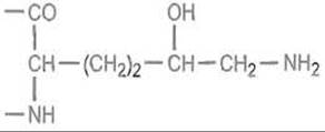
Galactose and glucose are sequentially attached to them. These modifications take place within fibroblasts and initiate a whole cascade of subsequent transformations of the collagen molecule. "Immature" collagen that has undergone no further deep modifications is referred to as procollagen and can be isolated from the skin of young animals.
Tropocollagen is a rod-shaped molecule 3000 Å in length and 15 Å in diameter, consisting of three unidirectional
polypeptide chains wound into helices (Fig. 14.1). Each of these resembles the helix of the well-studied model polypeptide poly-L-proline. The latter differs from the a-helix primarily because stabilization by Hydrogen Bonds along the axis of the helix is impossible due to the absence of NH donor groups. Consequently, the conformation of poly-L-proline is dictated almost entirely by the steric constraints imposed by the pyrrolidine rings of the proline residues. There are precisely three amino acid residues per turn of the helix. Subsequently, three parallel poly-L-proline-type helices twist together in collagen to form a superhelix.
The three helices are linked together by transverse hydrogen bonds. The Role of glycine, which invariably occupies every third position in the peptide chain, is to allow the three collagen chains to come into close proximity; lacking a side chain, it creates no steric hindrance at the center of the helix. The bulky pyrrolidine rings of proline and hydroxyproline are directed outward (Fig. 14.2).
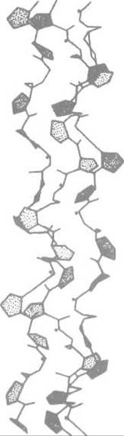
Fig. 14.1. The collagen triple helix. Dots indicate the NH groups of glycine residues occupying every third position.
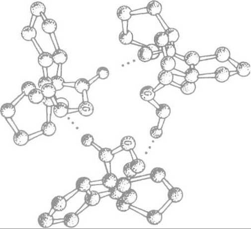
Fig. 14.2. Interaction of collagen polypeptide chains through The formation of hydrogen bonds between glycine (G) residues.
The necessary close alignment of the chains is ensured by regularly spaced glycine residues that lack side chains.
The Stability of the triple helix is determined by cooperative interactions. Its Denaturation—the transition into gelatin—typically occurs abruptly within a narrow Temperature range, the midpoint of which is conventionally called the melting temperature of collagen. Interestingly, the melting temperature correlates with the total content of proline and hydroxyproline residues and is somehow related to the animal's body temperature, exceeding it by a few degrees.
|
Sum of proline and hydroxyproline residues |
Collagen melting temperature, °C |
Animal body temperature, °C |
|
|
Calf skin |
232 |
39 |
37 |
|
Shark skin |
191 |
29 |
24-28 |
|
Cod skin |
155 |
16 |
10-14 |
Such "fine-tuning" of the melting temperature to body temperature is evidently necessary to ensure the elasticity of collagen fibers. Proline hydroxylation increases both the stability and the melting temperature of collagen; therefore, impaired hydroxylation—such as in scurvy due to Vitamin C Deficiency—leads to lesions of the skin and blood vessels, tooth loss, and other disorders.
The Formation of the collagen triple helix can occur spontaneously, but it is a slow process that would be insufficiently rapid in vivo. Therefore, collagen is synthesized as a precursor known as procollagen, which has a molecular mass of 140 kDa, whereas the molecular mass of the mature a-chain is 95 kDa. In the precursor, the N- and C-terminal Regions of the three polypeptide chains—whose primary structures lack the characteristic "collagen" motif—form globular structures that subsequently associate through mutual interactions, with the contact between the C-terminal globules being additionally stabilized by interchain Disulfide Bonds.
As a result, the procollagen molecule acquires a dumbbell-like shape in which the future collagen peptide chains are brought close together and aligned in parallel, dramatically simplifying and accelerating the folding of the triple helix. Thus, the terminal globules act as "assembly devices," facilitating the correct folding and alignment of the procollagen molecule.
Procollagen is synthesized and secreted into the extracellular matrix by fibroblasts. Specific procollagen peptidases then cleave off the globular domains that have completed their function, leaving behind the triple helix formed by polypeptide chains in each of which the Gly—Хаа—Yaa structural motif is repeated 338 times (Fig. 14.3).
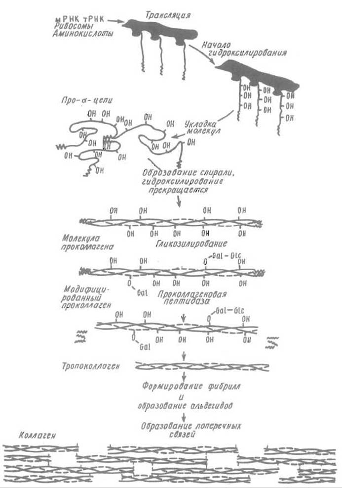
Fig. 14.3. Scheme of Biosynthesis, triple helix assembly, fiber formation, and post-translational modification of collagen (for other details, see the text)
Tropocollagen molecules pack together into collagen fibers in a parallel arrangement, yet staggered relative to one another by approximately 1/4 of the molecular length. A gap of 400 Å is formed between the end of one molecule and the beginning of the next, which serves as the nucleation site for hydroxyapatite formation in Bone tissue.
Subsequently, the collagen fiber is stabilized by an extensive system of cross-links. Among the post-translational modification processes that govern collagen maturation, the formation of allysine (an aldehyde derivative of lysine) mediated by lysyl oxidase is of particular importance:
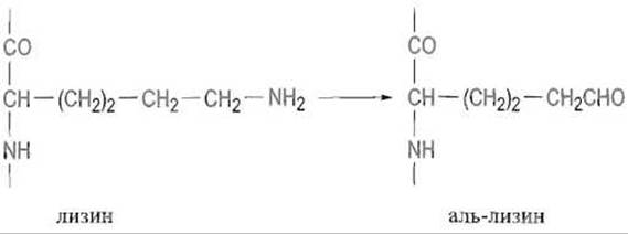
Hydroxylysine undergoes a similar conversion into an aldehyde. Lysyl oxidase is a copper-containing enzyme that utilizes pyridoxal as a cofactor. Next, a Schiff base is spontaneously formed, without enzyme participation, with the spatially proximate amino group of a lysine residue located in an adjacent polypeptide chain:
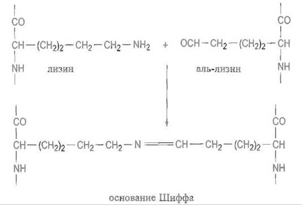
The —N=CH— bond, which is inherently unstable, is reduced to —NH—СН2—, yielding a highly stable cross-link known as lysinonorleucine:
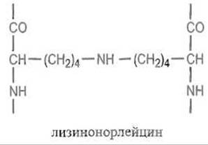
Due to their high reactivity, the aldehyde groups of allysine readily participate in aldol Condensation reactions, followed by crotonic condensation. Further reactions may involve The addition of a Histidine imidazole group to the activated double bond, along with even more complex transformations:
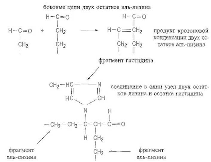
The number and Nature of the resulting cross-links make it possible to regulate fiber elasticity.
Thus, collagen biosynthesis involves a whole cascade of post-translational modifications. Procollagen is synthesized and assembled in fibroblasts, where the formation of hydroxyproline and hydroxylysine residues as well as their glycosylation already take place. Following secretion, procollagen undergoes Limited proteolysis, which results in the Cleavage of terminal globular domains and the formation of tropocollagen. This is followed by the maturation of this still Water-soluble protein, leading to the establishment of a more or less extensive network of intermolecular cross-links. The rate of this final process depends on the age of the animal and The Nature of the tissue where collagen is localized, thereby allowing for the modulation of the Physical Properties of structures containing it.
The Biological Significance of this sequence of transformations is underscored by the fact that the disruption of even a single step leads to severe defects in connective tissue formation. For instance, the inhibition of lysyl oxidase by the biogenic amine lathyrine, NH2CH2CH2CN (ß-aminopropionitrile), which enters the animal's body upon ingestion of certain species of vetch, prevents the formation of allysine and thereby halts the subsequent cross-linking required to stabilize collagen fibers, resulting in profound skin lesions. Cross-link formation is likewise impaired in the event of a deficiency in copper ions, which form the active center of lysyl oxidase.
Interestingly, plant Cell walls contain hydroxyproline-rich Glycoproteins that act as structural proteins. Another structural component of Plant Cell Walls is a glycine-rich protein found in legumes, where glycine accounts for 60% of the Amino Acid Sequence. It is hypothesized that this protein adopts a ß-Structure.
Last update: 13/08/2026
Editorial and Educational Adaptation: This material has been compiled based on the primary/original source text. The project team performed an editorial review, corrected technical inaccuracies, structured sections, and adapted the content for an educational format.
What was processed:
- elimination of formatting defects (OCR errors, structural breaks, corrupted characters);
- editorial organization of content;
- standardization of terminology in accordance with academic sources;
- verification of factual statements against the original source text.
All mentions of the author, publication year, and origin of the primary text have been preserved in accordance with the source.