Biochemistry - Chemical Reactions in Living Cells, Volume 1 - D. Metzler 1980
The Molecules We Are Made Of
Proteins
Globular Proteins
Unlike Fibrous Proteins, most living Cell proteins are characterized by a significantly more complex chain-folding pattern3. The first protein whose complete three-dimensional Structure was determined by X-Ray Diffraction Analysis was Myoglobin, a small (mol. wt. 17,500) oxygen-binding protein found in Muscles. Myoglobin's 153 amino acid residues are distributed mainly among 8 a-helical regions of varying length, containing from 7 to 26 residues. These helical regions, shaped like rectilinear rods, are arranged in space in a highly irregular manner, as shown in Fig. 2-8. The figure does not provide a complete picture of the protein's architecture because the space between the rods and inside the molecule is actually entirely filled with amino acid side chains, almost all of which are hydrophobic. Conversely, most of the polar side chains project outward from the helices into the surrounding aqueous environment.
3 The folding pattern of a peptide chain (The formation of a helix or ß-Structure) is often referred to as the Introduction/11.html">Secondary structure of a protein. Further folding of the molecule, driven by the interaction of groups located far apart along the chain, leads to the Formation of the tertiary structure. The aggregation of monomeric protein subunits into oligomers (Chap. 4) determines The quaternary structure of the protein.
Class="center">

FIG. 2-8. a-Helix. A. Right-handed helix with vertical Hydrogen Bonds represented by dashed lines. The positions of amino acid side chains are indicated by numbers in order of increasing sequence from the C-terminus to the N-terminus of the polypeptide chain (the reverse direction—from the N-terminus to the C-terminus—is conventional for peptide chains. — Trans.) B. Conformation of the myoglobin peptide backbone (Kendrew J. C., Sci. Am., 205, 96–110, Dec. 1961). Five long a-helices are depicted as cylindrical rods. Several additional, shorter helices are also visible. The overall Molecular dimensions are approximately 4.4 x 4.4 x 2.5 nm.
As the structures of various proteins have been deciphered (particularly in recent years), it has become increasingly evident that Globular proteins, much like myoglobin, maintain their structural integrity primarily through interactions between hydrophobic residues. Within the protein interior, side groups are packed with exceptional compactness. If any unoccupied space remains within the structure, it is typically filled with Water [24, 25]. For instance, the packing density (The ratio of the volume bounded by the Van der Waals envelope to the total volume) of Lysozyme and Ribonuclease molecules is ~0.75; for comparison, the theoretical packing density for closely packed spheres is 0.74. Polar groups generally reside On the surface, though they are occasionally «buried» internally, forming hydrogen bonds with other groups inside the protein molecule. Non-polar side chains also appear in isolated patches on the surface, occasionally clustering into hydrophobic domains. These clusters can mediate interactions with other proteins or with lipid regions of membranes.
Myoglobin is, in a sense, an exceptional protein, because in most other globular proteins the proportion of a-helical regions is relatively low. For example, the 129-residue chain of lysozyme (Fig. 2-9), one of the smallest Enzymes (mol. wt. 14,600), contains only a few short helices. The lysozyme chain is folded for the most part in a complex and irregular fashion. Note the region containing the antiparallel pleated ß-sheet. It begins with the segment between residues 42 and 45, after which the chain turns back to form a hairpin-like loop, establishing hydrogen bonds between residues 51–54 and 42–45. This pleated architecture is also discernible in other PARTS OF THE chain.
A characteristic feature of larger protein molecules is the presence of a distinct ß-structure in their core. A prime example is the enzyme Carboxypeptidase A, which consists of 307 residues (Fig. 2-6, bottom) [26]. The ß-structure, curved like a left-handed propeller blade, forms a rigid «scaffolding» to which the remaining parts of the molecule attach. Many proteins contain both parallel and antiparallel ß-structural regions. Several large proteins, such as glyceraldehyde-3-phosphate dehydrogenase (334 residues), are characterized by clearly distinguishable domains (two or more) linked by flexible «hinge» regions [27]. The predominant motif in each of the two domains is a pleated ß-sheet (Fig. 2-10). Note that the NAD+-binding domain is almost entirely composed of a parallel ß-structure, whereas the «catalytic» domain contains both parallel and antiparallel chains. Both domains feature a-helical regions flanking either side of the central sheet.
While three-dimensional structures are best illustrated using stereopairs, the distribution pattern of internal hydrogen bonds is most clearly revealed through two-dimensional diagrams, one of which is presented in Fig. 2-11.
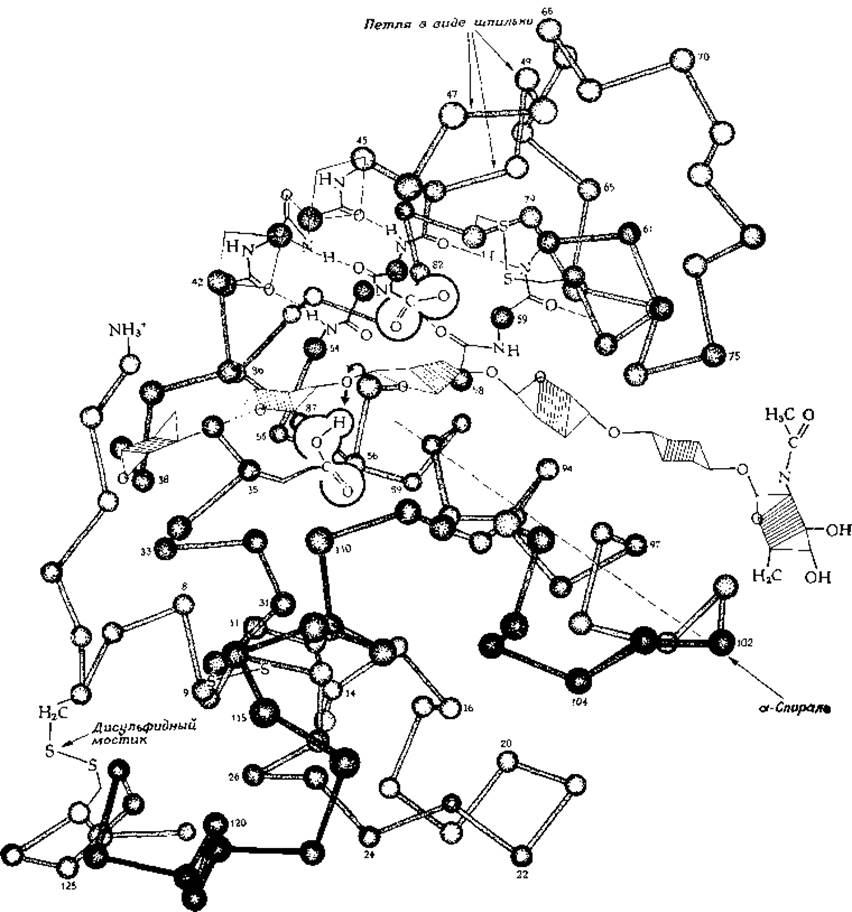
FIG. 2-9. Polypeptide chain folding in the enzyme lysozyme. (Dickerson R. E., Geis I., The Structure and Action of Proteins, p. 71. Benjamin, New York, 1969.) For the greater part of the molecule, only the attachment points of the side chains (a-carbon atoms) are shown. One region also displays the atoms participating in the peptide bond, illustrating the arrangement of hydrogen bonds within the antiparallel ß-structure at the core of the molecule. One of the a-helices is indicated. The hatched hexagons in the middle denote the backbone of the polysaccharide substrate bound at the «Active Site». It is suggested that the carboxyl group of residue 35 facilitates the Cleavage of the O–C bond (bold arrows) (Chap. 7, Sec. B.4.a). The arrow on the left points to one of the three disulfide bridges. (In reality, the lysozyme molecule contains four disulfide bridges. Aside from the three indicated in the figure, there is an additional bridge between residues 76 and 94—which, in the drawing, would run beneath the substrate molecule. — Trans.) The remaining two bridges are also visible—one located up and to the right, and the other down and to the left of the molecular center.
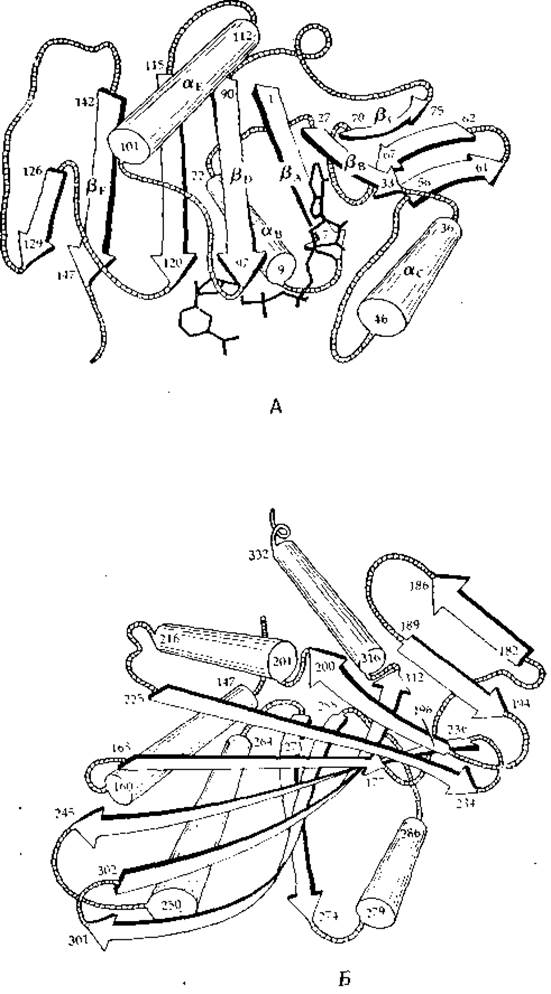
FIG. 2-10. Schematic representation of the two domains in the glyceraldehyde-3-phosphate dehydrogenase molecule (from lobster Muscle). A. NAD+-binding domain. Numbers indicate amino acid residue positions in the sequence; aB, aC, etc., ßA, ßB, etc., designate a-helices and pleated ß-sheet strands, respectively. B. Catalytic domain, oriented to provide a clearer view of the pleated sheet in the subunit interface region [27].
Investigations of synthetic Polypeptides, alongside analyses of known protein structures derived via X-ray crystallography, have demonstrated that Certain Amino Acids—such as glutamic acid, Alanine, and leucine—promote a-helix formation. Other Amino Acids, notably Methionine, valine, and isoleucine, are more commonly
found within ß-structures, whereas Glycine, Proline, and asparagine typically localize to chain bends. This strongly suggests that a polypeptide's folding pattern is directly dictated by its Amino Acid Sequence. For instance, if several helix-promoting residues cluster in a particular region of the chain, a helix will form. This helix propagates in both directions until it encounters residues (such as proline) that disrupt its formation. Similarly, the clustering of appropriate residues triggers the assembly of a ß-structure. The irregular folding of other chain segments generates additional Hydrogen bonds and a high frequency of hydrophobic interactions, which are especially characteristic of the interior domains of globular proteins.
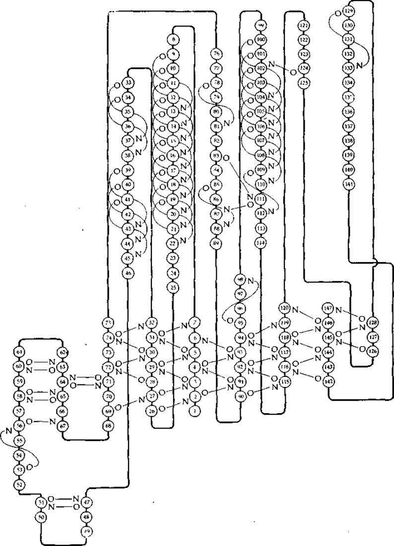
FIG. 2-11. Schematic diagram of the hydrogen bonding network between main-chain groups in the NAD+-binding domain (residues 1–149) of glyceraldehyde-3-phosphate dehydrogenase [27].
Now that the three-dimensional structures of many proteins are known, attempts are underway to predict the Folding Pathways of other protein molecules based solely on their Amino acid sequences [28–30]. One such method relies on calculating numerical «conformational parameters» that express the probability of a given residue occurring within a helix, a ß-structure, or a ß-bend (Table 2-4). According to Chou and Fasman [29], if four out of six clustered residues favor helix formation, that group can be considered a nucleation site for helicity. The helix extends outward from this center in both directions until it encounters a tetrapeptide composed of helix-disrupting residues. Likewise, three out of five residues favoring ß-structure act as a seed for ß-sheet formation. In this case, the capacity to form bonds with other chain segments—separated from the growing ß-sheet by ß-bends or irregular Conformations (random coils)—is critically important. Researchers have even organized predictive competitions wherein various scientific groups attempt to forecast protein folding patterns immediately prior to the experimental determination of their structures by crystallographic Methods [31].
Table 2-4 Classification of amino acid residues according to their capacities for a-helix, ß-structure, and ß-bend formationa
|
Amino Acid |
Pa |
Helix-forming tendency |
ß-structure-forming tendency |
Pt |
|
|
Glu- |
1 ,53 |
++ |
0,26 |
anti.+ |
0,44 |
|
Ala |
1 ,45 |
++ |
0,97 |
weak |
0,57 |
|
Leu |
1 ,34 |
1,22 |
+ |
0,53 |
|
|
His+ |
1,24 |
+ |
0,71 |
anti. |
0,69 |
|
Met |
1,20 |
+ |
1 ,67 |
++ |
0,67 |
|
Gin |
1 ,17 |
+ |
1 ,23 |
+ |
0,56 |
|
Trp |
1,14 |
+ |
1,19 |
+ |
1,11 |
|
Val |
1,14 |
+ |
1,65 |
++ |
0,30 |
|
Phe |
1,12 |
+ |
1 ,28 |
+ |
0,71 |
|
Lys+ |
1 ,07 |
weak |
0,74 |
anti. |
1,01 |
|
Ile |
1,00 |
» |
1 ,60 |
++ |
0,58 |
|
Asp- |
0,98 |
indif. |
0,80 |
++ |
1,26 |
|
Thr |
0,82 |
» |
1 ,20 |
+ |
1,00 |
|
Ser |
0,79 |
» |
0,72 |
anti. |
1,56 |
|
Arg+ |
0,79 |
» |
0,90 |
indif. |
1,00 |
|
Cys |
0,77 |
1,30 |
+ |
1,17 |
|
|
Asn |
0,73 |
anti. |
0,65 |
anti- |
1,68 |
|
Tyr |
0,61 |
» |
1,29 |
+ |
1,25 |
|
Pro |
0,59 |
anti. + |
0,62 |
anti. |
1,54 |
|
Gly |
0,53 |
» |
0,81 |
indif. |
1,68 |
a Conformational parameters Pa, Pβ, and Pt represent the frequencies of occurrence of a given amino acid within an a-helix, ß-structure, or ß-bend (based on data from 15 proteins of known structure), divided by the average frequency of occurrence of that amino acid in the molecule. The amino acid residues in the table are arranged in order of decreasing helix-forming capacity. The symbols «++», «+», «weak», «indif.», «anti.», and «anti.+» indicate that the amino acid exhibits, respectively, a strong tendency to form the structure, a moderate tendency, a weak tendency, indifference to structural context, a propensity to break the structure, or a strong propensity to disrupt a structure of that type [29].
Recently, Levitt and Chothia classified a group of proteins comprising 31 proteins of known structure, yielding four distinct Structural classes: (I) a-proteins, in which a-helices predominate, as in Hemoglobin and myoglobin. (II) Proteins built from stacked ß-sheets forming a multi-layered architecture; this category includes the small protein rubredoxin (Fig. 10-4) and concanavalin A (Fig. 5-7), among others. (III) (a + ß)-proteins, containing regions composed entirely of a-helices and regions consisting entirely of ß-sheets (typically antiparallel) within the same polypeptide chain. Examples include Papain and Thermolysin (Chap. 7). (IV) a/ß-proteins, in which a-helices and ß-strands alternate along the polypeptide chain. Typically, these feature a central ß-sheet flanked on both sides by helical segments; this Organization is observed, for instance, in carboxypeptidase (Fig. 2-6), glyceraldehyde-3-phosphate dehydrogenase (Fig. 2-10), as well as hexokinase and phosphoglycerate kinase (Fig. 7-5).
Last update: 06/08/2026
Editorial and Educational Adaptation: This material has been compiled based on the primary/original source text. The project team performed an editorial review, corrected technical inaccuracies, structured sections, and adapted the content for an educational format.
What was processed:
- elimination of formatting defects (OCR errors, structural breaks, corrupted characters);
- editorial organization of content;
- standardization of terminology in accordance with academic sources;
- verification of factual statements against the original source text.
All mentions of the author, publication year, and origin of the primary text have been preserved in accordance with the source.