Principles of Biochemistry Volume 1 - A. Lehninger 1985
Biomolecules
Fibrous Proteins
Chapter Summary
There are four types of Fibrous Proteins that play a protective or structural role in animal organisms: a-keratin, ß-keratin, Collagen, and Elastin. Their study has yielded important insights into the relationship between the Structure and function of protein molecules. a-Keratins are insoluble, tough proteins found in Hair, wool, feathers, scales, horns, hooves, and tortoise shells. X-Ray Diffraction Analysis shows that a-keratin fibrils have a repeat period of about 0.54 nm and that their polypeptide chains are coiled into a helix. X-ray data also indicate the rigidity and planar configuration of peptide groups. This is because the C—N bonds of the polypeptide backbone possess partial double-bond character. Based on these observations, it was concluded that The polypeptide chains of a-keratin exist as right-handed a-helices, with 3.6 amino acid residues per turn and a pitch of 0.54 nm. All peptide groups participate in The formation of intrachain Hydrogen Bonds that stabilize the a-helix. A destabilizing effect on the a-helix is exerted by adjacent R-groups bearing electrical charges of the same sign or having large sizes, as well as by Proline residues, which bend the chain and distort the a-helix. Hair is a rope-like, multistranded structure formed by a-helical polypeptide chains wound around one another in a superhelix. A-keratins contain many cross-links involving cystine residues.
ß-Keratins (Silk Fibroin being the most typical example) have a periodicity of about 0.70 nm. Steam-treated and stretched a-keratin acquires the same periodicity. In ß-keratins, the polypeptide chain is extended along a single axis into a pleated sheet structure. Adjacent polypeptide chains of ß-keratins are linked by hydrogen bonds; they are antiparallel, i.e., oriented in opposite directions, and form a pleated sheet with R-grоuрs protruding from both sides. ß-Keratins contain high proportions of Glycine and Alanine residues.
Collagen is the most abundant of all vertebrate proteins. It is found in tendons, Fibrous Connective Tissue of the Skin, Blood Vessels, bones, and Cartilage. Collagen fibrils consist of three intertwined polypeptide chains, each forming a specialized type of staggered helix containing about 21% proline and hydroxyproline residues. Collagen fibrils are inelastic and possess an extremely high tensile strength. Upon partial Hydrolysis, collagen is converted into gelatin, a soluble and digestible mixture of Polypeptides. Elastin, a specific protein of elastic connective tissue, exists as a network of polypeptide chains cross-linked by desmosine residues. It exhibits high elasticity. Myosin, Actin, and tubulin are Examples of intracellular filamentous proteins involved in energy-dependent ATP processes of Muscle contraction and Cell motility.
Books
See also the bibliography for chapters 6 and 8. Cantor, C. R., and Schimmel, P. R., Biophysical Chemistry, part I: The Conformation of Biological Macromolecules, Freeman, San Francisco, 1980.
Dickerson, R. E., and Geis, I., Proteins: Structure, Function, and Evolution, Benjamin/Cummings, Menlo Park, Calif., 1983.
Schultz, G. E., and Schirmer, R. H., Principles of Protein Structure, Springer-Verlag, New York, 1979.
Selected Papers
Eyre, D. R., Collagen: Molecular Diversity in the Body’s Protein Scaffold, Science, 207, 1315, March (1980).
Fraser, R. D. B., Keratins, Sci. Am., 221, 86–96, August (1969).
Gross, J., Collagen, Sci. Am., 204, 120–130, May (1961).
1. Properties of the peptide bond. In X-ray diffraction studies of crystalline Peptides, Linus Pauling and Robert Corey discovered that the length of the C—N bond in the peptide group (0.132 nm) is intermediate between those of a typical single C—N bond (0.149 nm) and a double C=N bond (0.127 nm). Furthermore, they established that the peptide group has a planar configuration—that is, all four atoms attached to the C—N group lie in the same plane, with the two a-carbon atoms linked to the C—N group always in the trans configuration, i.e., on opposite sides of the peptide bond.
Class="center">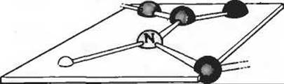
a) What Conclusion can be drawn from the length of the C—N bond in the peptide group regarding the bond's strength and multiplicity (i.e., whether it is single, double, or triple)?
b) Expanding on the answer to the previous question, explain why such a C—N bond has a length intermediate between double and single bonds.
c) What can be inferred from Pauling and Corey's data regarding the possibility of rotation around the peptide C—N bond?
2. Early data on wool structure. William Astbury was the first to notice that the X-ray diffraction pattern of wool indicates the presence of a structural unit repeating along the fiber with a period of about 0.54 nm. After stretching steam-treated wool, the X-ray pattern revealed A change in periodicity: the new structural unit repeated every 0.70 nm. When the steamed wool was allowed to shorten, the periodicity of about 0.54 nm reappeared. Although these observations provided a clue to understanding the molecular structure of wool, Astbury was unable to interpret them at the time. Based on modern knowledge of wool structure, explain these observations.
3. Rate of a-keratin synthesis in hair. By human standards, hair grows relatively slowly—at a rate of 15–20 cm per year. The growth zone is located at the Base of the hair, where epidermal Cells synthesize a-keratin filaments, which then twist into rope-like structures (see Fig. 7-9). The main structural element of a-keratin is the a-helix, which has a pitch of 0.54 nm and 3.6 amino acid residues per turn (see Fig. 7-6). Assuming that the rate-limiting factor in hair growth is The Biosynthesis of a-helical keratin chains, calculate The rate of peptide bond formation in a-keratin chains (number of peptide bonds per second) required to produce the observed hair elongation over 1 year.
4. Effect of pH on the conformation of Polyglutamic acid and polylysine. The unfolding of a polypeptide chain in an a-helical conformation to form a random coil is accompanied by a sharp decrease in specific optical rotation. Polyglutamic acid—a polypeptide consisting solely of glutamic acid residues—adopts an a-helix conformation at pH 3. However, when the pH is raised to 7, the specific optical Rotation of the solution drops markedly. A similar phenomenon is observed for polylysine, which assumes an a-helix conformation at pH 10, whereas lowering the pH to 7 leads to a sharp decrease in its specific optical rotation, as shown in the graph.
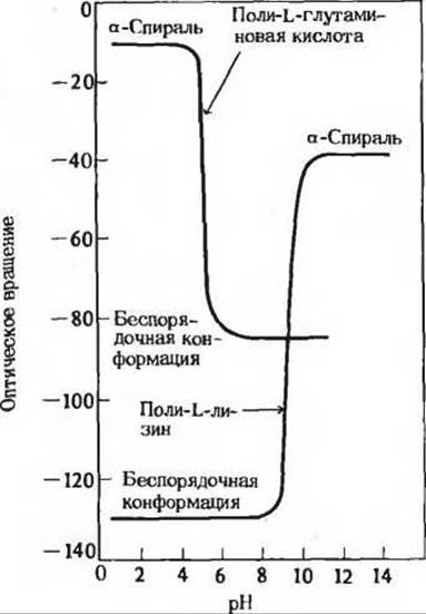
How can this effect of pH on the conformation of polyglutamic acid and polylysine be explained? Why does the transition from one conformation to the other occur within such a narrow pH range?
5. Cystine content determines the mechanical properties of many proteins. A large number of natural proteins contain a high proportion of cystine residues. A correlation is observed between the mechanical properties of these proteins (tensile strength, viscosity, hardness, etc.) and their cystine content. For example, glutenin (a cystine-rich wheat protein) determines the viscosity and elasticity of wheat flour dough. Similarly, the hard and tough shell of a turtle owes its properties to the high cystine content of the a-keratin of which it is composed. What is the Molecular Basis of the observed correlation between cystine content and protein mechanical properties?
6. Why does wool shrink? If a wool sweater or wool socks are washed in hot Water and then dried in an electric tumble dryer, they become smaller. Based on what is known about The structure of a-keratin, how can this phenomenon be explained? At the same time, silk does not shrink under the same conditions. Explain why.
7. Heat stability of cystine-containing proteins. Most Globular proteins undergo Denaturation (unfolding of their polypeptide chains) upon brief heating to 65°C, accompanied by a complete loss of activity. However, globular proteins rich in cystine residues denature only upon prolonged heating to significantly higher temperatures. One such protein is Ribonuclease, which consists of 124 amino acid residues in a single polypeptide chain cross-linked by four Disulfide Bonds formed by cystine residues. For the polypeptide chain of ribonuclease to unfold, the solution containing it must be heated to a high Temperature; if the solution is then rapidly cooled, its enzymatic activity is restored. Can you explain the molecular basis for this behavior?
8. Cleavage of cystine cross-links. Cross-linking —S—S— bonds are formed in proteins when two Cysteine residues within the same chain or in different chains are oxidized. When determining the Amino Acid Sequence of a protein, it is practically necessary to cleave all —S—S— bonds beforehand. Since this is the reverse of oxidation, it is carried out using a reducing agent.
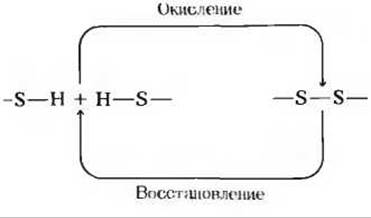
a) One standard method for cleaving disulfide bridges involves treating the protein with an excess of 2-mercaptoethanol (HSCH2CH2OH). Explain the chemical basis of this method.
b) One disadvantage of this method is that after the cystine cross-links are cleaved, they can reform. Why does this happen?
9. Periodicity of ß-sheets in silk fibers. Chemical Analysis of the product obtained from the partial hydrolysis of silk fibroin produced by the silkworm Bombyx mori showed that the polypeptide chain of this protein repeatedly contains a six-residue segment
(—Gly—Ser—Gly—Ala—Gly—Ala—)n.
At the same time, X-ray diffraction data indicate that the main structural unit repeats in fibroin with a periodicity of 0.70 nm (see text). However, two additional repeating units with periods of 0.35 and 0.57 nm were also detected, corresponding to the distances between the ß-sheets. Propose an arrangement of these six-residue segments that accounts for these inter-sheet distances in silk fibroin.
10. Bacteriorhodopsin, the purple membrane protein. Under favorable environmental conditions, the bacterium Halobacterium halobium, growing in high salt concentrations, synthesizes a membrane protein (mol. wt. 26,000) known as bacteriorhodopsin. The molecules of this purple protein, whose color is due to bound retinal, form aggregate patches in The Cell membrane known as purple patches. Bacteriorhodopsin Functions as a light-driven proton pump and thereby supplies the cell with energy. It has been shown that this protein consists of seven parallel a-helical segments spanning the bacterial cell membrane, which is 4.5 nm thick. Calculate the minimum number of Amino Acids required in a single a-helical segment to completely span the membrane. Estimate what fraction of The amino acid residues in bacteriorhodopsin is involved in forming the a-helical segments (the average Molecular Weight of an amino acid residue is 110). Provide the rationale for your calculations.
11. Collagen biosynthesis. Collagen, which is more abundant in mammalian Tissues than any other protein, has an unusual Amino acid composition. Unlike most other proteins, it is very rich in proline and hydroxyproline (see Fig. 7-13). Because hydroxyproline is not one of the 20 standard Amino Acids Commonly Found in Proteins, its incorporation into collagen can occur via two possible pathways: 1) enzymatic hydroxylation of proline to hydroxyproline prior to its incorporation into collagen, or 2) hydroxylation of proline residues already incorporated into the collagen chain. To distinguish between these two possibilities, the following experiments were performed. Rats were fed 14C-labeled proline, and collagen was subsequently isolated from their tails. The newly synthesized collagen was found to be radioactive. In a separate experiment, 14C-labeled hydroxyproline was administered in the same manner, but in this case, no radioactivity was detected in the newly synthesized collagen. How do these experiments allow you to choose between the two proposed pathways?
12. Pathogenic mechanism of gas gangrene Bacteria. The pathogenic anaerobic bacteria Clostridium perfringens, which cause tissue-destroying gas gangrene, secrete an enzyme that efficiently catalyzes the hydrolysis of the peptide bond (shown in red) in the following sequence:
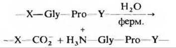
where X and Y represent any of the 20 amino acids. How does this secreted enzyme help the bacterium invade human tissues? Why is this enzyme harmless to the bacterium itself?
Last update: 06/08/2026
Editorial and Educational Adaptation: This material has been compiled based on the primary/original source text. The project team performed an editorial review, corrected technical inaccuracies, structured sections, and adapted the content for an educational format.
What was processed:
- elimination of formatting defects (OCR errors, structural breaks, corrupted characters);
- editorial organization of content;
- standardization of terminology in accordance with academic sources;
- verification of factual statements against the original source text.
All mentions of the author, publication year, and origin of the primary text have been preserved in accordance with the source.