Protein Chemistry - Part 1 - General Protein Chemistry - Ashmarin I. P. 1968
Spatial Organization of the Protein Molecule
Methods for Studying the Secondary Structure of Proteins and Polypeptides
Rotation of the Plane of Light Polarization (Optical Activity)
It is well known from experience that all Proteins and synthetic Polypeptides rotate the plane of polarization of light; that is, they are optically active. The observed optical activity is composite in nature. First and foremost, it is the sum of the contributions made by the asymmetric alpha-carbon atoms of the Amino Acids. Since all Natural Amino Acids are levorotatory, the alpha-C atoms in the polypeptide chain also rotate light to the left. Consequently, the sign of this term in the total optical activity is negative, and any polypeptide existing in a random coil conformation will exhibit negative activity.
However, in the case of proteins or polypeptides with a high degree of helicity, a second major contribution to optical rotation arises from the intrinsic Asymmetry of the alpha-helix itself. As already mentioned, polypeptides built from L-amino acids presumably form exclusively right-handed helices, which rotate the plane of polarization to the right. Thus, the second increment of optical activity is opposite in sign to the first; in absolute value, these two components are quite close to each other, and overall, the optical activity of a polypeptide helix approaches zero. How, then, can these composite optical activities be evaluated and used to calculate the degree of helicity of a polypeptide chain?
The rotatory power of a compound in the form of a pure solid, liquid, or solution is expressed in terms of the specific rotation [a]D (the specific activity measured for the sodium D-line, 589.3 mμ), determined by the equation
Class="center">
where a is the measured angle of rotation; I is the path length in dm; and c is the concentration of the optically active substance in g/100 mL.
When dealing with polypeptides and other long-chain compounds, it is more convenient to express optical activity as the rotation per chain link ("residue rotation"):

where M0 is the Molecular Weight of the link, i.e., The amino acid residue.
When comparing the rotation of polypeptides in various Solvents, It is important to account for the nonspecific effect of the refractive index n. A dependence of specific rotation on the refractive index exists even when the substance's Structure is insensitive to the solvent. Therefore, it is more appropriate to adjust the specific rotation or residue rotation value to a hypothetical medium with n = 1. In this case, we obtain the corrected residue rotation, or the "effective chain link rotation":

The value of [m] for a polypeptide in a helical or random coil conformation can be easily calculated if we know the molecular weight of the amino acid residue and the specific rotation [ah for the helical and coil forms. The latter is determined through melting experiments on alpha-helices of synthetic polypeptides. For instance, if the side-chain carboxyl groups of Polyglutamic acid are titrated with alkali, the neutral COOH groups convert into charged COO- groups. As a result, powerful electrostatic repulsion forces begin to operate along the molecule, capable of breaking Hydrogen Bonds and completely melting the alpha-helix (Fig. 2I). This destruction of the alpha-helix occurs within a narrow pH range and is accompanied by a sharp change in optical activity. The specific rotation loses the large positive increment generated by the alpha-helix, dropping from +6 to —80°. Since the helix is completely disrupted, this value of —80° represents the specific rotation of the coil conformation caused by the alpha-carbon atoms of L-amino acids. Correspondingly, THE CONTRIBUTION OF the alpha-helix is much larger and opposite in sign, amounting to +86°. The algebraic sum of both values, i.e., [a]D of the helical configuration, is close to zero (+6°).
Quite similar values were obtained for [m] as well. Thus, for synthetic polypeptides in a random coil conformation and for denatured proteins, the effective rotation of the average amino acid residue ranged from —80 to —110°, whereas for fully helical polypeptides and native proteins, this value was close to zero. Consequently, the positive increment of [a]D or [m] associated with the ideal alpha-structure is 80—100°. According to data for synthetic polypeptides and paramyosin, whose structure is considered fully helical, this contribution is 90°. Thus, by placing a polypeptide in various solvents (for example, ranging from pure dichloroethane to its mixtures with dichloroacetic acid and the acid itself), we can approximately estimate the percentage of alpha-helicity in the chain from The change in [a]D or [m].
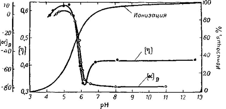
Fig. 21. Melting of polyglutamic acid helices upon Changes in the degree of ionization of carboxyl groups (after Doty, 1961).
Does this mean that optical activity can be reliably used not only to infer the presence of helical segments in a protein molecule, but also to estimate the degree of helicity? Unfortunately, no. Definitive proof of the presence of an alpha-helix can only be obtained through X-Ray Diffraction Analysis, whereas a large positive contribution to optical activity can be reliably attributed solely to the presence of some ordered structure in a polypeptide or globular protein. For instance, transferring the polypeptide poly-O-acetylserine from chloroform to dichloroacetic acid also induces a transition toward more negative [a]D values. However, infrared spectrophotometry has shown that this polypeptide adopts exclusively beta-structures. Consequently, the ordered structure responsible for the positive [a]D increment may be not only an alpha-helix but also a beta-structure. Estimating the degree of helicity from optical activity is also highly conditional. For example, upon the thermal melting of the Introduction/11.html">Secondary structure of Trypsin and one of its fragments, the [a]D values change from —35 to —55° and from —75 to —40°, respectively. This indicates that the trypsin fragment consists predominantly of left-handed helices, whereas the trypsin macromolecule is dominated overall by right-handed structures. It is thus clear that the helical regions in trypsin can generate contributions of opposite signs, resulting in an absolute optical activity smaller than that of a protein with the same degree of helicity but composed entirely of right-handed helices. Similar right-handed (chain B) and left-handed (chain A) helices have also been discovered in the Insulin molecule.
Thus, the change in optical activity accompanying The breakdown of an ordered structure is not, in itself, proof of the existence of helices within a polypeptide or protein. It must be supplemented by data on The rate of imide hydrogen exchange, ultraviolet absorption, and other measurements.
Last update: 06/08/2026
Editorial and Educational Adaptation: This material has been compiled based on the primary/original source text. The project team performed an editorial review, corrected technical inaccuracies, structured sections, and adapted the content for an educational format.
What was processed:
- elimination of formatting defects (OCR errors, structural breaks, corrupted characters);
- editorial organization of content;
- standardization of terminology in accordance with academic sources;
- verification of factual statements against the original source text.
All mentions of the author, publication year, and origin of the primary text have been preserved in accordance with the source.