Human Biochemistry Volume 2 - Murray R. 1993
Biochemistry of Intra- and Intercellular Communications
Pancreatic Hormones
Insulin
Background History
Pancreatic islets were discovered in 1860 by Langerhans, who, however, had no idea of their function; nor did von Mering and Minkowski, who established in 1889 that pancreatectomy leads to Diabetes Mellitus. The hypothesis of a close link between the islets and diabetes was proposed by de Meyer in 1909 and Sharpey-Schafer in 1917, but it was not until 1921 that Banting and Best proved it. By extracting pancreatic tissue with acidified ethanol, they isolated a factor possessing a potent hypoglycemic effect. This factor was named insulin. It was soon established that insulin extracted from the pancreatic islets of cattle and pigs is also active in humans. Within less than a year, this preparation came into widespread and successful clinical use for the Treatment of diabetes.
Bovine and porcine insulin can be readily obtained in large quantities, which is a crucial prerequisite for successful biochemical research. Insulin turned out to be the first protein with proven hormonal activity, the first protein obtained in crystalline form (Abel, 1926), the first protein whose Amino Acid Sequence was determined (Sanger et al., 1955), and the first protein synthesized by chemical Methods (Du et al.; Zahn; Katsoyanis, 1964). Furthermore, insulin was the first protein shown to be synthesized as a larger precursor (Steiner et al., 1967). In addition, it was the first protein produced for commercial purposes using Recombinant DNA technology. Yet, despite these impressive "firsts," The Mechanism of insulin action at THE MOLECULAR LEVEL is less well understood than that of most Other Hormones.
Chemical Properties
The insulin molecule is a polypeptide consisting of two chains, A and B, linked together by two Disulfide Bonds connecting residue A7 to residue B7, and residue A20 to residues B19. A third disulfide bond links residues 6 and 11 of the A-chain. The localization of all three disulfide bonds is invariant, and the A- and B-chains in most species consist of 21 and 30 Amino Acids, respectively. The Covalent Structure of human insulin (mol. mass 5734) is shown in Fig. 51.1, and data on Amino Acid Substitutions in insulins of various species are given in Table 51.2. Substitutions that do not affect the biological activity of the hormone occur at many positions in both chains, but they are most frequent at positions 8, 9, and 10 of the A-chain. This implies that this region of the molecule is not critical for the biological activity of insulin. Nevertheless, certain segments and domains of the insulin molecule exhibit a high degree of conservation.
Class="center">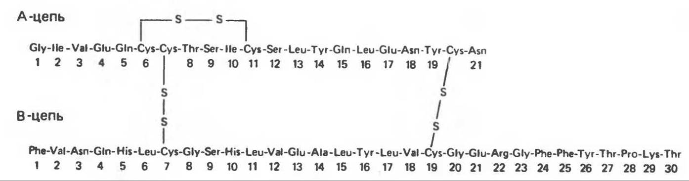
Fig. 51.1. Covalent structure of human insulin. (Reproduced, with permission, from Ganong W.F. Review of Medical Physiology, 13th ed., Appleton and Lange, 1987.)
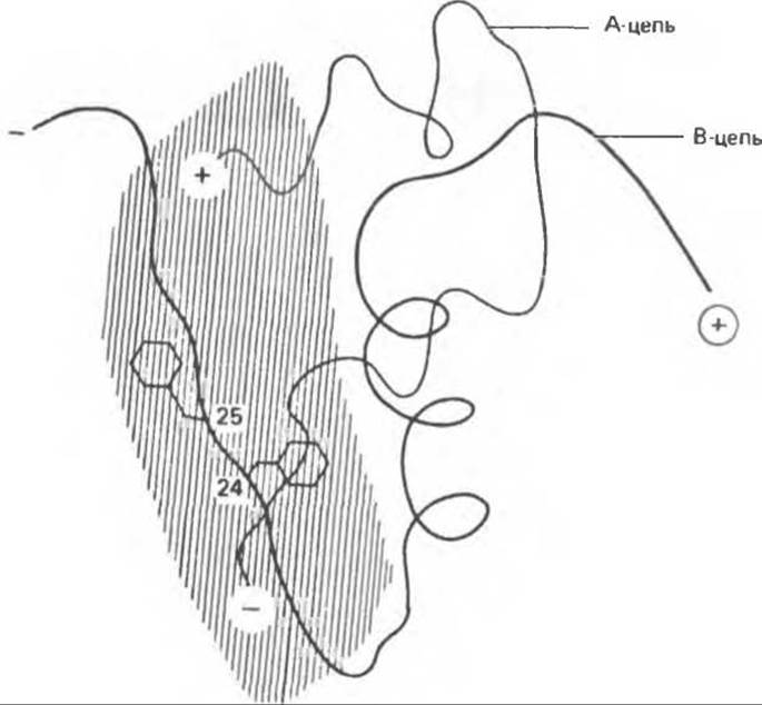
Fig. 51.2. Region of the insulin molecule responsible for its biological activity. This schematic model of the insulin molecule is based on X-ray crystallographic data. The hatched area corresponds to the part of insulin assigned the primary role in mediating the biological activity of the hormone. Phe residues at positions B24 and B25 are the sites where Mutations affect insulin biological activity. The N-termini of the insulin A- and B-chains are indicated by "+", whereas the C-termini are indicated by "—". (Redrawn and reproduced, with permission, from Tager H.S. Abnormal products of the human insulin Gene, Diabetes, 1984, 33, 693.)
These include 1) the positions of the three disulfide bonds, 2) hydrophobic residues in the C-terminal segment of the B-chain, and 3) the C- and N-terminal segments of the A-chain. Chemical modifications and individual amino acid substitutions in these six regions have helped identify a complex active center (Fig. 51.2). The hydrophobic region located at the C-terminus of the B-chain is also involved in insulin dimerization.
Table 51.2. Differences in insulin structure among mammalian species. (Modified and reproduced, with permission, from Ganong W. F.: Review of Medical Physiology. 13th ed., Appleton and Lange, 1987.)
|
Species |
Amino acid substitutions relative to human insulin |
|||
|
A-chain position |
B-chain position |
|||
|
8 |
9 |
10 |
30 |
|
|
Human |
Thr |
Ser |
Ile |
Thr |
|
Pig, dog, sperm whale |
Thr |
Ser |
Ile |
Ala |
|
Rabbit |
Thr |
Ser |
Ile |
Ser |
|
Cattle, goat |
Ala |
Ser |
Val |
Ala |
|
Sheep |
Ala |
Gly |
Val |
Ala |
|
Horse |
Thr |
Gly |
Ile |
Ala |
|
Sei whale |
Ala |
Ser |
Thr |
Ala |
As evident from Table 51.2, There is a striking similarity among human, porcine, and bovine insulins.
Porcine insulin differs from human insulin by a single amino acid substitution: Alanine replaces Threonine at position 30 of the B-chain. In bovine insulin, In addition to this change, threonine A8 is replaced by alanine, and isoleucine A10 by valine. These substitutions have virtually no effect on the biological activity of the hormone and very little impact on its antigenic properties. Although most patients treated with heterologous insulin develop low-titer circulating Antibodies against the administered hormone, some patients exhibit antibody titers of clinically significant magnitude. Until human insulin could be produced using Introduction/32.html">Genetic Engineering techniques, bovine and porcine insulins were routinely used for therapeutic purposes. Despite significant differences in Primary Structure, all three insulins exhibit similar biological activity (25–30 IU/mg dry weight).
Insulin forms very interesting complex structures. Zinc, whose concentration in B-Cells reaches high values, forms complexes with insulin and proinsulin. The insulins of all vertebrates form isologous dimers via Hydrogen Bonds between the peptide groups of residues B24 and B26 of two monomers, which, at high concentrations, further reorganize into hexamers containing two zinc atoms each. The presence of such a highly ordered structure has greatly facilitated the elucidation of the crystal structure of insulin. At physiological concentrations, insulin is likely present in monomeric form.
A. Insulin precursors. Insulin is synthesized as a preprohormone (mol. mass 11,500). It serves as a prime example of a peptide generated through various Processing steps from a larger precursor molecule. The sequence and subcellular localization of the corresponding biochemical transformations are shown in Fig. 51.3. A 23-amino-acid hydrophobic leader sequence (pre-fragment) directs the precursor molecule into the cisternae of The Endoplasmic reticulum, where it is cleaved. This yields the proinsulin molecule (mol. mass 9,000), which adopts the conformation necessary for The formation of correct disulfide bonds. As shown in Fig. 51.4, the proinsulin molecule has the following structure starting from the amino terminus:

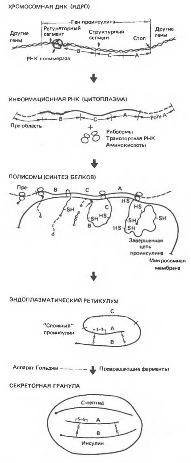
Fig. 51.3. Biosynthesis of insulin via a short-lived precursor. Letters A, B, and C designate the A- and B-chains of insulin and the connecting (C) peptide. The 23-amino-acid leader sequence encoded in the mRNA segment adjacent to the segment determining the B-chain (dashed line) is cleaved off following synthesis, likely even before the completion of the rest of the proinsulin molecule. (Reproduced, with permission, from Steiner D.F., Errors in insulin biosynthesis, N. Engl. J. Med., 1976, 294, 952.)
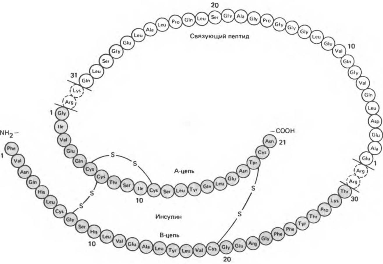
Fig. 51.4. Structure of human proinsulin. The insulin molecule and the C-peptide are linked together by two dipeptide linkers located on either side of the C-peptide. (Slightly modified and reproduced, with permission, from Karam J.H., Salter P.R., Forsham P.H. Pancreatic hormones and diabetes mellitus. In: Basic and Clinical Endocrinology, 2nd ed., Greenspan F.S., Forsham P.H. [eds.], Appleton and Lange, 1986.)
The proinsulin molecule is cleaved at several specific sites to yield equimolar amounts of mature insulin and C-peptide. These enzymatic transformations, schematically illustrated in Fig. 51.5, are initiated by a proteinase with Trypsin-like activity—an enzyme that cleaves two basic amino acids from each side of the C-peptide: the Arg31-Arg32 dipeptide at the N-terminus of the C-peptide and the Lys64-Arg65 dipeptide at the C-terminus of the C-peptide2.
1 C-peptide, from "connecting peptide." — Transl.
2 The indicated positions of the dipeptides correspond to their positions in the full proinsulin molecule counting from the N-terminus. — Transl.
B. Precursors of other islet Cell hormones. The Synthesis of Other islet cell hormones also requires the enzymatic conversion of higher-molecular-weight precursor molecules. The structures of Pancreatic Polypeptide, Glucagon, and Somatostatin compared to that of insulin are schematically shown in Fig. 51.6. Various combinations of endoproteolytic (trypsin-like) and exoproteolytic (carboxypeptidase B-like) Enzymes participate in the formation of these hormones, since the sequences possessing hormonal activity can be located in different Regions of the precursor molecule: somatostatin at the carboxyl terminus of the molecule, pancreatic polypeptide at the amino terminus, insulin at both termini, and glucagon in the middle portion.
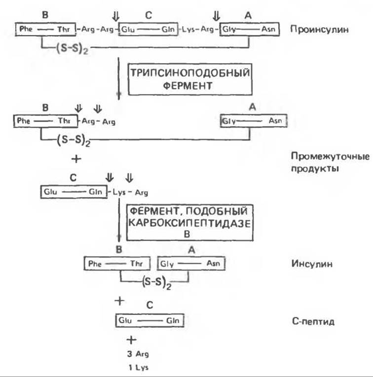
Fig. 51.5. Stages of human proinsulin Cleavage under the combined action of trypsin-like proteinases and carboxypeptidase B. The arrows indicate the sites of molecular cleavage. (Redrawn and reproduced, with permission, from Steiner D. F., Tager H. S. p. 927. In: Endocrinology, Vol. 2., DeGroot L. J. (ed.), Grune and Stratton, 1979.)
C. Subcellular localization of insulin synthesis and granule formation. The synthesis of insulin and its packaging into granules occur in a specific order (Fig. 51.7). Proinsulin is synthesized on the Ribosomes of the rough endoplasmic reticulum. Then, within the cisternae of this organelle, Enzymatic cleavage of the leader sequence (pre-segment) occurs, followed by the formation of disulfide bonds and molecule folding (Fig. 51.3). Afterward, the proinsulin molecule is transported to the Golgi apparatus, where proteolysis and packaging into secretory granules begin. Granule maturation continues as they move through the Cytoplasm toward The Plasma Membrane. Both proinsulin and insulin bind to zinc to form hexamers, but since about 95% of proinsulin is converted into insulin, it is the crystals of the latter that impart the morphological features to the granules. Along with insulin, the granules also contain equimolar amounts of C-peptide; however, these molecules do not form crystalline structures. Upon appropriate stimulation, mature granules fuse with the plasma membrane, releasing their contents into the extracellular fluid via emocytosis (exocytosis).
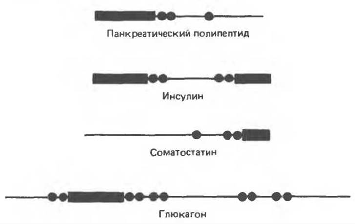
Fig. 51.6. Cytology/cytology/92.html">SCHEMATIC STRUCTURE OF the four main products of pancreatic endocrine cells. The black bars represent the portion of the precursor molecule corresponding to the hormone indicated in the label, while the thin line denotes the remaining regions of the precursor molecule's peptide chain. The locations of basic amino acids (Arginine or Lysine) where Cleavage of the precursor molecule occurs are indicated by black circles. The proinsulin molecule is depicted as a linear structure in which disulfide bonds are not shown. In reality, the proinsulin molecule has the sequence: B-chain — C-peptide — A-chain. (Redrawn and reproduced, with permission, from Tager H.S. Abnormal products of the human insulin gene. Diabetes. 1984. 33. 693.)
D. Properties of proinsulin and C-peptide. The length of proinsulins ranges from 78 to 86 amino acids, with these differences being attributed to the length of the C-peptide. Proinsulin has the same solubility and isoelectric point as insulin. It also forms hexamers with zinc crystals and reacts with insulin antiserum. The biological activity of proinsulin is less than 5% of that of insulin. It follows that a large part of the active center of insulin is masked in the precursor molecule. A certain portion of proinsulin is secreted along with insulin, and in certain situations (islet cell tumors), it is released in larger quantities than normal. Since the plasma half-life of proinsulin is significantly longer than that of insulin, and proinsulin exhibits strong cross-reactivity with insulin antiserum, the level of "insulin" determined by radioimmunoassay may in some cases exceed the content of the biologically active hormone.
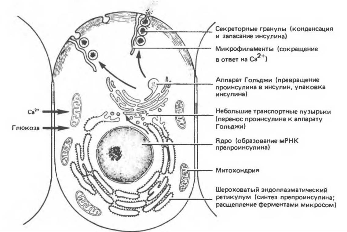
Fig. 51.7. Structural components of the pancreatic B-cell involved in glucose-induced hormone biosynthesis and secretion. In the diagram, secretory granules are adjacent to microfilaments, which contract under METABOLISM/18.html">The Influence of calcium. (Based on data presented by Orci L. A portrait of the pancreatic B cell, Diabetologia, 1974, 10, 163.) (Modified and reproduced, with permission, from Junqueira L. C., Carneiro J., Long J. A., Basic Histology. 5th ed., Appleton and Lange, 1986.)
No biological activity of the C-peptide has been detected. This molecule possesses different antigenic properties than insulin and proinsulin; therefore, the immunological Determination of the C-peptide allows endogenous insulin to be distinguished from the administered hormone and makes it possible to estimate The amount of endogenous insulin in cases where its direct determination is impossible due to the presence of insulin antibodies. C-Peptides from various species are characterized by a high frequency of amino acid substitutions, which Supports the premise that this fragment likely lacks biological activity.
E. Precursors of insulin-related peptides. The Structural Organization of a prohormone molecule is not unique to the insulin precursor. Precursors of Peptide Hormones closely related to insulin (relaxin and Insulin-like Growth Factors) share the same organization (Fig. 51.8). In all these hormones, the A- and B-chain sequences in the precursor molecule have highly homologous regions at their carboxyl and amino termini, connected to each other by a linking peptide. In the peptide precursors of insulin and relaxin, two basic Amino acids are located on either side of the connecting peptide, joining it to the A- and B-chains. After disulfide bonds are formed between the A- and B-chains, the connecting peptide is excised by endoproteolysis, and the molecule is converted into a two-chain peptide hormone (A and B). Insulin-like growth factors, while highly homologous to insulin and relaxin in their primary structure, nevertheless have one important difference: their precursor molecule lacks the sites for the cleavage of the connecting peptide, and therefore the active hormones retain The structure of a single polypeptide chain.
F. The human insulin gene. The human insulin gene (Fig. 51.9) is localized on the short arm of chromosome 11. In most mammals, a single insulin gene is expressed, organized similarly to the human gene, but rats and mice have two non-allelic genes. Each of them encodes a distinct proinsulin, giving rise to two different active insulin molecules. Currently, a method for producing human insulin in bacterial Expression Systems Using recombinant DNA technology has been developed. Thus, The problem of obtaining this hormone in quantities necessary for diabetic patients can be considered solved.
G. Abnormal products of the human insulin gene. Knowledge of the insulin gene and insulin molecule structure makes it possible to identify abnormal gene products, which in turn provides additional information about the function of this hormone. Three mutations of this gene have been identified, and for each of them, the Molecular Basis of the defect has been elucidated. In one case, a single base mutation resulted in the substitution of Serine for phenylalanine-B24, and in another (again resulting from a single mutation), phenylalanine-B25 was replaced by leucine. In the third case, the processing of proinsulin into the active hormone was altered: a mutation disrupted the cleavage of the 3'-end of the C-peptide at the boundary with the A-chain. This defect is based on the replacement of the Lys-Arg dipeptide at this site of the polypeptide chain with Lys-X, making trypsin-like cleavage impossible. The identification of these mutations was facilitated by their localization in the active center of the insulin molecule, resulting in the respective carriers exhibiting: 1) hyperinsulinemia, 2) no signs of insulin resistance, 3) reduced biological activity of circulating insulin, and 4) a normal response to exogenous insulin. At least four other nucleotide substitutions have been identified in "healthy" individuals. These mutations are localized in intervening (i.e., non-coding) sequences and have no effect on the functional activity of the insulin molecule.
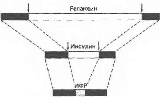
Fig. 51.8. Schematic representation of the structure of insulin-related peptide precursors. Homologous regions of relaxin, insulin, and insulin-like growth factor are depicted as black bars. The Amino acid sequences connecting the B- and A-chains in the relaxin and insulin precursor molecules are indicated by light bars. During the processing of precursors to form the corresponding two-chain products, these connecting sequences are removed (vertical arrows). The amino acid sequence of the insulin-like growth factor that corresponds to such connecting peptides but is not removed during processing is shown as a dotted region. Insulin-like growth factor consists of only a single peptide chain. (Redrawn and reproduced, with permission. from Tager H. S. Abnormal products of the human insulin gene, Diabetes, 1984, 33, 693.)

Fig. 51.9. Schematic representation of the human insulin gene structure. Areas hatched with diagonal lines correspond to untranslated mRNA regions. Light areas correspond to intervening sequences, and dotted areas correspond to coding sequences. The letters L, B, C, and A denote the sequences encoding the leader (signal) peptide, the insulin B-chain, the C-peptide, and the insulin A-chain, respectively. It should be noted that the coding sequence for the C-peptide is interrupted by an intervening sequence. The diagram is drawn to scale. (Redrawn and reproduced, with permission. from Tager H. S. Abnormal products of the human insulin gene. Diabetes, 1984, 33, 639.)
Regulation of Insulin Secretion
The human Pancreas secretes up to 40–50 IU of insulin per day, which corresponds to 15–20% of the total hormone content in the gland. Insulin secretion is an energy-dependent process involving the microtubule and microfilament system of islet B-cells and A number of mediators.
A. Glucose. An increase in Blood glucose concentration is the primary physiological stimulus for insulin secretion. The threshold glucose concentration for insulin secretion is a fasting level of 80–100 mg%, and the maximal response is achieved at a glucose concentration of 300–500 mg%. Insulin secretion in response to elevated glucose concentration is biphasic (Fig. 51.10). The immediate response, or The first phase of the reaction, begins within 1 min after the increase in glucose concentration and lasts for 5–10 min. This is followed by a slower and more prolonged second phase, which terminates immediately after the removal of the glucose stimulus. According to current concepts, the presence of two Phases of the insulin response reflects the existence of two distinct intracellular compartments, or pools, of insulin. Absolute plasma glucose concentration is not the sole determinant of insulin secretion; B-cells also respond to The rate of change in plasma glucose concentration.
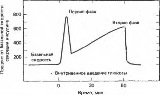
Fig. 51.10. Biphasic pattern of insulin secretion in response to an increase in plasma glucose concentration.
Oral administration of glucose produces a much stronger stimulation of insulin secretion than its intravenous administration. It follows that various Gastrointestinal Hormones, such as secretin, cholecystokinin, gastrin, and enteroglucagon, also influence insulin secretion in addition to glucose. However, the most important role in this process belongs to the gastric inhibitory polypeptide (GIP).
Two different mechanisms are hypothesized for the glucose-regulated secretion of insulin. According to one hypothesis, glucose interacts with a receptor, presumably localized On the surface membrane of the B-cell, which leads to the activation of the secretory mechanism. The second hypothesis assumes that intracellular metabolites or the rate of metabolic pathways such as The pentose phosphate shunt, The Citric Acid Cycle, or Glycolysis participate in stimulating insulin secretion. Both hypotheses have found experimental support.
B. Hormonal factors. Numerous hormones influence insulin release. alpha-Adrenergic agonists, especially epinephrine, inhibit insulin secretion even when this process is stimulated by glucose. beta-Adrenergic agonists stimulate insulin secretion, likely by increasing the intracellular cAMP concentration (see below). This mechanism presumably underlies the action of the gastric inhibitory polypeptide, which increases insulin secretion, as well as the effects of high concentrations of TSH, ACTH, gastrin, secretin, cholecystokinin, and enteroglucagon.
Chronic exposure to excessive levels of Growth Hormone, cortisol, placental lactogen, estrogens, and progestins also increases insulin secretion. Therefore, it is hardly surprising that insulin secretion rises significantly during late Pregnancy.
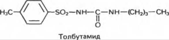
B. Pharmacological agents. Although many drugs stimulate insulin secretion, sulfonylureas are most commonly used for therapeutic purposes. Agents such as tolbutamide are widely employed in the treatment of type II (non-insulin-dependent) diabetes; tolbutamide stimulates insulin secretion through a mechanism distinct from that of glucose.
C. Intracellular mediators of secretion. The glucose-stimulated secretion of insulin is accompanied by increased O2 consumption and ATP utilization. This process is coupled with K+-induced membrane depolarization, which leads to a rapid influx of Ca2+ into The Cell via voltage-gated channels. The fusion of insulin-containing secretory granules with the plasma membrane—and the resulting insulin secretion—is a calcium-dependent process. Furthermore, glucose-stimulated insulin secretion involves phosphatidylinositol metabolites (Ch. 44).
cAMP is also involved in the insulin secretion process, where it potentiates the effects of glucose and amino acids. This nucleotide may stimulate the release of Ca2+ from intracellular Organelles or activate a kinase that phosphorylates a component of the microfilament-microtubule system (thereby conferring Ca2+ sensitivity and contractility). Replacing extracellular Na+ with another monovalent cation attenuates the effects of glucose and other insulin secretagogues; Na+ may regulate intracellular Ca2+ concentration via a cotransport system.
Insulin Metabolism
Unlike insulin-like growth factors, insulin lacks a carrier protein in plasma. Consequently, its normal half-life is less than 3–5 minutes. The metabolic degradation of insulin occurs primarily in the Liver, Kidneys, and Placenta. Approximately 50% of the hormone is cleared from the plasma during a single passage through the liver. Two enzyme systems are involved in insulin metabolism. The first is an insulin-specific proteinase found in many Tissues, with highest concentrations in the aforementioned Organs. This proteinase has been isolated from Skeletal Muscle and purified; its activity is known to be sulfhydryl-dependent and operates at physiological pH values. The second system is Glutathione-insulin transhydrogenase. This enzyme reduces disulfide bonds, after which the separated A and B chains are rapidly degraded. It remains unclear which of these two mechanisms is more active under physiological conditions, nor is it established whether either mechanism is subject to regulation.
Physiological effects of Insulin
The critical role of insulin in carbohydrate, protein, and Lipid Metabolism is most clearly demonstrated by the consequences of human insulin deficiency. The hallmark of diabetes mellitus is hyperglycemia, which develops As a result of (1) reduced cellular glucose uptake, (2) decreased glucose utilization by various tissues, and (3) increased hepatic glucose production (Gluconeogenesis). These processes are examined in greater detail below.
Polyuria, polydipsia, and weight loss despite adequate caloric intake are the primary symptoms of insulin deficiency. How can these be explained? While normal human plasma glucose levels rarely exceed 120 mg%, they are typically much higher in patients with insulin deficiency. When plasma glucose exceeds a certain threshold (usually above 180 mg% in humans), the maximum reabsorption capacity of the renal tubules is overwhelmed, and sugar spills into the urine (glucosuria). This results in increased urine volume due to osmotic diuretic effects, invariably leading initially to fluid loss (polyuria), followed by dehydration, thirst, and excessive Water intake (polydipsia). Glucosuria causes significant caloric loss (4.1 kcal per gram of excreted glucose), which, combined with the loss of muscle and adipose tissue, leads to profound weight loss despite an increased appetite (polyphagia) and normal or elevated caloric consumption.
In the absence of insulin, Protein Biosynthesis declines, partly due to diminished Amino Acid Transport into muscle (as amino acids serve as substrates for gluconeogenesis). Consequently, human insulin deficiency is accompanied by a negative nitrogen balance. The characteristic lack of insulin's antilipolytic action, alongside its impaired lipogenic effect, leads to elevated plasma fatty acid levels. When these levels exceed the liver's capacity to oxidize Fatty acids to CO2, β-hydroxybutyric and acetoacetic acids accumulate in the blood (Ketosis). Initially, the body compensates for this organic acid accumulation by increasing the rate of expired CO2. However, if ketosis is not halted by insulin administration, severe metabolic acidosis develops, and the patient succumbs to diabetic coma. The mechanism of insulin deficiency is schematically illustrated in Fig. 51.11.
A. Effect on membrane glucose transport.
The intracellular concentration of free glucose is significantly lower than its extracellular concentration. A wealth of evidence indicates that the rate of glucose Transport Across the plasma membrane of muscle and adipose cells determines the rate of glucose phosphorylation and subsequent metabolism. D-glucose and other sugars with a similar configuration at C1–C3 (galactose, D-xylose, and L-arabinose) enter cells via carrier-mediated Facilitated Diffusion. In many cell types, insulin enhances this process (Fig. 51.12) by increasing the number of transporters ($V_{max}$ effect) rather than by altering binding affinity ($K_m$ effect). Evidence suggests that in fat cells, this occurs via the mobilization of glucose transporters from an inactive intracellular pool in the Golgi apparatus to the active plasma membrane domain. This transporter translocation is a Temperature- and energy-dependent process that does not require de novo Protein Synthesis (Fig. 51.13).
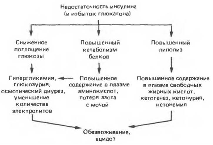
Fig. 51.11. Pathophysiology of insulin deficiency. (Courtesy of R. J. Havel.)
Hepatic cells represent a notable exception to this scheme. Insulin does not stimulate facilitated diffusion of glucose into hepatocytes, but rather enhances its influx indirectly by inducing glucokinase, the enzyme that converts glucose to glucose-6-phosphate. Due to this rapid phosphorylation, the concentration of free glucose within hepatocytes is maintained at a very low level, which facilitates glucose entry into the cells via simple diffusion down a concentration gradient.
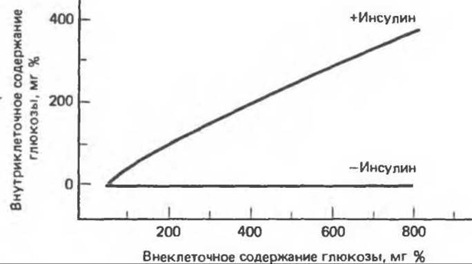
Fig. 51.12. Glucose entry into muscle cells.
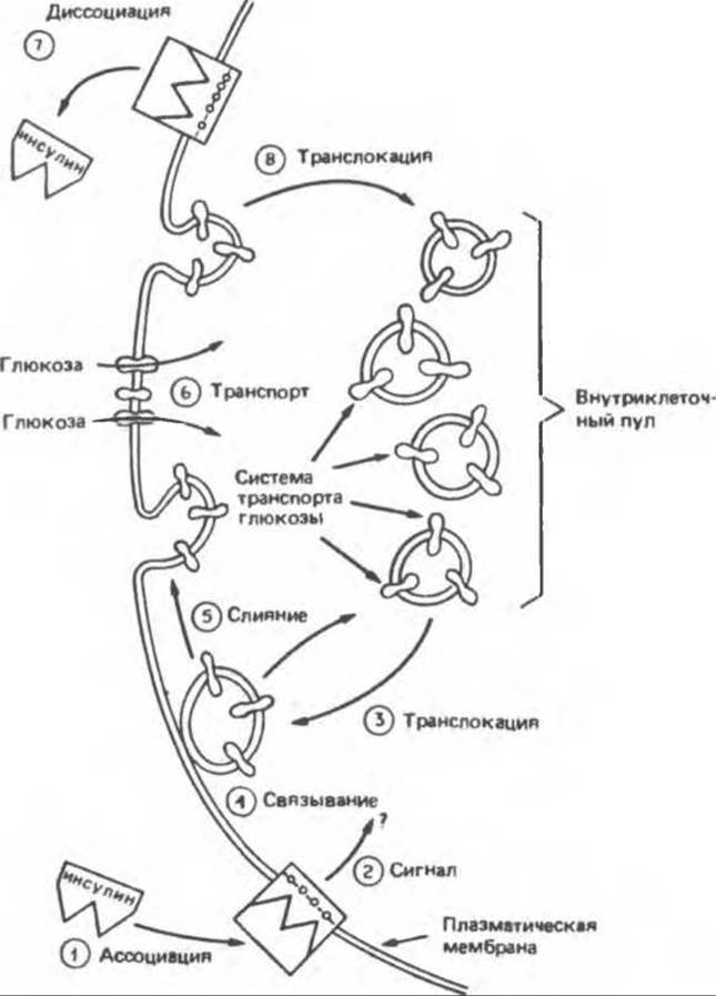
Fig. 51.13. Insulin-stimulated translocation of glucose transporters. (Reproduced, with permission, from Karnieli E. et al. Insulin-stimulated translocation of glucose transport systems in the isolated rat adipose cell, J. Biol. Chem., 1981, 256, 4772. Courtesy of S. Cushman.)
Insulin also promotes the cellular Uptake of Amino acids (particularly in muscle cells) and stimulates the transmembrane fluxes of K+, Ca2+, nucleosides, and organic phosphate. These effects are independent of insulin's action on cellular glucose uptake.
B. Effect on glucose utilization. As outlined below, insulin influences intracellular glucose utilization through multiple pathways.
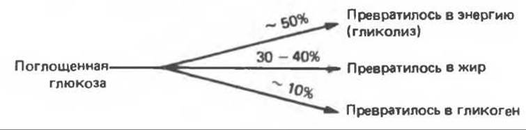
Under normal conditions, roughly half of the absorbed glucose enters The Glycolytic Pathway to be converted into energy, while the other half is stored as fat or Glycogen. In the absence of insulin, glycolytic flux decreases, and the anabolic processes of Glycogenesis and Lipogenesis slow down. Indeed, in insulin-deficient diabetes, as little as 5% of absorbed glucose is converted into fat.
Insulin enhances the rate of hepatic glycolysis by increasing the activity and concentration of several Key Enzymes, including glucokinase, Phosphofructokinase, and Pyruvate kinase. Enhanced glycolysis is accompanied by increased glucose utilization and thereby indirectly helps reduce glucose output into the plasma. Furthermore, insulin suppresses The activity of glucose-6-phosphatase, an enzyme found in the liver but absent in muscle. As a result, glucose is retained within the liver, since the plasma membrane is impermeable to glucose-6-phosphate.
In adipose tissue, insulin stimulates lipogenesis by 1) providing an influx of acetyl-CoA and NADPH, which are required for fatty acid synthesis; 2) maintaining normal levels of the enzyme acetyl-CoA carboxylase, which catalyzes The conversion of acetyl-CoA to malonyl-CoA; and 3) providing an influx of glycerol, which participates in the synthesis of triacylglycerols. Under conditions of insulin deficiency, all of these processes are impaired, resulting in a decreased rate of lipogenesis. Another reason for reduced lipogenesis in insulin deficiency is that fatty acids—released in large quantities by the Action of Certain hormones unopposed by insulin—suppress their own synthesis by inhibiting acetyl-CoA carboxylase. Consequently, the overall effect of insulin on fat metabolism is anabolic.
The mechanism by which insulin influences glucose utilization encompasses another anabolic process. In The Liver and Muscles, insulin stimulates the conversion of glucose to glucose-6-phosphate, which then undergoes isomerization to glucose-1-phosphate and is incorporated into glycogen via the action of glycogen synthase (an enzyme whose activity is also stimulated by insulin). This effect is both dual and indirect. Insulin lowers intracellular cAMP levels by activating phosphodiesterase. Because cAMP-dependent phosphorylation inactivates glycogen synthase, a low level of this nucleotide keeps the enzyme in its active form. Insulin also activates a phosphatase that catalyzes the dephosphorylation of glycogen synthase, thereby turning the enzyme on. Finally, insulin inhibits phosphorylase through a mechanism involving cAMP and phosphatase, as described above. As a result, the release of glucose from glycogen is diminished. Thus, The Effect of insulin on Glycogen Metabolism is similarly anabolic.
B. Effect on glucose production (gluconeogenesis). The influence of insulin on glucose transport, glycolysis, and glycogenogenesis manifests within seconds or minutes, since the primary reactions of this effect involve the activation or inactivation of enzymes via phosphorylation or dephosphorylation. The more prolonged effect of insulin on plasma glucose levels is associated with the inhibition of gluconeogenesis. The formation of glucose from non-carbohydrate precursors proceeds through a series of enzymatic reactions, many of which are stimulated by glucagon (whose action is mediated by cAMP), glucocorticoid hormones, and to a lesser extent by a- and ß-adrenergic agents—angiotensin II and vasopressin. Insulin, by contrast, suppresses these enzymatic reactions. The key gluconeogenic enzyme in the liver is phosphoenolpyruvate carboxykinase (PEPCK), which catalyzes the conversion of oxaloacetate to phosphoenolpyruvate. Recent studies (see below) indicate that insulin decreases the amount of this enzyme by selectively inhibiting the Transcription of the gene encoding mRNA for phosphoenolpyruvate carboxykinase.
C. Effect on glucose metabolism. The net result of all the aforementioned effects of insulin is a reduction in blood glucose levels. This Action of Insulin is counteracted by the effects of numerous hormones, which undoubtedly reflects one of the body's vital protective mechanisms, as prolonged hypoglycemia can cause life-threatening damage to the Brain and must therefore be avoided.
D. Effect on lipid metabolism. The lipogenic action of insulin has already been discussed in the section concerning its effect on glucose utilization. In addition, insulin is a potent inhibitor of lipolysis in the liver and adipose tissue, thereby exerting an indirect anabolic effect. This is partly due to insulin's ability to lower cAMP levels (which rise in tissues under the Influence of the lipolytic hormones glucagon and epinephrine), as well as its ability to inhibit hormone-sensitive lipase activity. This inhibition is apparently mediated by the Activation of a phosphatase that dephosphorylates and thereby inactivates the lipase or cAMP-dependent protein kinase. Consequently, insulin lowers blood fatty acid levels. In turn, this contributes to insulin's effect on Carbohydrate Metabolism, since fatty acids suppress glycolysis at multiple steps and stimulate gluconeogenesis. This example demonstrates that when discussing Metabolic Regulation, one cannot account for the action of a single hormone or metabolite in isolation. Regulation is a complex process in which transformations along a specific metabolic pathway represent the outcome of intricate interactions among a wide array of hormones and metabolites.
In patients with insulin deficiency, lipase activity increases, leading to enhanced lipolysis and elevated concentrations of fatty acids in the plasma and liver. Glucagon levels are also elevated in such patients, which further promotes the release of free fatty acids into the blood. (Glucagon counteracts many of insulin's effects, and the metabolic status in diabetes reflects The ratio of glucagon to insulin levels.) A portion of the free fatty acids is metabolized to acetyl-CoA (the reverse of lipogenesis) and subsequently oxidized in The Citric Acid cycle to CO2 and H2O. In insulin deficiency, the capacity of this pathway is quickly exceeded, and acetyl-CoA is converted into acetoacetyl-CoA and then into acetoacetic and ß-hydroxybutyric acids. Insulin prompts the reverse transformations.
Insulin appears to influence the formation or clearance of very-low-density Lipoproteins (VLDL) and low-density lipoproteins (LDL), since patients with poorly controlled diabetes often exhibit elevated levels of these particles and, consequently, increased Cholesterol levels. This particular metabolic defect is evidently the underlying cause of accelerated atherosclerosis—a severe complication observed in many diabetic patients.
The effect of insulin on metabolic processes is illustrated in Fig. 51.14, which depicts several major Metabolic Transformations in the absence of insulin.
E. Effect on Protein metabolism. Insulin generally exerts an anabolic effect on protein metabolism by stimulating PROTEIN SYNTHESIS AND reducing protein degradation. Insulin stimulates the uptake of neutral A-type amino acids by muscle—an effect independent of glucose uptake or the subsequent Incorporation of Amino acids into Proteins. The influence of insulin on Protein synthesis in skeletal and cardiac muscle appears to be manifested at the level of mRNA Translation.
In recent years, it has been demonstrated that insulin affects the Synthesis of specific proteins by inducing changes in their corresponding mRNAs. This may well explain the hormone's action on the activity or Abundance of individual proteins. (This topic is discussed in more detail below.)
F. Effect on cell proliferation. Insulin stimulates the proliferation of various cells in culture and may also participate in The regulation of growth in vivo. Fibroblast cultures are most commonly used in studies of growth regulation. In such cells, insulin enhances the ability of fibroblast growth factor (FGF), platelet-derived growth factor (PDGF), epidermal growth factor (EGF), tumor-promoting phorbol esters, prostaglandin F2a (PGF2a), vasopressin, and cAMP analogs to stimulate the proliferation of cells arrested in the G1 phase following serum deprivation.
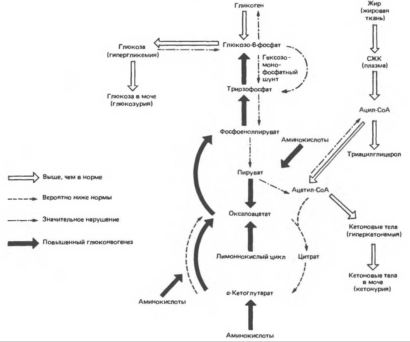
Fig. 51.14. Metabolic Consequences of Insulin deficiency. FFA — free fatty acids.
The transient requirement for various growth factors underlies THE CONCEPT OF two classes of such factors. One of these, which includes TGF, FGF, PGF2, and phorbol esters, apparently induces certain Biochemical changes in the early G-phase that, once triggered, eliminate the cell's further need for these factors and enable its Replication. Growth factors of the second class (which includes insulin) promote the "progression" of the cell into and through the S-phase and must be present continuously. This model describes processes occurring in 3T3 fibroblasts, and its universality has not been proven. It is also unknown whether the effect of insulin is related to its interaction with its own receptor or with the insulin-like growth factor (IGF) receptor (especially since IGF-1 is also a "progression" factor).
Insulin supports the growth and replication of many epithelial-derived cells, including hepatocytes, hepatoma cells, adrenal cortex tumor cells, and breast carcinoma cells. Very low concentrations of insulin stimulate replication (presumably via the Insulin Receptor), often in the absence of other peptide growth factors. Indeed, insulin is an essential component of all known tissue culture media, underscoring its undeniable importance for cell growth and replication.
The biochemical mechanism by which insulin influences cell replication remains unclear; it is hypothesized to rely on the anabolic action of the hormone. Its effects on the uptake of glucose, phosphate, neutral amino acids of type A, and cations may play a role here. The hormone can stimulate replication by utilizing its ability to activate or inactivate enzymes through the Regulation of the rate and degree of protein phosphorylation, or by regulating enzyme synthesis.
A highly promising new area of research involves The Study of Tyrosine kinase activity. The insulin receptor, like the receptors for many other growth factors including TGF and EGF, exhibits tyrosine kinase activity. Notably, at least 10 oncogenic products (many of which likely participate in stimulating malignant cell replication) are also tyrosine Kinases. Mammalian cells contain analogs of these oncogenes (Proto-oncogenes), whose products could be involved in normal cell replication. The hypothesis regarding The Role of proto-oncogenes is supported by recent studies showing that the expression of at least two proto-oncogene products — c-fos and c-myc — increases after serum is added to growth-arrested cell cultures. It has also been demonstrated that TGF stimulates The production of specific mRNAs. It remains to be determined whether the mechanism of insulin action is analogous.
Mechanism of Insulin Action
A. Insulin receptor. The action of insulin begins with its binding to a specific glycoprotein receptor on The surface of the target cell. The various effects of this hormone (Fig. 51.15) can manifest either within a few seconds or minutes (transport, protein phosphorylation, enzyme Activation and inhibition, RNA Synthesis) or over several hours (protein and DNA Synthesis AND cell growth).
The insulin receptor has been studied in detail using biochemical and recombinant DNA technologies. It is a heterodimer consisting of two subunits ($\alpha$ and $\beta$) in an $\alpha_2$-$\beta_2$ configuration, linked by disulfide bridges (Fig. 51.15). Both subunits contain numerous glycosyl residues. The removal of sialic acid and galactose reduces both the insulin-binding capacity and the hormone's activity. Each glycoprotein subunit possesses a distinct structure and a specific function. The $\alpha$-subunit (mol. mass 135,000) is located entirely extracellularly, and insulin binding likely occurs via a cystine-rich domain. The $\beta$-subunit (mol. mass 95,000) is a transmembrane protein performing the second major receptor function (Chap. 44), namely, signal Transduction. The cytoplasmic portion of the $\beta$-subunit exhibits tyrosine kinase activity and contains an autophosphorylation site. Both of these features are believed to be crucial for signal transduction and insulin action (see below). The striking similarity among three receptors with different Functions is illustrated in Fig. 51.16. Indeed, the sequences of certain regions of the $\beta$-subunit are homologous to those in the EGF receptor.
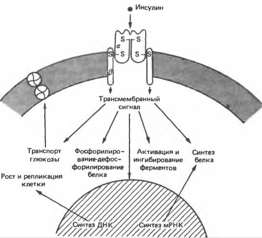
Fig. 51.15. Relationship between the insulin receptor and its action. (Courtesy of C. R. Kahn.)
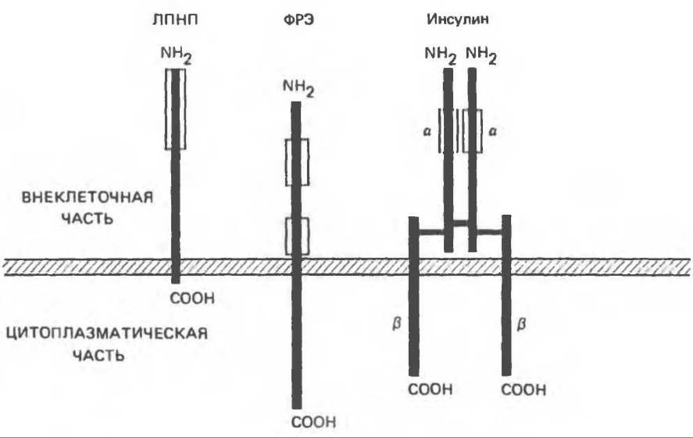
Fig. 51.16. Schematic STRUCTURE OF THE low-density lipoprotein (LDL), epidermal growth factor (EGF), and insulin receptors. In each of these receptors, the amino termini are located in the extracellular portion of the molecule. Boxed regions indicate cystine-rich domains believed to be involved in Ligand binding. Each receptor contains a short domain spanning the plasma membrane (hatched bar) of approximately 25 Amino Acids and an intracellular domain of varying length. The EGF and insulin receptors possess tyrosine kinase activity localized in the cytoplasmic domain; in addition, this domain contains sites where autophosphorylation occurs. The insulin receptor is a heterotetramer, with individual chains (vertical bars) linked together by disulfide bridges.
The insulin receptor undergoes continuous Synthesis and degradation, with a half-life of 7–12 h. The receptor is synthesized as a single-chain peptide in the rough Endoplasmic reticulum and is rapidly glycosylated in the Golgi apparatus. The human insulin receptor precursor consists of 1,382 amino acids with a molecular mass of 190,000, and upon cleavage, it yields mature $\alpha$- and $\beta$-subunits. In humans, the insulin receptor gene is localized to chromosome 19.
Insulin receptors are found on the surface of most mammalian cells. Their concentration reaches up to 20,000 per cell, and they are frequently detected even on cells not typically classified as insulin targets. THE SPECTRUM OF insulin's metabolic effects is well established. However, insulin is also involved in such processes as cell growth and replication (see above), fetal Organogenesis and differentiation, as well as tissue healing and regeneration. The structure of the insulin receptor, the ability of various insulins to bind to receptors, and their capacity to elicit biological responses are virtually identical across all cell types and species. For instance, pork insulin is almost always 10 to 20 times more effective than pork proinsulin, which in turn is 10 to 20 times more effective than guinea pig insulin, even in the guinea pig itself. The insulin receptor appears to possess a highly conserved structure—even more conserved than the structure of insulin itself.
When insulin binds to its receptor, the following events occur: 1) the receptor undergoes a conformational change, 2) the receptors associate with one another to form microaggregates, patches, or clusters, 3) the receptor is internalized, and 4) a signal is generated. The Significance of the Conformational Changes in the receptor remains unknown, but internalization likely serves as a mechanism to regulate receptor number and turnover. Under conditions of high plasma insulin, such as in obesity or acromegaly, the number of insulin receptors decreases, and target tissue sensitivity to insulin is diminished. This down-regulation results from the loss of receptors through internalization—that is, the process by which insulin-receptor complexes enter the cell via endocytosis mediated by clathrin-coated vesicles (see Chapter 41). Down-regulation partially explains insulin resistance in obesity and type II diabetes mellitus.
B. Intracellular Mediators. Although the mechanism of insulin action has been studied for over 60 years, some of the most critical questions, such as The Nature of the intracellular signal, remain unresolved, and insulin is no exception in this regard. Intracellular messengers have not been identified for a great many hormones (Table 44.1). A multitude of different molecules have been considered as potential intracellular second messengers or mediators. These include insulin itself, calcium, Cyclic NUCLEOTIDES (cAMP, cGMP), H2O2, membrane-derived peptides, membrane Phospholipids, monovalent cations, and tyrosine kinase (the insulin receptor). None of these hypotheses have been confirmed.
The focus of modern research is centered on the fact that the insulin receptor is itself an insulin-sensitive enzyme, as it undergoes autophosphorylation upon insulin binding. This function is carried out by the ß-subunit, which, acting as a protein kinase, transfers a y-phosphate from ATP to a tyrosine residue within the ß-subunit. Insulin increases the Vmax of this enzymatic reaction, while divalent cations, particularly Mn2+, decrease the Km for ATP.
Tyrosine phosphorylation is atypical for mammalian cells (phosphotyrosine accounts for only 0.03% of the phosphoamino acids found in normal cells), and it is quite possible that the presence of tyrosine kinase activity in the receptors for EGF, TGF, and IGF-1 is not coincidental. It has been suggested that tyrosine kinase activity is an important factor in the action of products from a number of Viral Oncogenes. Their relationship with cellular oncogene analogs, which exhibit similar properties in malignant and normal cell growth, was discussed above. Structural studies of these components have revealed a high degree of Homology between receptors and oncogenes—for example, between the EGF receptor and erb-B, the TGF receptor and v-sis, and the insulin receptor and v-ros.
The involvement of tyrosine kinase in insulin-receptor signal transduction has not been formally proven, but it could consist of phosphorylating a specific protein that initiates insulin action, triggering a phosphorylation-dephosphorylation cascade, altering certain properties of The cell membrane, or generating a membrane-associated product, such as a phospholipid.
C. Protein Phosphorylation-Dephosphorylation.
Many of insulin's metabolic effects, particularly those that occur rapidly, are mediated by its influence on protein phosphorylation-dephosphorylation reactions, which in turn affect the enzymatic activity of the given protein. A List of enzymes whose activity is regulated in this manner is provided in Table 51.3. In some cases, insulin lowers the intracellular cAMP content (by activating cAMP phosphodiesterase), leading to a decrease in cAMP-dependent protein kinase activity. Such effects are characteristic of Glycogen synthase and phosphorylase. In other instances, insulin action is independent of cAMP and results from the activation of other protein kinases (for example, in the case of the insulin receptor tyrosine kinase), the inhibition of still other protein kinases (Table 44.4), or (much more frequently) The stimulation of phosphoprotein Phosphatases. Dephosphorylation increases the activity of a number of key enzymes (Table 51.3). Such covalent modifications ensure almost instantaneous changes in enzyme activities.
Table 51.3. Enzymes whose phosphorylation state and activity are regulated by insulin. (Modified and reproduced, with permission, from Denton R. M. et al.: A partial view of the mechanism of insulin action. Diabetologia 1981, 21, 347.)
|
Enzyme |
Activity change |
Possible mechanism |
|
cAMP metabolism Phosphodiesterase (low Km) |
Increased |
Phosphorylation |
|
Protein kinase (cAMP-dependent) |
Decreased |
Association of R and C subunits |
|
Glycogen metabolism Glycogen synthase |
Increased |
Dephosphorylation |
|
Phosphorylase kinase |
Decreased |
" |
|
Glycolysis and gluconeogenesis |
||
|
Pyruvate dehydrogenase |
Increased |
" |
|
Pyruvate kinase |
" |
" |
|
6-Phosphofructo-2-kinase |
" |
" |
|
Fructose-2,6-bisphosphatase |
Decreased |
" |
|
Lipid metabolism Acetyl-CoA carboxylase |
Increased |
Phosphorylation |
|
HMG-CoA reductase (hydroxymethylglutaryl-CoA reductase) |
" |
Dephosphorylation |
|
Triacylglycerol lipase |
Decreased |
" |
|
Other processes Tyrosine kinase (insulin receptor) |
? |
Phosphorylation |
D. Effect on mRNA Translation. Insulin is known to influence the abundance and activity of at least 50 proteins across various tissues, with many of these effects boiling down to covalent modification. The concept of insulin's role in mRNA translation is based primarily on data regarding the ribosomal S6 protein, a component of the 40S ribosomal subunit. Such a mechanism could account for insulin's generalized effect on protein synthesis in the liver, skeletal muscle, and Heart.
E. Effect on Gene Expression. All the described effects of insulin are realized at the plasma membrane level or within the cytoplasm. However, insulin is also capable of influencing (presumably via its intracellular mediator) certain specific nuclear processes. The enzyme phosphoenolpyruvate carboxykinase (PEPCK) catalyzes the rate-limiting step of gluconeogenesis. PEPCK synthesis is decreased under the influence of insulin, and consequently, the rate of gluconeogenesis is also reduced. Relatively recently, it was demonstrated that when insulin is added to hepatoma cell cultures, the transcription rate of the PEPCK gene selectively declines within just a few minutes (Fig. 51.17). As a result, the levels of both the primary transcript and mature PEPCK mRNA diminish, which in turn leads to a reduction in PEPCK synthesis. This effect manifests at physiological insulin concentrations (10-12 to 10-9 mol/L), is mediated by the insulin receptor, and appears to stem from a decreased synthesis rate of PEPCK mRNA.
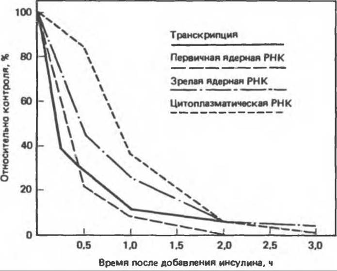
Fig. 51.17. Effect of insulin on specific gene transcription. Upon The addition of insulin to H4IIE hepatoma cell culture, the transcription rate of the PEPCK gene rapidly drops, which is accompanied by a decrease in the amount of primary transcript in the mature PEPCK mRNA. As cytoplasmic PEPCK mRNA levels decline, the synthesis rate of the PEPCK protein decreases as well. (Reproduced, with permission, from Sasaki K. et al. Multihormonal regulation of phosphoenolpyruvate carboxykinase gene transcription, J. Biol. Chem., 1984, 259, 15242.)
The influence of insulin on gene transcription was first discovered during studies of PEPCK regulation, but other Examples are now well known. Furthermore, it appears probable that the regulation of mRNA synthesis is a major action of insulin. Insulin affects the synthesis of numerous specific mRNAs (Table 51.4), including as-yet-unidentified mRNAs in the liver, adipose tissue, and muscle (both skeletal and cardiac). Insulin's action on the transcription of Ovalbumin, albumin, and casein genes has been firmly established.
Insulin's action extends to intracellular enzymes, secreted enzymes and proteins, reproductive proteins, and structural proteins (Table 51.4). These effects are recorded in many organs and tissues and across numerous species. The Regulation of Specific mRNA transcription by insulin is now beyond doubt. This pathway of modulating enzymatic activity is every bit as important as the phosphorylation-dephosphorylation mechanism. It is precisely insulin's effect on gene transcription that likely accounts for its role in Embryogenesis, cellular differentiation, and Cell Growth and Division.
Table 51.4. Proteins whose mRNAs are regulated by insulin
|
Intracellular enzymes Tyrosine aminotransferase1) Phosphoenolpyruvate carboxykinase1) Fatty acid synthase Pyruvate kinase Glycerol-3-phosphate dehydrogenase1) Glyceraldehyde-3-phosphate dehydrogenase1) Glucokinase |
|
Secreted Proteins and Enzymes Albumin1) Amylase a2-Globulin Growth hormone1) |
|
Proteins involved in reproduction Ovalbumin1) Casein1) |
|
Structural proteins 5-Crystallin |
|
Other proteins In the liver (p33, etc.) In adipose tissue In cardiac muscle In skeletal muscle |
1) Insulin regulates the transcription rate of the corresponding genes.
Pathophysiology
Diabetes mellitus develops as a result of insulin deficiency or resistance to its action. Approximately 90% of diabetic patients suffer from type II non-insulin-dependent diabetes mellitus (NIDDM). These patients are typically characterized by obesity, elevated plasma insulin levels, and a reduced number of insulin receptors. The remaining 10% of patients have type I diabetes, i.e., insulin-dependent diabetes mellitus (IDDM). The metabolic disturbances discussed above are more typical specifically of type I diabetes.
A number of rare conditions illustrate important features of insulin action. Some individuals develop antibodies against insulin receptors. These antibodies prevent insulin from binding to its receptor, resulting in The Development of severe insulin resistance syndrome in these patients (see Table 43.2). B-cell tumors give rise to hyperinsulinemia and a syndrome characterized by severe hypoglycemia. The critical role of insulin (or possibly IGF-1 or IGF-2) in organogenesis is evidenced by rare cases of dwarfism. This syndrome is characterized by low birth weight, reduced muscle mass, scanty subcutaneous fat, very delicate facial features, insulin resistance with a marked elevation of biologically active plasma insulin, and early death. In some such patients, insulin receptors were either entirely absent or defective.
Last update: 06/08/2026
Editorial and Educational Adaptation: This material has been compiled based on the primary/original source text. The project team performed an editorial review, corrected technical inaccuracies, structured sections, and adapted the content for an educational format.
What was processed:
- elimination of formatting defects (OCR errors, structural breaks, corrupted characters);
- editorial organization of content;
- standardization of terminology in accordance with academic sources;
- verification of factual statements against the original source text.
All mentions of the author, publication year, and origin of the primary text have been preserved in accordance with the source.