Molecular Biology of the Cell - Volume 2 - Alberts B., Bray D., Lewis J., Raff M., Roberts K., Watson J. 1993
Intracellular sorting of macromolecules and maintenance of cellular compartments
Endoplasmic reticulum
All Eukaryotic Cells contain an Endoplasmic Reticulum (ER). Its highly convoluted membrane typically accounts for more than half of the total cellular membrane (see Table 8-2). Although the ER membrane features numerous folds and curves permeating the entire Cytoplasm, it is believed to form a continuous surface that encloses a single internal space. This internal space, known as the ER lumen, frequently occupies over 10% of the total Cell volume (see Table 8-1). The ER lumen is separated from the Cytosol by a single membrane (the ER membrane), which acts as a connecting link between these two compartments. Conversely, the lumen of the ER and that of each Golgi cisterna are separated from one another by two membranes and the cytosol; therefore, macromolecular transport between these Organelles is mediated by transport vesicles (Fig. 8-36). The ER plays a crucial role in cellular Biosynthesis. The synthesis of transmembrane Proteins and Lipids for the ER, the Golgi apparatus, the Lysosomes, and The Plasma Membrane begins on the ER membranes. Most of the lipids for mitochondrial and peroxisomal membranes are also produced here (see Section 8.6.14). Furthermore, all Newly synthesized proteins—regardless of their destination (ER lumen, Golgi apparatus, lysosomes, or extracellular space)—initially enter the ER lumen. Since the ER serves as the starting point for the synthesis of all secreted proteins, it is also the site where The formation of the Extracellular matrix begins.
Class="center">
Fig. 8-36. Connection of the ER lumen with other intracellular compartments that contact the ER. The ER lumen is separated from both The Nucleus and the cytosol by a single membrane, whereas it is separated from stacked Golgi cisternae by two membranes. In most cases, the ER and the Golgi apparatus can be viewed as a single functional unit whose parts are linked by transport vesicles.
8.6.1. Ribosomes attached to the ER define the boundaries of its rough regions [32]
The ER removes certain proteins from the cytosol immediately after their synthesis. These are proteins of two types: (1) transmembrane proteins, which are only partially translocated across the ER membrane and remain embedded in it, and (2) Water-soluble proteins, which are completely translocated across the ER membrane and released into its lumen. Transmembrane proteins are destined for the plasma membrane or the membranes of other organelles, whereas water-soluble proteins are directed either to the lumen of organelles or secreted. All these proteins are translocated across the ER membrane via the same mechanism and the same type of signal peptide.
In mammalian cells, the import of proteins into the ER begins even before the polypeptide chain is fully synthesized—that is, it occurs concurrently with Translation (co-translationally). This distinguishes it from import into Mitochondria, METABOLISM/14.html">Chloroplasts, nuclei, and Peroxisomes, which is post-translational and requires different signal Peptides. Because a protein is translocated into the ER immediately as the polypeptide chain is formed, the ribosomes synthesizing it must be attached to the ER membrane. Regions of the ER studded with membrane-bound ribosomes are referred to as the rough endoplasmic reticulum (Fig. 8-37).
Thus, There are two spatially isolated populations of ribosomes in the cytoplasm. Some of these (membrane-bound ribosomes) are located on the cytosolic surface of the ER membrane and are engaged in synthesizing proteins that are immediately translocated into the ER. Others (free ribosomes) are not attached to any membrane and produce all other nucleus-encoded proteins. Bound and free ribosomes are identical in Structure and function; they differ solely in the proteins being synthesized on them at any given moment. If a ribosome is tasked with synthesizing a protein bearing an ER signal peptide, this signal directs the ribosome to the ER membrane. Because many ribosomes can bind to a single mRNA molecule, a polyribosome typically forms and attaches to the membrane via multiple growing polypeptide chains simultaneously. Individual ribosomes bound to such an mRNA molecule can return to the cytosol as soon as they finish translation near its 3' end. The mRNA itself, however, tends to remain bound to the ER membrane through a turnover of membrane-bound ribosomes (Fig. 8-38). Conversely, if the mRNA encodes a protein lacking an ER signal peptide, the resulting polyribosome remains in the cytosol, and its protein product remains there as well. Consequently, only mRNAs encoding proteins that carry ER signal peptides bind to the membranes of the rough ER; mRNA molecules encoding all other proteins remain in the cytosol. Individual ribosomes are thought to move randomly between these two distinct populations of mRNA molecules.

Fig. 8-37. A. Electron micrograph showing prominent differences in the Morphology of rough and smooth ER. The Leydig cell shown here produces Steroid Hormones in the Testis and therefore features an unusually well-developed smooth ER. Part of a large spherical lipid droplet is also visible. B. Three-dimensional reconstruction of smooth and rough ER regions in a Liver cell. The rough ER derives its name from the numerous ribosomes studded on its cytoplasmic surface; it forms polarized stacks of flattened cisternae, each with a lumen (cavity) 20 to 30 nm wide. Connected to these cisternae are the membranes of the smooth ER, which form a network of fine tubules 30 to 60 nm in diameter. The ER membrane is believed to be continuous and encloses a single lumen. (A, courtesy of Daniel S. Friend; B, after R. Krstic, ULTRASTRUCTURE OF THE Mammalian Cell. New York: Springer-Verlag, 1979.)
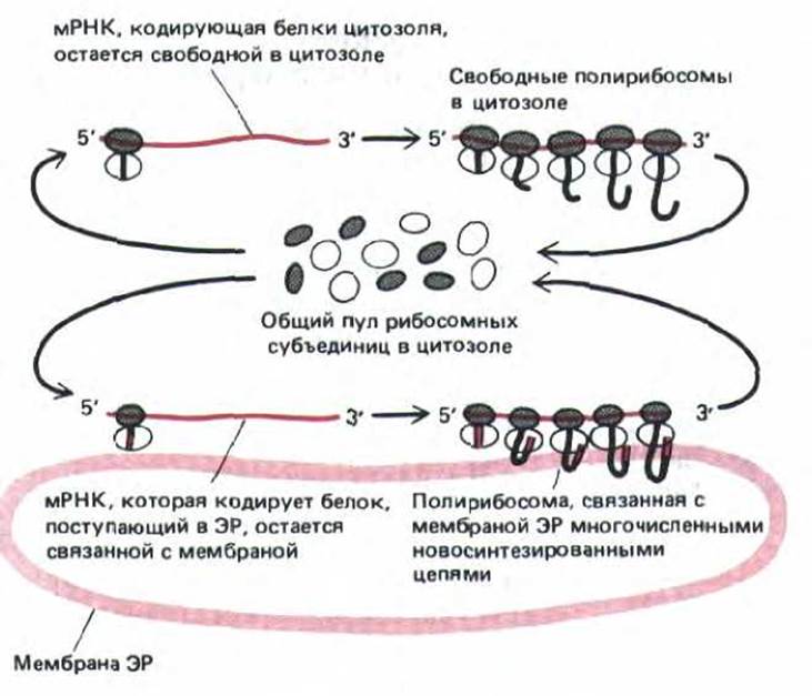
Fig. 8-38. The same ribosomes are used for the synthesis of cytosolic proteins and proteins entering the ER. If a signal peptide is present on a newly synthesized protein molecule, it directs the associated ribosome to the ER membrane. The mRNA molecule can remain bound to the ER membrane throughout, while ribosomes moving along it continuously recycle; at the end of each Protein Synthesis cycle, the ribosomal subunits are released and return to the general cytosolic pool of ribosomes.
8.6.2. Certain specialized cells abound in smooth ER [33]
Regions of the ER that lack attached ribosomes are called the smooth ER. As a rule, if cells contain a true smooth ER, it is present in very small amounts; in fact, most ER regions are partially smooth and partially rough, and are termed the transitional ER. It is from these regions that transport vesicles bud off to carry newly synthesized proteins to the Golgi apparatus (see Fig. 8-9). However, there are specialized cells in which the smooth ER is well developed and performs specific functions. In particular, the smooth endoplasmic reticulum predominates in cells specialized for Lipid Metabolism. For example, cells that synthesize steroid hormones from Cholesterol possess an extensive smooth ER dedicated to housing the Enzymes involved in cholesterol synthesis and its conversion into hormones (see Fig. 8-37A).
Another example of cells rich in smooth ER is hepatocytes, which are the primary site for The production of lipoprotein particles destined for export. The enzymes that synthesize the Lipid Components of Lipoproteins are localized on the membranes of the smooth ER. Situated on these same membranes are enzymes that catalyze a series of detoxification reactions, neutralizing both pharmaceutical drugs and harmful metabolic by-products. The best-studied detoxification reactions are those catalyzed by the cytochrome P450 family of enzymes. These proteins utilize high-energy electrons derived from NADPH to add hydroxyl groups to any of A wide variety of potentially harmful, water-insoluble Hydrocarbons entering the bilayer. Other Enzymes in the ER membrane then add negatively charged, water-soluble molecules (such as sulfate or glucuronic acid) to these hydroxyl groups. After several such reactions, a water-insoluble drug (or metabolite) that might otherwise accumulate in cell membranes becomes sufficiently soluble to leave The Cell and be excreted in the urine. Because the rough ER alone cannot accommodate all these and other essential enzymes, a significant portion of a hepatocyte's membranes normally consists of smooth ER (see Table 8-2).
If large amounts of certain compounds, such as phenobarbital, enter the bloodstream, detoxification enzymes are synthesized in unusually large quantities in the liver, and the surface area of the smooth ER can double within a few days. Once the drug is cleared, the excess smooth ER membranes are degraded by lysosomes (via specialized structures called autophagosomes—see Section 8.8.3), and the smooth ER returns to its normal volume within 5 days. How all these changes are regulated remains unknown.
Muscle cells possess a specialized, smooth ER-like organelle called the sarcoplasmic reticulum, which sequesters Ca2+ from the cytosol. The primary membrane protein of the sarcoplasmic reticulum is the Ca2+-ATPase, which pumps Ca2+ ions into its lumen. The rapid contraction and relaxation of myofibrils in each Muscle contraction cycle are mediated by the release of Ca2+ from the sarcoplasmic reticulum followed by its reuptake from the cytosol. Smaller Ca2+-storing organelles are found in most eukaryotic cells, where they release Ca2+ into the cytosol in response to extracellular signals. They were previously thought to be part of the ER, but it now seems more likely that they constitute an independent membrane compartment of unknown origin.
We now turn to a Discussion of the two primary Functions of the ER: the synthesis and modification of proteins, and the synthesis of lipids.
8.6.3. Rough and smooth regions of the ER can be separated by centrifugation [34]
To study the functions and biochemistry of The endoplasmic reticulum, its membranes must first be separated from other cellular components. At first glance, this task appears impossible because the ER effectively interleaves with all other cytoplasmic components (see Fig. 8-35). Fortunately, when Tissues or cells are disrupted by homogenization, the ER breaks down into numerous small (~100 nm in diameter) closed vesicles called microsomes, which are relatively easy to purify.
Microsomes derived from the rough ER are studded with ribosomes and are termed rough microsomes. The ribosomes are always located on their outer surface, indicating that the space inside the microsomes is biochemically equivalent to the ER lumen (Fig. 8-39). Such homogenates also contain numerous vesicles similar in size to rough microsomes but devoid of ribosomes. These smooth microsomes form partly from smooth regions of the ER and partly from fragments of the plasma membrane, Golgi apparatus, endosomes, and mitochondria (their exact proportion depends on the tissue from which the microsomal fraction is prepared). Thus, while rough microsomes can be definitively identified with the rough ER, THE ORIGIN OF smooth microsomes cannot be established with the same ease. An exception is the liver: because hepatocytes contain an exceptionally large amount of smooth ER, the majority of smooth microsomes in liver homogenates originate from the smooth ER.

Fig. 8-39. When cells are disrupted by homogenization, rough ER cisternae (A) break down into small closed vesicles called rough microsomes (B). Similarly, the smooth ER breaks down into small vesicles lacking ribosomes, termed smooth microsomes. (A, electron micrograph courtesy of Daniel S. Friend; B, electron micrograph courtesy of George Palade.)
Ribosomes, containing a high amount of RNA, confer a higher density on rough microsomes compared to smooth ones. Consequently, smooth and rough microsomes can be separated from each other by sedimenting their mixture through a sucrose density gradient (Fig. 8-40). When rough and smooth liver microsomes are compared in parameters such as enzymatic activity or polypeptide composition, they prove to be remarkably similar (though not identical). This implies that most Components of the ER membrane can move freely between its smooth and rough domains, as would be expected given the fluidity and continuity of the membrane system.
Because rough microsomes are easily purified while retaining their functional activity, they are exceptionally useful for studying the multitude of processes that take place within the ER.

Fig. 8-40. Procedure used to isolate rough and smooth microsomes from the ER.
These organelles are topologically organized in the same way as the rough ER; their cytoplasmic surface is readily accessible to ingredients that can be added in vitro. To the biochemist, rough microsomes are nothing more than a scaled-down version of the rough endoplasmic reticulum, capable of protein synthesis, glycosylation, and lipid synthesis.
8.6.4. Granular (rough) regions of the ER contain proteins responsible for ribosome binding [35]
Since the ER membrane, like all membranes, is a two-dimensional fluid, most proteins and lipids should be free to distribute within it (barring any special restrictions) between rough and smooth regions. Nevertheless, rough microsomes isolated from the liver were found to contain more than 20 proteins absent from smooth microsomes. This fact points to the existence of specific limiting mechanisms. Some of these "nonequilibrium" Proteins of the rough ER membrane
help bind ribosomes, while others presumably determine its flattened shape (see Fig. 8-37). It remains unclear how these proteins are retained in the membrane: whether they form large two-dimensional aggregates or interact with a network of structural proteins on either surface of the ER membrane (see Section 6.2.10).
Ribosomes of the rough ER are held on the membrane partly thanks to growing polypeptide chains that advance through the membrane as they are synthesized (see below). However, if polypeptide chain formation is interrupted by an inhibitor (such as puromycin), the ribosomes still remain bound to the rough microsome membrane. This affinity increases significantly in solutions with low salt concentrations. If purified ribosomes are mixed under such conditions with rough microsome membranes previously stripped of ribosomes, these "peeled" membranes regain the same number of ribosomes they had upon isolation from cells. The membrane-binding site is located on the large ribosomal subunit, but it is still unclear which of the numerous rough ER Membrane Proteins the ribosome binds to. It has been established, however, that under physiological conditions, binding of ribosomes to the ER membrane requires additional, more specific attachment that necessitates a newly synthesized protein bearing a signal peptide.
8.6.5. Signal peptides were first discovered in proteins entering the ER [36]
Signal peptides (and The Mechanism of protein transfer mediated by them) were discovered in the early 1970s while studying secretory proteins, which enter the ER before being transferred to the Golgi apparatus and secreted from the cell. The Essence of the experiment was as follows: mRNA encoding a secretory protein was translated in an in vitro system. When microsomes were omitted from this cell-free system, the synthesized protein turned out to be slightly larger than the normal secretory product. This excess length could be explained by the presence of an N-terminal leader peptide. However, in the presence of microsomes obtained from the rough endoplasmic reticulum, the protein was of normal size. These results were explained using the signal hypothesis. It postulates that the leader region serves as a signal peptide that directs the secretory protein to the ER membrane and is subsequently cleaved by a specific protease on the ER membrane even before the polypeptide chain is fully synthesized (Fig. 8-41).

Fig. 8-41. Simplified scheme of protein translocation into the ER, consistent with the original "signal hypothesis." As soon as the signal peptide is synthesized on the ribosome, it directs the ribosome to a receptor protein on the ER membrane. It is believed that as synthesis proceeds, the polypeptide chain is transferred across the membrane through a protein pore associated with this receptor. During translation, the signal peptide is cleaved off, and immediately after synthesis, the mature protein is released into the ER lumen.
In accordance with the signal hypothesis, a secretory protein should penetrate the lumen of microsomes during its in vitro synthesis. To verify this, protease Treatment was applied. It turned out that the newly synthesized protein generated in the absence of microsomes was degraded upon The addition of protease. The same protein synthesized in the presence of microsomes remained intact due to the Protection of the microsomal membrane. When proteins lacking a signal peptide were translated in the cell-free system, these proteins failed to enter the microsomes and therefore remained sensitive to protease treatment.
The signal hypothesis has been tested in both genetic and biochemical experiments. Its validity has been proven for both PLANT AND ANIMAL cells. This hypothesis is also supported in the case of protein translocation across the prokaryotic plasma membrane. Moreover, N-terminal leader peptides were found not only in secretory proteins but also in precursors of plasma membrane and lysosomal proteins, which are likewise translocated via the ER. As noted earlier, the signal role of these leader peptides was directly demonstrated using recombinant DNA techniques by attaching signal sequences to proteins normally lacking them; the resulting hybrid proteins were targeted to the ER.
The in vitro cell-free system for protein translocation provided a powerful experimental framework for studying the Molecular Mechanism of protein import into the ER.
8-25
8.6.6. A signal-recognition particle targets the ER signal peptide to a specific receptor in the ER membrane [37]
Targeting the signal peptide to the ER membrane involves 1) a signal-recognition particle (SRP) that binds the signal peptide, and its receptor, also known as the docking protein. Signal-recognition particles were discovered in experiments showing that washing microsomes in salt solutions destroys their ability to import secretory proteins. This ability can be restored by adding a supernatant containing a salt extract. Subsequently, the "translocation factor" was isolated. It proved to be a complex particle consisting of six distinct polypeptide chains bound to a single 7SL-RNA molecule (Fig. 8-42).

Fig. 8-42. Highly simplified diagram of the signal-recognition particle (SRP). It is an elongated complex consisting of six polypeptide chains (shaded areas) and one 7SL-RNA molecule. One end of this particle binds to the ribosome, and the other to the signal peptide of the newly synthesized polypeptide chain. It has been suggested that part of the 7SL-RNA can fold into a tRNA-like structure that competes at the ribosomal A-site with incoming aminoacyl-tRNAs, thereby causing a pause in translation. (After V. Siegel and P. Walter, Nature 320: 170-173, 1986.)
The signal-recognition particle binds to the signal peptide as soon as it emerges from the ribosome. This leads to a temporary arrest of protein synthesis, and sometimes completely halts it. The resulting pause in translation presumably allows the ribosome to bind to the ER membrane before Synthesis of the polypeptide chain is completed. Consequently, unnecessary release of the protein into the cytosol is prevented.
The SRP consists of two groups of proteins held together by a single RNA framework (see Fig. 8-42). According to one model, this particle tightly grips the ribosome by attaching both to the signal peptide (as soon as it appears on the large ribosomal subunit) and to the ribosomal aminoacyl-tRNA binding site. As a result, translation stops because the binding of the next aminoacyl-tRNA to the ribosome is blocked (Fig. 8-43).
The pause in translation lasts until the ribosome-bound particle attaches to the SRP receptor located on the cytoplasmic face of the rough ER membrane. The receptor, like the particle itself, was initially identified in vitro as an essential component for protein translocation into the ER. It is now known to be an integral membrane protein composed of two chains. It interacts with SRP-bound ribosomes in such a way that the particle repositions and translation resumes. Simultaneously, the ribosome binds to the ER membrane, and the polypeptide chain growing on it is transferred to the translocation system within the membrane. This system is poorly understood; it is known only to include a second signal peptide receptor protein distinct from SRP (see Fig. 8-43). Its role appears to be the attachment of the ribosome synthesizing the ER signal peptide to the ER membrane, as well as participating in the subsequent translocation of the protein across the membrane.

Fig. 8-43. The signal-recognition particle and the SRP receptor are believed to act in concert to direct a protein bearing an ER signal peptide to the ER. The SRP binds to the exposed signal peptide and to the ribosome, possibly blocking the A-site. Since The entry of the next aminoacyl-tRNA is blocked, translation is interrupted. The SRP receptor in the ER membrane binds the SRP-ribosome complex; then, in a complex and poorly understood reaction, the SRP is released, and translation resumes, now on a ribosome positioned on the ER membrane. The mechanism by which the polypeptide chain is initially inserted into the membrane requires a separate transmembrane protein that binds to the signal peptide (the signal peptide receptor), as well as other protein components involved in translocation that are not yet fully characterized.
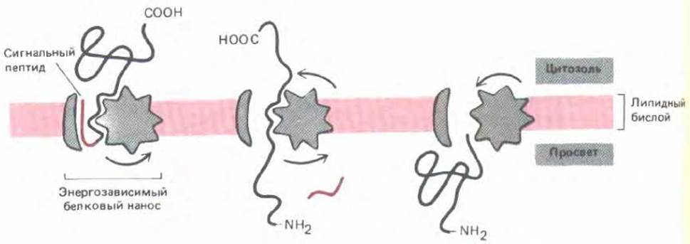
Fig. 8-44. One concept of Protein Transport Across the membrane. After the receptor recognizes the N-terminal signal peptide, an energy-dependent protein pump is activated, which pushes the entire protein through the membrane while temporarily unfolding the polypeptide chain. An alternative possibility is that protein unfolding occurs on the cytosolic side of the membrane and is ATP-dependent, with the protein being driven across the membrane solely by The energy released during refolding.
8-26
8.6.7. Translocation across the ER membrane does not always proceed concomitantly with Polypeptide chain elongation [38]
As discussed above, the import of proteins into mitochondria, chloroplasts, and peroxisomes occurs post-translationally, after the protein has been fully synthesized and released into the cytosol. In contrast, translocation across the ER membrane takes place concurrently with translation (co-translationally). This is precisely why ribosomes bind to the ER membrane rather than to the cytoplasmic surface of other organelles. For many years, it was believed that rough ER ribosomes could harness the energy released during protein synthesis to "push" growing polypeptide chains through the ER membrane. However, recent in vitro studies have shown that precursors of certain proteins can enter the ER even after their synthesis is complete. This translocation requires ATP Hydrolysis, but not the continuation of protein synthesis (Fig. 8-44). Similar to mitochondrial import, ATP hydrolysis is thought to be necessary for unfolding the protein as it crosses the membrane—a Conclusion supported by both genetic and biochemical experiments in Yeast.
Most precursor proteins fail to enter the ER if they are synthesized elsewhere. Either they fold in a way that masks the signal peptide, or the ER translocation machinery is unable to unfold the given protein. It is possible that co-translational translocation allows these proteins to mature without undergoing the premature folding typical of proteins targeted to other organelles.
8-27
8.6.8. The diverse spatial structures of transmembrane proteins can be determined by combinations of peptides that dictate the start and stop of translocation [39]
Most signal peptides are cleaved by a specific signal peptidase associated with the ER membrane. However, the mere presence of a signal peptide is insufficient for this peptidase to function: it requires an adjacent Cleavage site, which is not strictly necessary for translocation itself. Research has shown that in some proteins, signal peptides are located internally within the polypeptide chain and are never cleaved.

Fig. 8-45. Topology of protein translocation across the ER membrane, illustrated for two simple cases. The translocation intermediate is thought to contain a loop of the polypeptide chain, where the signal peptide (also referred to as a start-transfer signal) forms one half of the vertical segment of the loop, while the other half at any given moment is formed by the translocated region of the polypeptide. When only a start-transfer peptide is present without a stop-transfer peptide, the entire polypeptide is translocated across the membrane, and following the Cleavage of the start peptide, a mature soluble protein is released into the ER lumen (A). If both a start-transfer peptide and a stop-transfer peptide are present, translocation halts when the stop peptide reaches the vertical segment of the loop, while protein synthesis continues on the cytosolic side of the membrane; after the start-transfer signal is cleaved, the mature protein remains embedded in the membrane, spanning The Lipid Bilayer of the ER with a domain protruding on each side (B).
It is believed that uncleaved signal peptides play a crucial role in establishing the various membrane-insertion topologies observed in transmembrane proteins. All these mechanisms can be viewed as variations of The sequence of events by which a soluble protein is translocated into the ER lumen. According to current models, the hydrophobic signal peptide of a soluble protein, besides its other functions, acts as a start-transfer signal and remains embedded in the membrane while the rest of the protein molecule is pulled through it as a large loop (Fig. 8-45, A). As the carboxyl terminus of the molecule crosses the membrane, the protein remains anchored to it solely by the signal peptide. Consequently, if this peptide is cleaved, the protein is released into the ER lumen.
The situation is more complex for membrane proteins, as only a portion of their polypeptide chain is translocated across the membrane, while the remainder is not. In the simplest case, such a protein is translocated via the mechanism just described for soluble proteins (except that its signal peptide lacks a cleavage site and is therefore not removed by signal peptidase). As a result, the protein becomes a single-pass transmembrane protein. The N-terminus is embedded in the membrane, where the signal peptide forms an a-helical segment consisting of 20–30 hydrophobic Amino Acids (see Fig. 8-48, A).
Transmembrane proteins that cross the membrane only once (single-pass proteins) and possess an ER lumenal N-terminus rather than a C-terminus require a more intricate translocation mechanism. In these proteins, translocation is also initiated by an N-terminal signal peptide, but an additional hydrophobic segment is present within the molecule to halt the process before the entire polypeptide chain crosses the membrane. In such proteins, it is this stop-transfer signal that anchors the protein in the membrane, while the start-transfer signal is cleaved (Fig. 8-45, B).
Many proteins are known whose polypeptide chain traverses the lipid bilayer multiple times in opposite directions (multipass transmembrane proteins). It is believed that in such proteins, an internal signal peptide serves as a start-transfer signal, which directs translocation until the next stop-transfer signal is encountered. Thus, the fundamental unit of translocation is the polypeptide loop situated between two hydrophobic segments (one start-transfer peptide and one stop-transfer peptide). In the mature protein, both of these segments form $\alpha$-helical membrane-spanning domains. A hypothetical mechanism by which such a loop can be inserted into the membrane is illustrated in Fig. 8-46. For a complex transmembrane protein whose lipid bilayer is traversed by numerous hydrophobic $\alpha$-helices, translocation must be re-initiated by a second start-transfer peptide and continue until the subsequent stop-transfer peptide halts it, and so forth for subsequent start- and stop-transfer peptides (see Fig. 8-48D).

Fig. 8-46. Hypothetical model for the insertion of an internal polypeptide loop into the ER lipid bilayer. It is proposed that the carrier protein can exist in two Conformations: "closed" and "open." Upon binding a start-transfer signal peptide, it transitions into the closed state and functions as a translocator. However, as soon as a stop-transfer signal peptide reaches its binding site, the translocator switches back to the inactive, open conformation and dissociates from the protein.
8-28
8.6.9. The overall conformation of a transmembrane protein can be predicted from the positions of its hydrophobic amino acids [40]
Typically, stop-transfer peptides are more hydrophobic than start-transfer peptides; however, altering their position within the protein can sometimes enable them to function as start signals. This implies that the distinction between hydrophobic start- and stop-peptides is partly determined by their Location within the polypeptide chain. Presumably, a mechanism exists that scans the unfolded polypeptide chain for hydrophobic segments in the direction of protein synthesis (from the NH2- to the COOH-terminus). The signal recognition particle identifies the first suitable segment and thereby establishes the "reading frame." The stretch of the polypeptide chain between this start segment and the subsequent stop-transfer signal is threaded through the membrane (see Fig. 8-46). This translocation process presumably continues until all hydrophobic regions are integrated into the membrane.
This mechanism of membrane insertion implies that the topology of a membrane protein can be predicted directly from its Amino Acid Sequence. To accomplish this, stretches of 20–30 amino acid residues with a high degree of Hydrophobicity are identified. These segments are sufficiently long to span the membrane as an $\alpha$-helix, and their positions can be mapped by analyzing a hydropathy profile (Fig. 8-47). Figure 8-48 presents four Examples of protein topologies reconstructed using this analytical approach.
Membrane proteins are always inserted into the ER from the cytosolic side. As a result, the ER membrane is asymmetrical: the Protein domains exposed on one side differ from those on the other. This Asymmetry is preserved during the numerous budding and fusion events by which proteins synthesized in the ER are delivered to other cellular membranes (see Fig. 8-10).
When proteins are extracted from the membrane and reconstituted into artificial lipid vesicles, some adopt the correct orientation while others are inverted. It is therefore believed that the asymmetry of proteins observed in cellular membranes is entirely attributable to The process of protein insertion into the ER membrane.
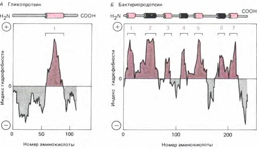
Fig. 8-47. Mapping potential hydrophobic (transmembrane) segments of a polypeptide chain using hydropathy profiles. The Free energy required to transfer successive segments of a polypeptide chain from a nonpolar solvent into water is calculated from the Amino Acid Composition using data derived from model compounds. Such calculations are performed for segments of a fixed length (typically about 10 amino acid residues), starting with each successive amino acid in the chain. The "hydropathy index" of a given segment is plotted on the Y-axis as a function of its position in the chain. Positive values indicate that free Energy is required for transfer into water (i.e., the segment is hydrophobic), and the peak height corresponds to the magnitude of the required energy. Peaks in the hydropathy index reveal the locations of hydrophobic segments within The amino acid sequence. Two examples are shown: Glycophorin has a single transmembrane hydrophobic domain and a corresponding single peak on its hydropathy profile (A); Bacteriorhodopsin contains seven transmembrane helical segments, which correspond to seven peaks on its hydropathy profile (B). (Modified from D. Eisenberg, Ann. Rev. Biochem. 53: 595–624, 1984.)
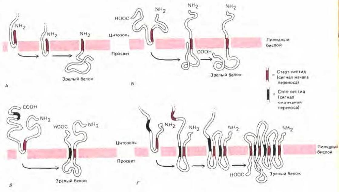
Fig. 8-48. The topology of a membrane protein is determined by the alternation of stop- and start-transfer signal peptides. In all the examples shown, the signal peptide is not cleaved. The hypothetical translocator protein presumably functions According to the mechanism depicted previously in Fig. 8-46. A.
When the N-terminal signal peptide is not cleaved and no stop-transfer peptide is present, the result is a membrane protein with a single C-terminal domain exposed to the ER lumen. B. When a signal peptide is located internally within the chain, it generates a protein with an N-terminal cytoplasmic domain and a C-terminal domain facing the ER lumen. B. When an internal signal peptide is followed by a stop-transfer peptide, the resulting membrane protein possesses three distinct domains projecting from the membrane. D. A membrane protein that spans the bilayer multiple times can be formed by a simple reiteration of this process, involving alternating signal peptides and stop-transfer peptides that interrupt protein translocation across the membrane.
8.6.10. Proteins translocated into the ER lumen undergo refolding [41]
Folding polypeptide chains within the confines of the ER presents a unique challenge not encountered by proteins that fold in the cytosol. The ER lumen is densely populated with folding proteins that are transient residents of this compartment, whereas the cytosol predominantly contains resident proteins that have already achieved their folded state. A folded protein hides its Hydrophobic core (see Section 3.3.1), but until folding is complete, the hydrophobic residues that make up this core are exposed to the aqueous phase. Even at low concentrations in solution, unfolded polypeptide chains have a strong tendency to aggregate with one another and with other proteins; the resulting aggregates precipitate out of solution. It is entirely plausible that such a complex mixture of unfolded proteins as that found in the ER lumen would readily form amorphous and heterogeneous precipitates.

Fig. 8-49. Because reducing agents such as the tripeptide Glutathione and the small protein thioredoxin are present in high concentrations in the cytosol and absent from the lumen of organelles like the ER, Disulfide Bonds can form in the ER lumen but not in the cytosol. Glutathione and thioredoxin are maintained in the cytosol in a strongly reduced state by enzymes that transfer electrons from NADPH, thereby converting any resulting disulfide bonds back into cysteines. The three-dimensional structure of E. coli thioredoxin shown here was determined by X-ray crystallographic analysis. It contains two closely spaced cysteines, depicted here linked by a disulfide bond.
The time a protein spends in the ER prior to export to the Golgi apparatus varies widely. These differences likely depend to a large extent on the rate at which a given protein escapes aggregation (becomes solubilized) and successfully folds.
The ER lumen contains a high concentration of binding protein (BiP), which appears to recognize misfolded proteins by binding to their exposed hydrophobic patches. At the carboxyl terminus of the BiP molecule lies a four-amino-acid signal peptide that ensures the protein is retained in the ER (see Table 8-3). One hypothesis suggests that BiP functions to keep misfolded proteins localized within the ER (thereby preventing them from reaching the Golgi apparatus). It is also possible that BiP acts as a catalyst for protein folding. This protein has been shown to bind ATP and is structurally related to heat-Shock proteins involved in protein import.
8.6.11. Protein disulfide isomerase promotes the formation of correct disulfide bonds in the ER lumen [42]
The cytosol contains a pool of reducing agents bearing SH groups. These substances prevent the formation of —S—S— bridges (disulfide bonds) by maintaining Cysteine residues in cytosolic proteins in their reduced (—SH) form (Fig. 8-49). Such reducing agents are absent from the ER lumen, allowing —S—S— bridges to form readily. Given the Abundance of folding proteins, this process occasionally goes awry. The ER lumen contains an enzyme dedicated to correcting such errors. Protein disulfide isomerase is an abundant protein located in the ER lumen and attached to the inner face of its membrane. It possesses the same ER retention signal as BiP. The MECHANISM OF ACTION of protein disulfide isomerase involves cleaving —S—S— bonds, thereby granting the protein the flexibility to rapidly sample numerous conformations until the conformation with the lowest overall free energy is attained (Fig. 8.50). At this juncture, the newly synthesized protein folds correctly. While the correct conformation might theoretically be found by chance, protein disulfide isomerase dramatically accelerates the search process.

Fig. 8-50. Within the ER lumen, protein disulfide isomerase repeatedly cleaves —S—S— bonds within the polypeptide chain until an arrangement possessing the minimal overall free energy is achieved. Under these conditions, the protein folds correctly. The enzyme thus facilitates the folding of newly synthesized proteins entering the ER.
8.6.12. Most proteins synthesized in the rough ER are glycosylated via an N-linked oligosaccharide [43]
One of the primary functions of the ER is the covalent attachment of sugars to proteins. The majority of proteins entering the ER lumen are converted into Glycoproteins before reaching the Golgi apparatus, lysosomes, plasma membrane, or extracellular space (Fig. 8-51). Conversely, very few cytosolic proteins are glycosylated, and those that are bear distinct sugar modifications (see Section 8.2.2).
A crucial breakthrough in understanding protein glycosylation was the discovery that proteins in the ER receive a single, uniform oligosaccharide composed of N-acetylglucosamine, mannose, and glucose, containing a total of 14 residues. Because this oligosaccharide is always attached to the NH2 group of an asparagine side chain, it is designated as N-linked or asparagine-linked (Fig. 8-52). Attachment is catalyzed by a membrane-bound enzyme whose Active Site faces the ER lumen. This explains why cytosolic proteins are not glycosylated in this manner. The preformed oligosaccharide precursor is transferred en bloc to the appropriate asparagine residue. This is a single-step reaction that occurs almost concurrently with The Emergence of the asparagine residue into the ER lumen during protein translocation across the membrane (Fig. 8-53). Because most proteins are imported into the ER cotranslationally, the N-linked oligosaccharide is almost invariably added during protein synthesis, ensuring optimal access to the target asparagine residues. The signals for N-linked glycosylation are the tripeptide sequences Asn-X-Ser or Asn-X-Thr (where X can be any amino acid except Proline). These sequences occur much less frequently in glycoproteins than in non-glycosylated cytosolic proteins. Evidently, selective pressure has operated against these sequences, likely because glycosylation at an excessive number of sites would interfere with proper protein folding.
The precursor oligosaccharide is anchored in the ER membrane by a specialized lipid molecule called dolichol. The oligosaccharide is linked to dolichol via a high-energy pyrophosphate bond, which provides the activation energy for the glycosylation reaction. Before being transferred to a protein, the oligosaccharide is assembled from Monosaccharides onto this membrane-bound lipid carrier. First, sugars are activated in the cytosol by forming nucleotide-sugar intermediates, which then (directly or indirectly) transfer their sugar moieties to the lipid molecule in a specific sequence. Once completed, the oligosaccharide precursor flips from the cytosolic face of the ER membrane into its lumen (Fig. 8-54). Dolichol is a long and highly hydrophobic molecule; its 22 isoprene units span the lipid bilayer multiple times, firmly anchoring the attached oligosaccharide within the membrane.

Fig. 8-51. Three-dimensional STRUCTURE OF THE Influenza virus hemagglutinin membrane glycoprotein, illustrating the positions of covalently attached Oligosaccharides (highlighted atoms). Glycoproteins are either incorporated into cellular membranes or secreted from cells; they can contain anywhere from 1 to 85% carbohydrate by weight. Some carbohydrate-rich glycoproteins contain dozens or even hundreds of attached oligosaccharide chains per molecule. The membrane-spanning segments of this viral envelope protein, composed of three identical polypeptide chains, serve as the base for the structure shown here. They are omitted from the drawing because this region of the protein is cleaved during Sample preparation for X-ray crystallographic analysis. (Photograph kindly provided by Richard J. Feldmann.)
The vast diversity of N-linked oligosaccharide structures arises from the Modification of the initial precursor molecule. While still in the ER, most glycoproteins undergo the removal of three glucose residues and one mannose residue from the oligosaccharide (see Fig. 8-63). Further trimming and Processing of the oligosaccharide continue in the Golgi apparatus (see Section 8.7.1).
N-linked oligosaccharides are the most prevalent type found in glycoproteins. Much less commonly, oligosaccharides are linked to the hydroxyl group of the side chain of a Serine, Threonine, or hydroxylysine residue. Such O-linked oligosaccharides are assembled in the Golgi apparatus via a pathway that is not yet fully understood (see Section 8.7.4).
8.6.13. In certain transmembrane proteins, shortly after their entry into the ER, the C-terminal transmembrane segment is exchanged for a covalently attached Inositol phospholipid [44]
As discussed previously, certain cytosolic enzymes catalyze the covalent attachment of a single fatty acid to specific proteins (see Section 8.2.3). It was recently discovered that a analogous process occurs in the ER: the carboxyl terminus of several plasma membrane proteins is covalently linked to a sugar residue of a glycolipid by specific enzymes. The mechanism of this linkage formation is illustrated in Fig. 8-55. Studies have shown that a glycosylated phosphatidylinositol molecule containing two Fatty acids is added to the protein in the process. This modification has been identified in A large number of plasma membrane proteins, including one isoform of the neural Cell Adhesion molecule and the major surface coat protein of trypanosomes. Because both of these proteins are anchored to the plasma membrane exclusively via this mechanism, they could in principle be released from the cell in a soluble form in response to a signal activating a specific phospholipase in the plasma membrane; however, this hypothesis has yet to be confirmed experimentally.
8.6.14. The bulk of membrane lipid bilayers is assembled in the ER [45]
The ER membrane synthesizes almost all the lipids required for the construction of new cellular membranes, including Phospholipids and cholesterol. The principal phospholipid produced is phosphatidylcholine (also known as lecithin), which can be synthesized in three enzymatic steps from two fatty acids, glycerophosphate, and Choline (Fig. 8-56). Each step is catalyzed within the ER membrane by enzymes whose active sites face the cytosol (where all the requisite metabolites reside). In the first step, an acyl transferase adds two fatty acids to glycerophosphate to yield phosphatidic acid, a compound sufficiently hydrophobic to remain in the lipid bilayer following synthesis. It is at this stage that the lipid bilayer expands. Subsequent steps fashion the polar HEAD group of the newly formed lipid molecule and thereby determine the chemical identity of the bilayer, but they do not contribute to bulk membrane growth (see Fig. 8-56). All major membrane phospholipids—phosphatidylcholine (PC), phosphatidylethanolamine (PE), phosphatidylserine (PS), and phosphatidylinositol (PI)—are synthesized in this fashion.

Fig. 8-52. Structure of the asparagine-linked oligosaccharide added to most proteins on the luminal face of the ER membrane. The sugars highlighted in color constitute the core of this oligosaccharide. In many glycoproteins, following extensive processing of the oligosaccharide in the Golgi apparatus, only the core sugars remain from the original structure (see Fig. 8-63). Note that the asparagine residue occurs within the consensus sequence Asp-X-Ser or Asp-X-Thr, where X can be any amino acid except proline.

Fig. 8-53. N-linked protein glycosylation in the ER. Almost immediately after the polypeptide chain enters the ER lumen, it becomes glycosylated at accessible asparagine residues. The oligosaccharide shown in Fig. 8-52 is transferred to asparagine as an intact unit; this reaction is catalyzed by a membrane-bound enzyme, glycosyl transferase.

Fig. 8-54. Synthesis of the lipid-linked oligosaccharide that is transferred to asparagine residues on the inner side of the ER membrane. This oligosaccharide is assembled sugar by sugar on a dolichol lipid molecule framework (polyisoprenoid—see Scheme 2-4). The first sugar is attached to dolichol by a pyrophosphate bridge. This high-energy bond then activates the oligosaccharide for transfer from the lipid molecule to the asparagine side chain. Oligosaccharide synthesis begins on the cytosolic side of the ER membrane. After the lipid-Man8GlcNAc2 intermediate "flips" across the membrane, synthesis continues on its inner side. Abbreviations: GlcNAc — N-acetylglucosamine, Man — mannose, Glc — glucose.

Fig. 8-55. Synthesis of membrane-anchored proteins via a phosphatidylinositol anchor. Immediately after synthesis is complete, the protein remains bound to the membrane solely through its hydrophobic C-terminal tail consisting of 15–20 amino acids, while the rest of the molecule resides in the ER lumen. In less than a minute, an ER enzyme cleaves the protein from its membrane-bound C-terminal segment, and simultaneously, the new carboxyl terminus attaches to a previously formed intermediate, glycosylphosphatidylinositol. Due to this covalently attached lipid anchor, the protein remains membrane-bound. All of its Amino acids are located on the inner side of the ER membrane, and if the protein is transported to the plasma membrane, they will face the extracellular space. The exact structure of this glycolipid head group remains unknown.
Both the initial formation of phosphatidic acid and its subsequent modifications into various types of phospholipid molecules occur in the half of the ER lipid bilayer facing the cytosol. This process could eventually convert the lipid bilayer into a monolayer were it not for a mechanism that transfers some of the newly formed phospholipid molecules to the other half of the ER bilayer. In artificial lipid bilayers, lipids do not undergo such "flip-flop" transitions. In the ER, however, The amount of phospholipids equalizes on both sides of the membrane within minutes, which is almost 100,000 times faster than the rate calculated for spontaneous "flip-flop." It is believed that such rapid movement across the bilayer occurs via phospholipid translocators that are specific for each lipid type (depending on the head group). Apparently, the ER membrane contains a translocator ("flippase") capable of transferring choline-containing phospholipids (but not ethanolamine-, serine-, or inositol-containing ones) from one half of the bilayer to the other. This means that PC reaches the inner surface of the bilayer much more easily than PE, PS, or PI. Thus, the translocator is responsible for the asymmetric arrangement of lipids in the bilayer (Fig. 8-57).
Cholesterol and ceramide are also known to be synthesized in the ER. Ceramide is exported to the Golgi apparatus, where it serves as a precursor for Two Types of lipids: oligosaccharide chains are added to some ceramide molecules to form glycosphingolipids, while the phosphocholine head group from phosphatidylcholine is added to others to yield sphingomyelin. Thus, both Glycolipids and sphingomyelin are formed relatively late during membrane biogenesis. They are located exclusively in the non-cytosolic half of the lipid bilayer because that is where the enzymes synthesizing them reside.

Fig. 8-56. Phospholipid synthesis takes place on the cytoplasmic face of the ER membrane. Each enzyme involved in this synthesis is an integral ER membrane protein with its active site facing the cytosol. The cytosol contains all the compounds required for phospholipid assembly. In the process depicted here, phosphatidylcholine (CDP-choline) is synthesized from a fatty acid–coenzyme A complex, glycerol-3-phosphate, and cytidine diphosphate-choline.

Fig. 8-57. Growth of both halves of the ER lipid bilayer requires the catalytic "flipping" of phospholipid molecules from one monolayer to the other. Because new lipid molecules are added exclusively to the cytoplasmic monolayer and lipids do not spontaneously jump from one monolayer to the other, membrane-bound phospholipid carriers ("flippases") are required to transfer specific lipid molecules to the inner leaflet of the membrane. As a result, the membrane grows evenly as a bilayer. Because these enzymes selectively recognize and transport only certain types of lipids, an asymmetric bilayer is formed in the ER. In particular, the inner leaflet (which becomes the outer half of the plasma membrane bilayer) is enriched in phosphatidylcholine.
8-29
8.6.15. Phospholipid transfer proteins can deliver phospholipids from the ER to mitochondria and peroxisomes [46]
The plasma membrane, as well as the membranes of the Golgi apparatus and lysosomes, are parts of a membrane system connected to the ER via transport vesicles that supply both proteins and lipids to it. Mitochondria and peroxisomes do not belong to this system and require alternative mechanisms for importing membrane proteins and lipids. We have already seen that most proteins of these organelles are delivered post-translationally from the cytosol. Although some lipids are modified within mitochondria, the mitochondria themselves must still acquire them either directly from the ER, where they are synthesized, or via other cellular membranes.
In vitro experiments have shown that specialized water-soluble proteins are capable of transferring individual phospholipid molecules from one membrane to another. These proteins are called phospholipid transfer proteins (or phospholipid exchange proteins). Transfer between membranes occurs as follows: the protein "extracts" a phospholipid molecule from a membrane and detaches from it, carrying the attached lipid within its binding pocket. When this protein reaches another membrane, it tends to "unload" the bound lipid molecule into the new lipid bilayer (Fig. 8-58). It is hypothesized that phosphatidylserine is transported to mitochondria in this manner, where it is subsequently decarboxylated to form phosphatidylethanolamine; phosphatidylcholine is most likely imported as an intact molecule.
Transfer proteins distribute phospholipids among organelles in a stochastic manner. In principle, such random exchange could result in lipid transport from a lipid-rich membrane to a lipid-depleted one, thereby transferring phosphatidylcholine and phosphatidylserine molecules from the ER, where they are synthesized, to the mitochondrial and peroxisomal membranes. It is possible that mitochondria and peroxisomes are the only "lipid-depleted" organelles in the cytoplasm, and such "random" transfer is sufficient, although specific mechanisms for phospholipid transfer to these organelles are also quite plausible.
Summary
The ER serves as a manufacturing plant for the protein and lipid components of many organelles. Its extensive membrane houses numerous biosynthetic enzymes, including those responsible for the synthesis of almost all cellular lipids and the attachment of N-linked oligosaccharides to a multitude of proteins. Newly synthesized proteins destined for secretion, as well as for the ER itself, the Golgi apparatus, lysosomes, and the plasma membrane, must first enter the ER from the cytosol. Only proteins possessing specific hydrophobic signal peptides are translocated into the ER. The signal peptide is recognized by the signal recognition particle (SRP), which binds to the nascent protein chain and ribosome and directs them to a receptor protein On the surface of the ER membrane. This membrane binding triggers an ATP-dependent translocation process in which a loop of the polypeptide chain is threaded across the ER membrane.
Soluble proteins destined for the ER lumen, secretion, or transport to other organelles penetrate fully into the ER lumen. Transmembrane proteins destined for the ER membrane or other cellular membranes are translocated across the membrane but are not released into the ER lumen. Instead, they remain anchored in the bilayer by one or more membrane-spanning $\alpha$-helical segments of the polypeptide chain. These hydrophobic protein segments can act as signal peptides that dictate the initiation or termination of translocation. If a polypeptide contains multiple alternating start- and stop-transfer peptides, it can traverse the bilayer in opposite directions multiple times.
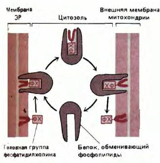
Fig. 8-58. Soluble phospholipid transfer proteins can redistribute phospholipids among membrane-bound organelles. Phospholipids are water-insoluble, so their transitions between membranes require a carrier protein. These proteins transfer one phospholipid molecule at a time, capable of capturing a lipid molecule from one membrane and releasing it into another. The transfer of phosphatidylcholine (PC) from the ER to mitochondria can, in principle, occur spontaneously because the concentration of PC is high in the ER membrane (where it is synthesized) and low in the outer mitochondrial membrane.
The asymmetry of protein insertion into ER membranes and their subsequent glycosylation ensures the polarity of membrane protein orientation in all other organelles.
8-32
Last update: 12/08/2026
Editorial and Educational Adaptation: This material has been compiled based on the primary/original source text. The project team performed an editorial review, corrected technical inaccuracies, structured sections, and adapted the content for an educational format.
What was processed:
- elimination of formatting defects (OCR errors, structural breaks, corrupted characters);
- editorial organization of content;
- standardization of terminology in accordance with academic sources;
- verification of factual statements against the original source text.
All mentions of the author, publication year, and origin of the primary text have been preserved in accordance with the source.