Molecular Biology of the Cell - Volume 2 - Alberts B., Bray D., Lewis J., Raff M., Roberts K., Watson J. 1993
Intracellular Macromolecular Sorting and Maintenance of Cellular Compartments
Cytosol
The cytosol is the portion of the Cytoplasm that occupies the space between membrane-bounded Organelles. It typically accounts for about half of the total Cell volume (see Table 8-1). The cytosol contains numerous Enzymes involved in Intermediary METABOLISM and is densely packed with Ribosomes synthesizing Proteins. About half of all proteins produced on ribosomes remain in the cytosol as its permanent constituents. In this section, we will discuss The Fate of these proteins, as well as some of the mechanisms that control their lifespan and direct them to specific locations within the cytosol.
8.2.1. The Organization OF THE cytosol is maintained by protein filaments [8]
The cytosol contains a wealth of protein filaments assembled into a filamentous Cytoskeleton (Chapter 11). It is the cytoskeleton that determines cell shape, drives cytoplasmic streaming, and provides a scaffolding that organizes enzymatic reactions. Furthermore, because proteins make up about 20% of the cytosolic mass, it is more accurate to think of the cytosol as a highly organized gel rather than a dilute enzyme solution. Diffusion rate measurements, however, show that small molecules and certain small proteins diffuse through the cytosol at rates nearly equal to those in distilled Water. Thus, from the perspective of intermediary metabolism (where both substrates and products are small molecules), we can view the cytosol as a simple solution.
Class="center">
Fig. 8-14. The only known form of glycosylation occurring in the cytosol of mammalian Cells is The addition of N-acetylglucosamine to Serine or Threonine residues within a protein. This modification is used to alter various Gene regulatory proteins and certain nuclear pore proteins; its exact function remains unknown. Much more complex glycosylation takes place in the ER and the Golgi apparatus (see Fig. 8-52).
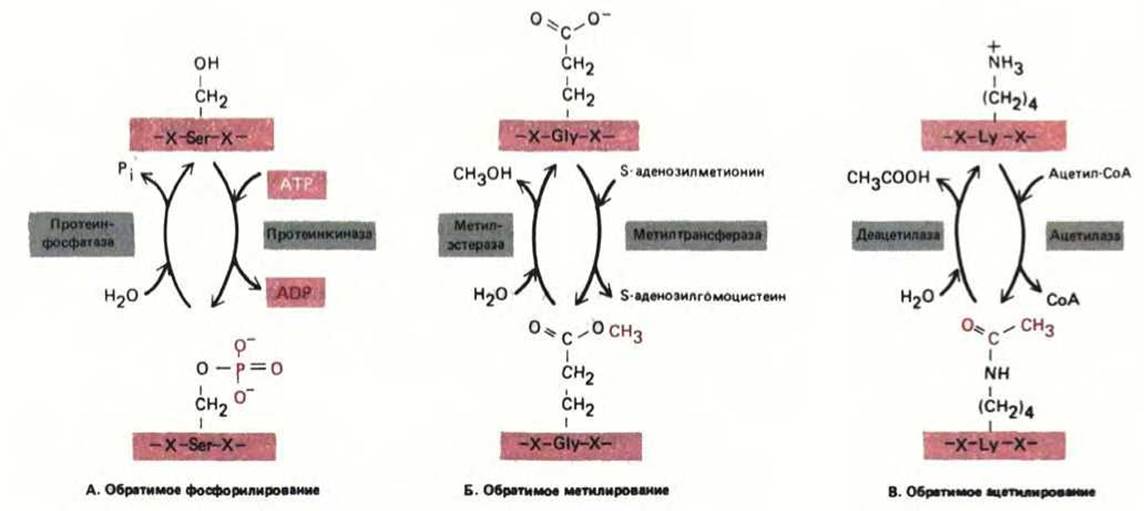
Fig. 8-15. Three types of reversible covalent modifications that proteins undergo to regulate their activity. Each depicted modification alters the charge of an amino acid side chain. The most prevalent modification is the phosphorylation of -OH groups on serine, threonine, and Tyrosine side chains in proteins. Estimates suggest that about 10% of cytosolic proteins in animal cells are modified in this manner.
On the other hand, large structures such as transport vesicles and organelles are known to move very slowly, partly due to frequent collisions with cytoskeletal components. To move at an acceptable rate, specialized protein "motors" hydrolyze ATP and use the released energy to propel large structures along microtubules or Actin filaments. In this case, specific filaments act as "tracks" that guide transport vesicles to their proper target membrane. However, most cell biologists consider this cytoskeletal function less specialized, believing that the Specificity of Vesicular Transport is primarily ensured by receptor systems located on the cytosolic surface of the vesicles themselves (see Section 8.8.6).
8.2.2. Many proteins undergo covalent modifications in the cytosol [9]
More than 100 different post-translational protein modifications have been described. The Role of most of these modifications remains unclear; some appear to be accidental and functionally insignificant, while others are vital for cell survival because they are tightly regulated by specific enzymes. As we will see later, certain modifications occur within the ER and the Golgi apparatus. In these organelles, for example, glycosylation enzymes attach complex chains of sugar residues to proteins to form Glycoproteins (see Section 8.6.12). The sole known instance of cytosolic glycosylation in mammalian cells is the addition of N-acetylglucosamine to proteins (Fig. 8-14). However, numerous other covalent modifications take place predominantly in the cytosol. Some of these are stable and essential for protein activity, such as the covalent attachment of Coenzymes (biotin, Lipoic Acid, or Pyridoxal phosphate). Certain covalent modifications occurring in the cytosol are reversible and serve to regulate The activity of many proteins (Fig. 8-15).
Among currently known modifications, one is crucial for proper protein targeting. The attachment of a fatty acid chain to a protein directs it to specific membranes facing the cytosol.
8.2.3. Some cytosolic proteins are attached to the cytoplasmic face of the membrane via a fatty acid chain [10]
Cells possess specialized mechanisms for transporting soluble proteins from the cytosol to membranes. Such proteins are covalently linked to a fatty acid chain, which subsequently inserts into The Lipid Bilayer from the cytoplasmic side, thereby anchoring the protein. The attachment of a protein to a membrane via a fatty acid can have important functional consequences. For example, the src oncogene of the Rous Sarcoma virus encodes a tyrosine-specific protein kinase that typically binds to the membrane through a covalently attached myristic acid chain (a 14-carbon saturated fatty acid). In this configuration, the protein kinase transforms a normal cell into a cancerous one. If, however, the fatty acid attachment is prevented by replacing the N-terminal Glycine with Alanine in the protein molecule, the src protein retains its kinase activity but remains in the cytosol and fails to transform The Cell. Apparently, efficient substrate binding requires this kinase to be tethered to the membrane. Similar experiments have shown that the product of another oncogene, the ras protein (see Section 12.3.1), must be anchored to the membrane via a covalently attached palmitic acid chain (a 16-carbon saturated fatty acid) in order to transform the cell.
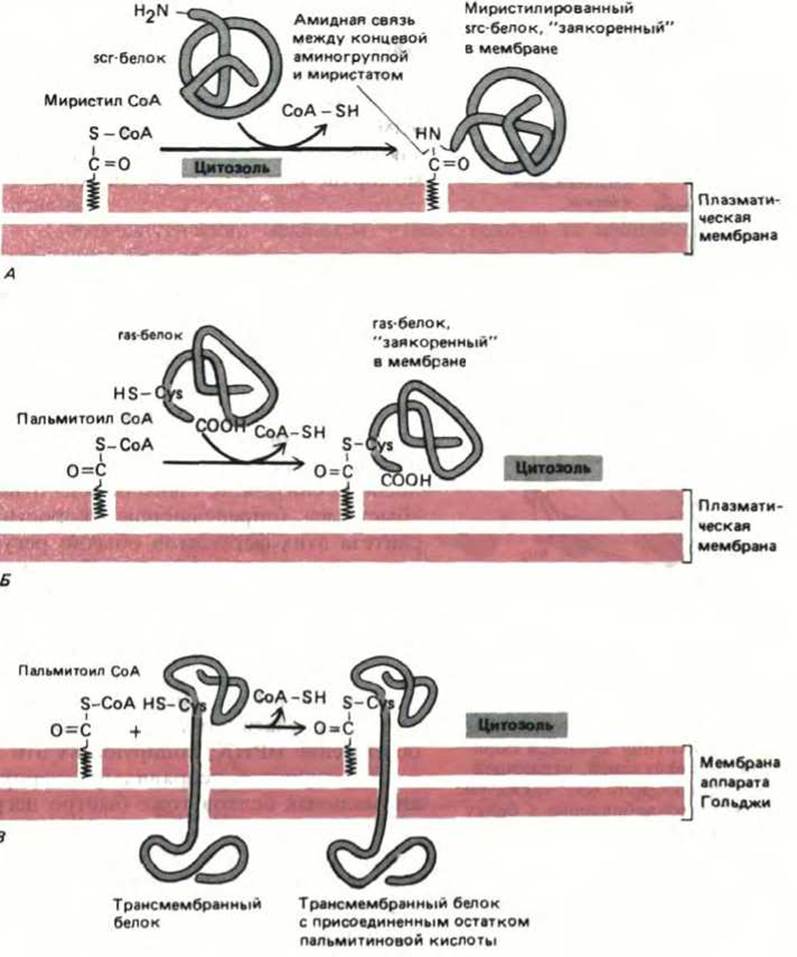
Fig. 8-16. The covalent attachment of a fatty acid to a protein can target a water-soluble protein to a membrane. A. The formation of an amide bond between the N-terminal glycine and myristic acid anchors the src protein (following its synthesis in the cytosol) to the cytoplasmic face of the membrane. B. A thioester bond between palmitic acid and a Cysteine near the carboxyl terminus anchors the ras protein in The Plasma Membrane after its synthesis in the cytosol. C. Palmitic acid and other Fatty acids are frequently attached via thioester bonds to specific cysteine residues located in the cytoplasmic domain of a transmembrane protein. This occurs as the protein travels from the ER to the Golgi apparatus on its way to the plasma membrane or elsewhere. The acetylated cysteine is usually preceded by three hydrophobic Amino Acids (X) in the sequence NH2-...-X-X-X-Cys-...-COOH. In example (C), unlike (A) and (B), the protein remains membrane-bound even without fatty acid attachment.
What determines whether a fatty acid chain is added to a given protein, and whether that chain is myristic or palmitic acid? The enzymes catalyzing these modifications recognize distinct signal Peptides within the protein: a myristic acid chain (Fig. 8-16A) is added to an N-terminal glycine residue embedded within a specific Amino Acid Sequence (see Table 8-3), whereas a palmitic acid chain is attached to a cysteine side chain located four residues away from the carboxyl terminus within a different signal peptide (Fig. 8-16B). Additionally, the cytosol hosts another enzyme-catalyzed reaction in which palmitic acid chains are added to the cytosolic tails of numerous transmembrane proteins as they pass from the ER through the Golgi apparatus en route to the plasma membrane or other destinations (Fig. 8-16C).
8-8
8.2.4. Some cytosolic proteins are short-lived [11]
In addition to localization signals, cellular proteins carry signals that dictate their lifespan. Proteins undergo continuous turnover: a fraction of their molecules randomly degrade and are replaced by new copies. Most permanent cytosolic proteins have relatively long half-lives of several days. Others, however, degrade much faster—sometimes within minutes of their synthesis. Such proteins include enzymes catalyzing rate-limiting steps in metabolism; the synthesis rates of these enzymes are typically regulated in response to environmental conditions to maintain metabolic efficiency. Other short-lived proteins include the products of cellular oncogenes such as fos or myc, which are thought to play critical roles in regulating Cell Growth and Division (see Section 13.4.6). Because these types of proteins are continuously and rapidly degraded, their concentrations can change swiftly in response to alterations in their synthesis rates (see Section 12.4.7). In most cases, such regulation also requires an unusually rapid turnover of the mRNAs encoding these proteins (see Section 10.4.12).
Most misfolded, denatured, and otherwise abnormal proteins are also rapidly degraded in the cytosol. They typically break down within minutes, whereas normal copies of the same proteins persist. Abnormal proteins arise from synthesis errors—such as the incorporation of an incorrect amino acid into the chain—or from chemical damage, such as The oxidation of Certain amino acid side chains. Various mutant forms of normal proteins are also recognized as abnormal. It is becoming increasingly clear that both abnormal proteins and those genetically programmed for rapid turnover are ultimately destroyed in the cytosol via the same proteolytic machinery.
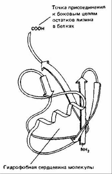
Fig. 8-17. Three-dimensional Structure of ubiquitin, a heat-stable protein consisting of 76 amino acid residues. The attachment of a single ubiquitin molecule to a protein is a reversible modification with regulatory Functions (see also Fig. 8-15). However, the addition of a branched ubiquitin chain to a protein triggers its immediate and complete degradation (see Fig. 8-18). (From S. Vijay-Kumar, C. E. Bugg, K. D. Wilkinson, W. J. Cook, Proc. Natl. Acad. Sci. USA 82: 3582-3585, 1985.)
8-6
8.2.5. Selective protein turnover in eukaryotes is mediated by ubiquitin-dependent proteolysis [12]
Those cytosolic proteins destined for rapid destruction carry signals that trigger the proteolytic machinery responsible for their degradation. One such signal is remarkably simple, consisting merely of the first amino acid in the polypeptide chain. The amino acids Met, Ser, Thr, Ala, Val, Cys, Gly, and Pro are stabilizing when located at the N-terminus, whereas the remaining 12 amino acids trigger proteolytic attack. These destabilizing Amino acids are virtually never found at the N-terminus of stable cytosolic proteins. However, they are frequently present at the N-terminus of proteins destined for other compartments, such as the ER. Because the cytosolic proteolytic machinery is absent from the lumen of the ER or the Golgi apparatus, such proteins are generally long-lived within "their" proper compartments. The destabilizing N-terminal amino acid in such non-cytosolic proteins may serve to eliminate erroneously mistargeted copies: molecules that fail to be rapidly translocated out of the cytosol are promptly degraded. A similar single-residue code apparently exists in Bacteria, where it also promotes the rapid degradation of specific proteins.
The proteolytic mechanism responsible for the selective degradation of proteins is complex and, once initiated, ensures the complete destruction of the protein. Eukaryotes possess a ubiquitin-dependent proteolytic pathway. In this system, multiple copies of the small protein ubiquitin become attached to the protein destined for destruction (Fig. 8-17). This conjugation of ubiquitin to the target protein is catalyzed by a multienzyme complex that is thought to bind to the N-terminus of the protein bearing a destabilizing N-terminal amino acid. This enzyme complex attaches a ubiquitin molecule to the nearest Lysine residue in the polypeptide chain and subsequently adds further ubiquitin molecules to the first, forming a branched ubiquitin chain (Fig. 8-18). Following this, a large ATP-dependent protease rapidly degrades such proteins. Apparently, the substrates for this protease are exclusively proteins containing branched polyubiquitin chains, whereas proteins bearing a single ubiquitin molecule linked to a lysine (such as Histones) are spared.
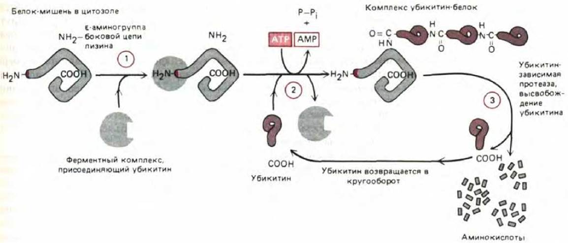
Fig. 8-18. Ubiquitin-dependent protein degradation. The target protein (containing a "destabilizing" N-terminal amino acid that serves as a degradation signal) is recognized by the enzyme complex responsible for ubiquitin attachment (Stage I). Then, through a sequential series of reactions (Stage 2), ubiquitin molecules are linked together to form a branched multiubiquitin chain attached to the $\varepsilon$-amino group in the side chain of the nearest lysine residue in the protein. This reaction requires mobility of the target protein and is accelerated if the protein is misfolded. Subsequently (Stage 3), a large protease—which specifically cleaves proteins marked with a branched ubiquitin chain—cuts the target protein into numerous small fragments.
8-7
8.2.6. Protein stability can be determined by enzymes that modify its N-terminus [13]
Since the N-terminal amino acid of cytosolic proteins dictates whether a given protein will be degraded by the ATP-dependent protease, it is crucial to understand how this key amino acid residue is attached to the protein. Presumably, in proteins genetically programmed for a short half-life, a destabilizing amino acid is attached to the N-terminus immediately upon completion of Protein Synthesis. As discussed in Chapter 5 (see Section 5.1.10), all proteins are initially synthesized with a Methionine at the N-terminus (or formylmethionine in bacteria). This methionine, which acts as a stabilizing residue, is often removed by a specific aminopeptidase shortly after its incorporation into the protein. In addition, aminoacyl-tRNA transferases can add a single destabilizing amino acid residue to the protein's N-terminus. The exact conditions governing these reactions remain poorly understood.
The primary targets for ubiquitin attachment and subsequent degradation are denatured or misfolded proteins, as well as those containing oxidized or otherwise aberrant amino acids, even when a stabilizing amino acid is present at their N-terminus. The destruction of misfolded or denatured proteins may be initiated by the recognition of hydrophobic amino acid patches that are normally buried within the interior of the protein globule but become exposed On the surface of an aberrant molecule (see Section 3.3.1). This is presumably followed by Cleavage or modification reactions that generate a novel, destabilizing N-terminal residue. Finally, through the action of the ubiquitin-dependent proteolytic machinery, the abnormal protein is degraded. A central question in formulating any hypothetical model for the recognition of denatured or misfolded proteins is how the cell distinguishes intact abnormal molecules from the myriad nascent polypeptide chains growing on ribosomes, which might transiently appear "misfolded." It has been shown, for example, that if the protein synthesis inhibitor puromycin is added to cells (see Section 5.1.15), incomplete Polypeptides are rapidly broken down via ubiquitin-dependent proteolysis. The metabolic stability of both long- and short-lived proteins during their Synthesis on Ribosomes is likely attributable to their being temporarily shielded by Introduction/26.html">The Translational Apparatus.
It has been established that the N-terminal residue is frequently resistant to Hydrolysis during the repetitive reactions utilized in amino acid sequenators (see Section 4.4.7). Proteins targeted for sequencing are usually N-terminally acetylated, which apparently renders this end "blocked." It is possible that certain proteins modified in this manner are exceptionally resistant to intracellular proteolysis and therefore exhibit unusually long half-lives; these include many cytoskeletal proteins and histones involved in nuclear DNA packaging. However, the precise mechanism by which proteins destined for Acetylation are selected remains unknown.
8.2.7. Heat-Shock proteins prevent the intracellular accumulation of protein aggregates [14]
Normal mammalian cells grow in culture at 37°C. When subjected to a brief "heat shock" by elevating the Temperature (typically to 43°C), they rapidly synthesize a large quantity of specific proteins. Most of these heat-shock proteins are also induced in response to Other forms of cellular stress, suggesting they play a vital role in helping cells survive adverse conditions. Similar proteins are synthesized in *Drosophila*, Yeast, and even bacteria. DNA sequence analyses reveal three major families of heat-shock proteins, with molecular masses of roughly 25, 70, and 90 kDa. Normal, unstressed cells have been found to contain a multitude of highly homologous proteins belonging to each of these families.
It is hypothesized that heat-shock proteins assist in solubilizing and refolding denatured or misfolded proteins. They also fulfill other functions (see Sections 8.4.4 and 8.6.7). For instance, members of the 90 kDa family have been shown to associate with inactive forms of steroid hormone receptor proteins and with tyrosine-specific protein Kinases, presumably participating in the functional regulation of these receptors. The 70 kDa family (Hsp70) is the most thoroughly characterized. These proteins bind to various other proteins as well as abnormal Protein Complexes and aggregates, subsequently releasing them upon ATP binding. Evidence indicates that they facilitate the solubilization and refolding of aggregated or misfolded proteins through repeated cycles of ATP binding and hydrolysis. Although aberrant proteins are present in any cell, certain stresses—such as heat shock—cause their intracellular levels to surge dramatically, creating an increased demand for heat-shock proteins. This demand is met by the transcriptional activation of specific genes. In Cells of the yeast *S. cerevisiae*, for example, there are eight Hsp70 genes; some are transcribed constitutively under all conditions, whereas others are induced solely by elevated temperatures or other extreme environmental factors.
The cytosol, which typically constitutes about half the volume of a Eukaryotic Cell, encompasses all intracellular space excluding the organelles. It is the site where the majority of intermediary metabolism and protein synthesis reactions take place. Newly synthesized proteins that lack organelle-targeting signals remain in the cytosol. Some of these proteins are degraded shortly after synthesis: a single "destabilizing" amino acid at their N-terminus promotes the attachment of multiple ubiquitin molecules to specific lysine residues on the target protein. Subsequently, a ubiquitin- and ATP-dependent protease breaks down the protein. Defective copies of most cytosolic proteins are degraded via this same ubiquitin-dependent pathway. Many proteins undergo covalent modifications within the cytosol. Some of these modifications are permanent, whereas others—such as phosphorylation—are reversible and play a crucial regulatory role in modulating protein activity. Fatty acids can also be covalently attached to specific proteins, endowing an otherwise soluble protein with The ability to bind to the cytoplasmic face of The cell membrane.

Fig. 8-19. Three-dimensional model of the double-membrane envelope surrounding The Nucleus. The nuclear envelope is perforated by nuclear pores and is continuous with The Endoplasmic reticulum.
Last update: 12/08/2026
Editorial and Educational Adaptation: This material has been compiled based on the primary/original source text. The project team performed an editorial review, corrected technical inaccuracies, structured sections, and adapted the content for an educational format.
What was processed:
- elimination of formatting defects (OCR errors, structural breaks, corrupted characters);
- editorial organization of content;
- standardization of terminology in accordance with academic sources;
- verification of factual statements against the original source text.
All mentions of the author, publication year, and origin of the primary text have been preserved in accordance with the source.