Molecular Biology of the Cell - Volume 2 - Alberts B., Bray D., Lewis J., Raff M., Roberts K., Watson J. 1993
Intracellular sorting of macromolecules and maintenance of cellular compartments
Compartmentation in cells of higher organisms
In this introductory section, we will provide a Brief Overview of cellular Organelles and their interrelationships. We will examine the Methods used to track the movement of Proteins between compartments and outline the primary pathways of protein translocation from one compartment to another.
8.1.1. All Eukaryotic Cells contain a set of core membrane-enclosed organelles [1]
Many essential biochemical processes take place within membranes or on their surfaces. For instance, Oxidative Phosphorylation AND Photosynthesis require a semipermeable membrane to couple proton transport with ATP synthesis. Furthermore, membranes serve as a structural framework for the synthesis of their own components. The internal membranes of a Introduction/5.html">Eukaryotic Cell enable the functional specialization of distinct membrane domains, which, as we will see, is crucial for compartmentalizing the multitude of cellular processes.
Class="center">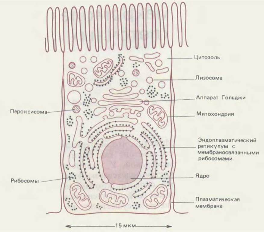
Figure 8-1. Schematic representation of the major intracellular compartments in a typical animal cell. The Cytosol, Endoplasmic reticulum, Golgi apparatus, Nucleus, mitochondrion, endosome, lysosome, and peroxisome represent distinct compartments bounded from the rest of The Cell by at least one selectively permeable membrane.
The intracellular compartments common to all Eukaryotic cells are shown in Figure 8-1. The Nucleus contains the bulk of The Genome and serves as the primary site for DNA and RNA Synthesis. The surrounding Cytoplasm consists of the cytosol and the cytoplasmic organelles suspended within it. The cytosol accounts for slightly more than half of the total cell volume. It is the site of Protein Synthesis AND the majority of Intermediary METABOLISM reactions—that is, pathways where small molecules are broken down or synthesized to provide the necessary building blocks for macromolecules. About half of all cellular membranes enclose the labyrinthine spaces of The endoplasmic reticulum (ER). The cytosolic face of the ER is studded with numerous Ribosomes, which synthesize integral Membrane Proteins and soluble proteins destined for secretion or other organelles. The ER also synthesizes Lipids for the entire cell. The Golgi apparatus consists of orderly stacks of flattened membrane-enclosed sacs called Golgi cisternae; it receives proteins and lipids from the ER and dispatches these molecules to various destinations within the cell, modifying them covalently along the way. Mitochondria and plant Chloroplasts generate most of the ATP utilized in Free energy-requiring biosynthetic reactions. Lysosomes contain digestive Enzymes that degrade worn-out organelles, as well as extracellular particles and molecules taken up by endocytosis. En route to lysosomes, internalized molecules and particles must pass through a series of organelles called endosomes. Finally, Peroxisomes (also known as Microbodies) are small vesicular bodies containing a variety of oxidative enzymes.
Table 8-1. Relative volumes of major intracellular compartments in a typical Liver cell (hepatocyte)
|
Intracellular compartment |
Percentage of total cell volume1) |
Approximate number per cell1) |
|
Cytosol |
54 |
1 |
|
Mitochondria |
22 |
1700 |
|
Cisternae of |
||
|
rough ER |
9 |
1 |
|
Cisternae of smooth |
||
|
ER plus Golgi cisternae |
6 |
|
|
Nucleus |
6 |
1 |
|
Peroxisomes |
1 |
400 |
|
Lysosomes |
1 |
300 |
|
Endosomes |
1 |
200 |
1) All rough and smooth ER cisternae are thought to be interconnected, forming a single large compartment. By contrast, the Golgi apparatus in each cell consists of numerous discrete sets of stacked cisternae, and the extent of interconnection between these stacks remains unclear.
In addition to the major membrane-enclosed organelles just listed, the cell contains numerous small transport vesicles that shuttle substances between organelles, as well as vesicles that fuse with The Plasma Membrane during Endocytosis and Exocytosis.
Overall, each membrane-enclosed organelle exhibits both general cellular properties and specialized features associated with the differentiated Functions of specialized cells. Collectively, membrane-enclosed organelles account for about half of the cell volume (Table 8-1). Naturally, their construction demands a vast amount of intracellular membrane. In the two mammalian cell types analyzed as Examples in Table 8-2, the total surface area of the endoplasmic reticulum membranes exceeds that of the plasma membrane by 25-fold and 12-fold, respectively. In terms of mass and surface area, the plasma membrane in most eukaryotic cells represents only a minor fraction of the total cellular membranes (Figure 8-2).
Table 8-2. Relative amounts of different membrane types in two eukaryotic cells
|
Membrane type |
Percentage of total cellular membranes |
|
|
Liver Hepatocyte1) |
Exocrine cell1) |
|
|
Plasma membrane |
2 |
5 |
|
Rough ER membrane |
35 |
60 |
|
Smooth ER membrane |
16 |
<1 |
|
Golgi membrane |
7 |
10 |
|
Mitochondria |
||
|
Outer membrane |
7 |
4 |
|
Inner membrane |
32 |
17 |
|
Nucleus |
||
|
Inner membrane |
0.2 |
0.7 |
|
Secretory vesicle membrane |
not determined |
~3 |
|
Lysosomal membrane |
0.4 |
not determined |
|
Peroxisomal membrane |
0.4 |
— |
|
Endosomal membrane |
0.4 |
— |
1) These cells vary significantly in size: the average volume of a hepatocyte is about 5000 µm3, whereas that of a pancreatic exocrine cell is about 1000 µm3. The total surface area of cellular membranes is approximately 110,000 µm2 and 13,000 µm2, respectively.
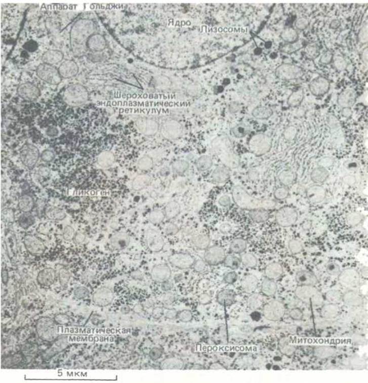
Figure 8-2. Electron micrograph of a liver cell cross-section, highlighting major intracellular compartments. (Courtesy of D. S. Friend.)
8.1.2. The topology of membrane-enclosed organelles reflects their evolutionary origin [2]
To understand the relationships between cellular compartments, it is helpful to consider how they might have arisen evolutionarily. Eukaryotic ancestors are believed to have been Bacteria-like organisms. Modern bacteria typically lack internal membranes; instead, their plasma membrane carries out analogous functions, such as ion transport, ATP synthesis, and Lipid Biosynthesis. However, contemporary eukaryotic cells are 10 to 30 times larger in linear dimensions than a typical bacterium (such as E. coli). The Abundance of internal membranes in eukaryotic cells can be viewed as an adaptation to this enlarged size, as the plasma membrane alone is simply insufficient to sustain many vital functions of a eukaryotic cell.
The evolution of internal membranes evidently proceeded in parallel with functional specialization. Certain modern bacteria possess specialized patches of plasma membrane where specific membrane proteins cluster to perform interrelated functions (Figure 8-3A). Examples include the "purple membranes" of Halobacterium containing Bacteriorhodopsin, and the chromatophores of photosynthetic bacteria. Both can be regarded as primitive organelles. In some photosynthetic bacteria, these membrane patches form deep invaginations of the plasma membrane (Figure 8-3B); in others, these invaginations have pinched off completely to form closed membrane vesicles dedicated to photosynthesis. The lumen of these vesicles is topologically equivalent to the cell exterior (Figure 8-3C).
It is reasonable to assume that a eukaryotic organelle originating through processes like those shown in Figure 8-3 would likewise possess an interior space topologically equivalent to the cell exterior. This is precisely the case for the ER, Golgi apparatus, endosomes, and lysosomes, as well as the numerous intermediate transport vesicles involved in endocytosis and exocytosis (see Section 8.6).
As discussed in Chapter 7, Mitochondria and chloroplasts differ from other membrane-enclosed organelles in possessing their own genomes. The Nature of these genomes and the close similarities between Mitochondrial and Chloroplast proteins and those of modern bacteria support the hypothesis that these organelles evolved from bacteria engulfed by other cells, with which they initially lived in Symbiosis (see Section 7.5.16). According to the hypothetical scheme in Figure 8-4A, the inner membrane of mitochondria and chloroplasts corresponds to the original bacterial plasma membrane, whereas the matrix of these organelles is derived from the bacterial cytoplasm. Consequently, these two organelles are isolated from the transport pathways connecting the lumens of most other organelles with one another and with the extracellular space.
The Water/144.html">Origin of the Cell Nucleus, with its uniquely structured double membrane, is more enigmatic. It is known that the single bacterial chromosome is anchored to specific sites on the inner surface of the prokaryotic plasma membrane. One hypothesis suggests that the double-layered nuclear envelope may have formed from a deep invagination of the plasma membrane, as depicted in Figure 8-4B. This hypothesis explains why the interior of the nucleus is topologically equivalent to the cytosol. Indeed, during mitosis in higher eukaryotes, the nuclear envelope breaks down and the nuclear contents mix completely with the cytosol—something that never occurs with any other membrane-enclosed organelle. Thus, during mitosis, the cell temporarily reverts to a prokaryotic-like state where Chromosomes lack a separate compartment.
This evolutionary framework divides the major eukaryotic intracellular compartments into five groups: (1) the nucleus and cytosol, which are interconnected by nuclear pores and are thus topologically continuous (though functionally distinct); (2) mitochondria; (3) chloroplasts (found exclusively in plants); (4) peroxisomes (whose evolutionary origin is discussed in Section 8.5); and (5) the remaining membrane-enclosed organelles (ER, Golgi apparatus, endosomes, and lysosomes).
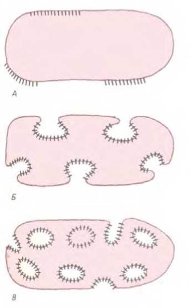
Figure 8-3. Specialized bacterial membranes. A. Regions of The cell membrane surface consisting of clusters of specialized membrane proteins. B. Invagination of such regions increases the surface area of membranes adapted for specific functions, such as photosynthesis. C. The inner surface of the resulting membrane vesicles is topologically equivalent to the cell exterior. Membrane-enclosed vesicles are found in certain species of photosynthetic bacteria. Their topological relationship to the cell surface resembles that of the ER, Golgi apparatus, endosomes, and lysosomes to The surface of eukaryotic cells.

Fig. 8-4. Hypotheses for the evolutionary origin of mitochondria, chloroplasts, the ER, and the cell nucleus, illustrating the topological relationships of these intracellular compartments in eukaryotic cells. A. Mitochondria and chloroplasts may have arisen when a eukaryotic cell engulfed bacteria. This hypothesis explains why the lumen of these organelles remains isolated from the extensive vesicular transport system that links the lumens of many other intracellular compartments. B.
A potential pathway for the Evolution of the ER and cell nucleus. In some bacteria, DNA is attached to an invagination of the plasma membrane called a mesosome. A similar invagination in a very ancient Prokaryotic Cell could have led to The formation of an envelope around the DNA while maintaining the DNA's access to the cytosol (since the DNA must direct protein synthesis). It is hypothesized that this envelope could eventually pinch off completely from the plasma membrane, forming a nuclear compartment surrounded by a double membrane. As shown in the figure, the nuclear envelope forms with the participation of a fibrous Structure—the nuclear lamina—and is pierced by channels known as nuclear pores. Because of these pores, the nuclear lumen is topologically equivalent to the cytosol. The lumen of the ER is continuous with the space between the outer and inner nuclear membranes and is topologically equivalent to the extracellular space.
The interior spaces of the organelles in the latter group are interconnected with each other and with the extracellular space via transport vesicles that bud off from one organelle and fuse with another. Notably, the lumens of these organelles are topologically equivalent to one another and to the extracellular space, functioning as interconnected parts of a unified complex.
8.1.3. Intracellular Protein Transport through the ER and Golgi apparatus can be tracked using autoradiography [3]
METHODS FOR STUDYING intracellular protein transport were developed in the 1960s and first applied to observe Transport from the ER to the extracellular space in pancreatic secretory cells.
Specialized secretory cells, such as pancreatic acinar cells, contain large amounts of proteinaceous secretion enclosed within secretory vesicles. Upon stimulation by an external signal, the contents of these vesicles are rapidly discharged into the extracellular space via exocytosis. This process is known as regulated secretion. It should be distinguished from constitutive secretion, another form of exocytosis that occurs continuously in the absence of a stimulating signal. Pancreatic acinar cells secrete various digestive enzymes (amylase, lipase, deoxyribonuclease, and Ribonuclease) as well as enzyme precursors called zymogens (e.g., trypsinogen and chymotrypsinogen). These precursors are activated through specific proteolytic Cleavage.
Because the group of proteins synthesized in pancreatic acinar cells is destined for secretion, we can trace their journey from the site of synthesis to the site of release. This pathway can be visualized by combining autoradiography with Electron Microscopy. The experimental setup is illustrated in Fig. 8-5. If cells are briefly incubated with [3H]-labeled Amino Acids (pulse labeling) and then chased for various times in a nonradioactive medium, Newly synthesized proteins are first detected in the ER and subsequently in the Golgi apparatus (Fig. 8-6). Later, [3H]-labeled proteins are found in large, immature secretory vesicles near the Golgi stack (Fig. 8-7), where they become increasingly concentrated, ultimately forming mature vesicles readily distinguishable in electron micrographs by their electron-dense contents (Figs. 8-7 and 8-8).
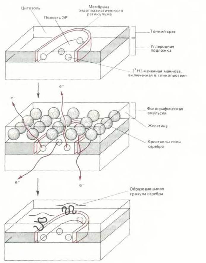
Fig. 8-5. Schematic diagram illustrating how electron microscopic autoradiography can be used to trace The pathway of secretory proteins containing [3H]-labeled amino acids. It is crucial that all free radioactive Amino acids are washed away before embedding so that proteins remain the sole radioactive source in the tissue. The tissue is then coated with a thin layer of photoemulsion. Electrons emitted by tritium expose silver grains at positions slightly displaced from the radiation source, meaning the localization of the radioactive molecule is determined with less precision than the resolution of the Electron microscope would otherwise allow.
Secretory vesicles accumulate in the apical region of the acinar cell (i.e., the region facing the duct system) between the Golgi apparatus
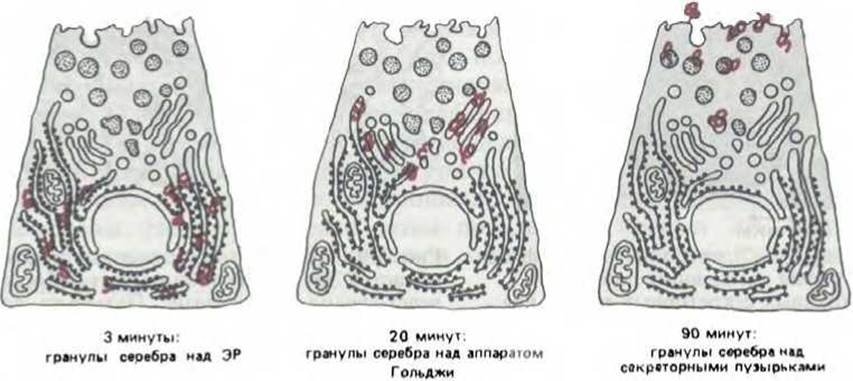
Fig. 8-6. Electron microscopic autoradiography of pancreatic acinar cells pulse-labeled with [3H]-Amino Acids and then chased for various times in an unlabeled medium ("pulse-chase" method). Over time, reduced silver grains (highlighted in color) migrate toward the extracellular space, mapping the route taken by newly synthesized secretory protein molecules. Acinar cells are unusual in that 85% of the proteins they synthesize are secreted; until the cell receives a secretory stimulus, these proteins are stored in secretory vesicles.
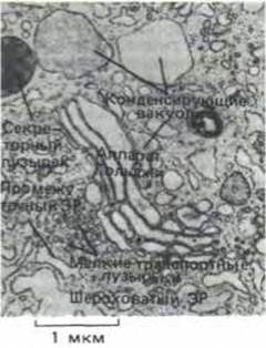
Fig. 8-7. Electron micrograph of the Golgi apparatus in a pancreatic acinar cell, showing secretory vesicles at various stages of maturation. Immature secretory vesicles (referred to in these cells as condensing vacuoles) bud off from the Golgi apparatus. The secretory proteins within these vesicles become progressively more concentrated (hence the term "condensing" vacuole), ultimately converting the condensing vacuoles into mature secretory vesicles (top left). In pancreatic acinar cells, these secretory vesicles are known as zymogen granules. (Courtesy of George Palade.)
and the plasma membrane. When a vesicle fuses with the plasma membrane, its contents are released into the extracellular space. These vesicles fuse exclusively with the apical domain of the plasma membrane, thereby preventing the wasteful and potentially hazardous release of secretions into the intercellular space or into the lumen of other organelles. Furthermore, exocytosis occurs only in response to specific extracellular chemical signals delivered by nerve or intestinal cells when pancreatic enzymes are required for Digestion. Numerous secretory proteins from various cell types have been studied, and all have been found to follow the same basic route: ribosome → ER → Golgi apparatus → extracellular space. Proteins destined to remain in the plasma membrane, lysosomes, or Golgi cisternae also first pass through the ER and are subsequently directed from there via the Golgi apparatus to their final destinations.
Elucidating the finer details of metabolic pathways originating in the ER requires more sophisticated techniques. Some proteins entering the ER remain there to perform enzymatic functions. However, the majority of proteins are packaged into transport vesicles (typically 50–100 nm in diameter), which then bud off from specialized Regions of the ER membranes known as transitional elements (Fig. 8-9) and specifically fuse with the nearest cisterna of the Golgi apparatus. Upon fusion, the vesicle membrane becomes part of the Golgi membrane, and the soluble proteins within the vesicle lumen are delivered into the Golgi cisternal space (Fig. 8-10). In this manner, soluble proteins are selectively transferred from one membrane-bounded compartment to another without crossing any membranes. It is believed that a similar mechanism of budding and fusion allows proteins to move from one Golgi cisterna to the next and ultimately to various final destinations according to their specific functions. Along this route, proteins pass through a series of enclosed spaces that are topologically equivalent to one another and to the extracellular space (Fig. 8-11). At each metabolic checkpoint, proteins make a series of choices: whether to remain in the cytosol or enter the ER, stay in the ER or proceed to the Golgi apparatus, become cargo in transport vesicles bound for lysosomes or instead enter vesicles moving toward the cell surface, and so on. Each decision depends on the Amino Acid Sequence of the protein molecule, which contains sorting signals that dictate its intracellular path.
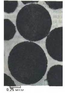
Fig. 8-8. Electron micrograph of a purified preparation of large secretory vesicles. Such vesicles are characteristic of specialized secretory cells. (Courtesy of Daniel S. Friend.)
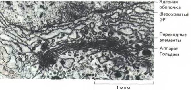
Fig. 8-9. Transport vesicles bud off from distinct regions of the ER that are largely devoid of ribosomes, known as transitional elements. These vesicles fuse with the nearest Golgi cisterna, transferring newly synthesized proteins and lipids primarily from the ER to the Golgi apparatus. (Courtesy of Brij J. Gupta.)
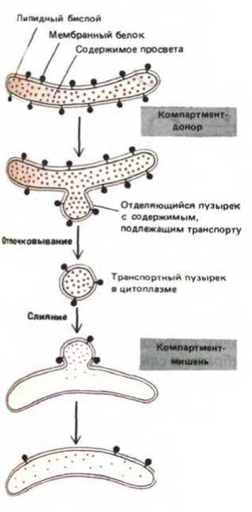
Fig. 8-10. The sidedness of membranes is preserved during vesicular transport. Note that the original orientation of both proteins and lipids in the target compartment membrane is maintained, while soluble Materials are transferred from lumen to lumen.
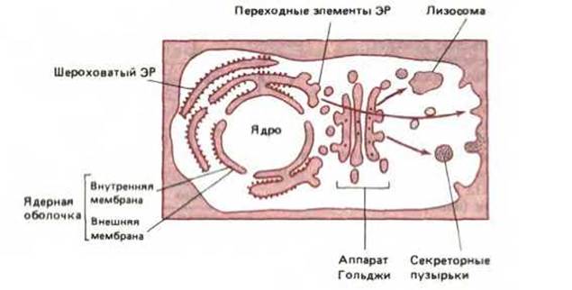
Fig. 8-11. Diagram of a cell with topologically equivalent regions highlighted in color. In principle, the cyclic budding and fusion of vesicles allows the lumen (cavity) of any organelle to communicate with any other lumen and with the extracellular space. However, certain organelles (such as mitochondria and chloroplasts) are not connected to other organelles via Vesicular Transport and thus remain isolated from the interorganellar transport pathways depicted here.
8-3
8.1.4. Protein transport occurs via two main pathways: through the cytosol and through the ER [4]
The Main Pathways of protein transport are shown in Fig. 8-12. Virtually all proteins are synthesized on ribosomes located in the cytosol (with a few exceptions synthesized on the mitochondrial ribosomes of chloroplasts). Their paths then diverge. Proteins belonging to one transport branch are released into the cytosol upon completion of their synthesis. Some of these contain sorting signals that direct them from the cytosol to mitochondria, chloroplasts (in plants), the nucleus, or peroxisomes; others—the majority—lack specific sorting signals and remain in the cytosol as permanent components.
Another major transport pathway is utilized during the synthesis of proteins destined for export from the cell, as well as proteins that are to become Components of the ER, the Golgi apparatus, the plasma membrane, or lysosomes. As they are formed, all these proteins are transferred to the ER using sorting signals usually located at the N-terminus. Ribosomes assembling such proteins remain bound to the ER membrane for a short time after the Synthesis of the polypeptide chain has begun. As soon as the next segment of the polypeptide chain is synthesized, it traverses The Lipid Bilayer of this membrane. Some proteins then enter the ER lumen, while others remain partially embedded in the membrane as transmembrane proteins.
Typically, no more than one or two minutes elapse from the moment a protein is released into the cytosol until its arrival at the appropriate organelle. Proteins destined for the nucleus, mitochondria, or peroxisomes complete their journey upon reaching these organelles, whereas proteins entering the ER are engaged in further transport, which is believed to occur via transport vesicles that bud off from one membrane and fuse with another. Apparently, several such cycles must take place to reach the final destination, and the journey from the ER membrane to the target site can take up to one hour.

Fig. 8-12. Simplified diagram of protein metabolic pathways. The signals directing a specific protein along a particular "road" and, consequently, determining the Intracellular Localization of that protein are contained within its amino acid sequence. The "journey" begins with protein synthesis on a ribosome and ends with its arrival at the final destination. At each intermediate stage (represented by rectangles), a decision is made—whether to retain the protein at that stage or transport it further. In principle, a signal may be required either to retain the protein in each of the indicated compartments or to propel it further, with the alternative event occurring "by default" (i.e., without A special signal). In this diagram, pathways most likely requiring special signals are highlighted in color; those likely chosen by default are shown with black arrows. Since endosome formation is poorly understood, no pathways leading to endosomes are depicted here.
8.1.5. Proteins can move between compartments by two fundamentally different mechanisms [5]
To understand the General Principles of sorting signal function, It is important to distinguish between two completely different pathways by which proteins move from one compartment to another. First, they can directly cross a membrane, passing from a space topologically equivalent to the cytosol to a space topologically equivalent to the extracellular space, or vice versa. This pathway requires a specialized translocator protein in the membrane; furthermore, the transported protein molecule must unfold to "snake" its way through the membrane. An example of this type of event is the translocation of certain proteins from the cytosol to the ER lumen. The second pathway of protein molecule movement is mediated by transport vesicles. These vesicles capture specific molecules in the lumen of one compartment (from which they pinch off) and transfer them to another compartment by fusing with it. This is precisely how soluble proteins are transferred from the ER to the Golgi apparatus (see Fig. 8-10). During such vesicular transport, proteins do not cross any membranes and are therefore transferred only between compartments that are topologically equivalent to each other.
Both types of transport processes are selectively controlled by specialized proteins that act as sorting signals. For a protein that is directly transferred across a membrane, these signals are recognized by a translocator in the membrane. Conversely, a protein enters a transport vesicle if its sorting signal binds to a receptor on the vesicle membrane. Presumably, There are also transport vesicles capable of capturing proteins that have lost specific sorting signals. In any case, newly formed vesicles transport only the proteins intended for them.
Currently, some sorting signals within proteins are known, whereas the majority of their complementary Membrane Receptors are not. Moreover, we know almost nothing about transport vesicles. We can only assume that they exist in a multitude of varieties, corresponding to the various transfers they must carry out. To perform its functions, each vesicle must capture only specific proteins and fuse only with a specific target membrane: for example, a vesicle transporting molecules from the ER to the Golgi apparatus must not include proteins that remain in the ER and must fuse exclusively with the Golgi apparatus rather than other organelles. In this chapter, we will examine how transport vesicles achieve such selectivity.
8.1.6. Signal Peptides and signal patches determine protein fate [6]
It is believed that proteins possess Two Types of sorting signals that guide them, step by step, along branching pathways (Fig. 8-12). For some stages, the sorting signals are an extended stretch of amino acid sequence 15–60 residues long. Once this stage is passed, such a signal peptide is cleaved off. The sorting signal for other stages is likely a specific three-dimensional structure formed by the surface atoms of a protein as its molecule folds. The amino acid residues forming such signal patches may be located very far apart in the linear sequence of the protein (Fig. 8-13). Signal peptides direct proteins from the cytosol to the ER, mitochondria, chloroplasts, and nucleus; they are also responsible for retaining certain proteins in the ER. Signal patches apparently play an important role in the recognition of specific lysosomal proteins by a specialized enzyme in the Golgi apparatus.
To determine the intracellular destination of a given protein, it is necessary to identify the type of its signal peptide (Table 8-3). Proteins destined for the ER typically bear an N-terminal signal peptide whose central core consists of 5–10 hydrophobic amino acid residues. Most of these proteins are directed from the ER to the Golgi apparatus; those that possess a specific four-amino-acid sequence at the C-terminus remain as permanent components. Many proteins destined for mitochondria have signal peptides in which positively charged amino acid residues alternate with hydrophobic ones. Among proteins directed to the nucleus, the majority have signal peptides formed by a cluster of positively charged amino acid residues. Finally, certain cytosolic proteins feature signal peptides to which a fatty acid is covalently attached—
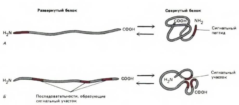
Fig. 8-13. Two ways of forming a transport signal on a protein. A. The signal is a single discrete segment of amino acid sequence called a signal peptide. In the folded protein, the signal peptide is located on the exterior. Signal peptides are most often found at the end of the polypeptide chain (as shown here), but can also be located elsewhere. Signal peptides are identified experimentally by their effect on intracellular protein sorting. To do this, recombinant DNA methods are used to "stitch" the signal peptide onto another protein. B. A signal patch can be formed by the apposition of amino acids from distant regions during molecule folding, or such a signal can be formed from separate "patches" On the surface of the folded protein that are separated from each other by a certain distance. In both cases, the transport signal depends on the three-dimensional conformation of the protein. For this reason, pinpointing the exact Location of such signals is extremely difficult.
Table 8-3. Typical Signal Peptide Sequences
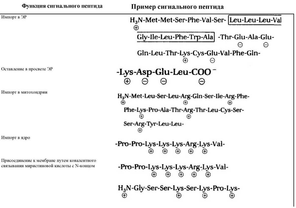
Charged residues are designated by + or —. A large block of hydrophobic residues is boxed. H3N- denotes the N-terminus of the protein; — COO' denotes the carboxyl terminus directing these proteins to membranes without penetrating the ER.
The Role of each such signal peptide in delivering a protein to its destination has been demonstrated in Genetic Engineering experiments. For example, placing an ER-binding N-terminal signal peptide at the beginning of a cytosolic protein caused that protein to be directed to the ER. Signal peptides of proteins with the same final destination are functionally interchangeable, although their Amino acid sequences may vary widely. Perhaps physical properties (such as Hydrophobicity) turn out to be more important for the signal recognition process than the exact amino acid sequence.
Signal patches are much more difficult to analyze than signal peptides, and consequently much less is known about their structure. Because they are formed from the complex three-dimensional configuration of a folded protein, they cannot simply be transferred from one protein to another. Moreover, experimental disruption of a signal patch frequently results in damage to the protein as a whole.
8.1.7. Cells cannot build their membrane-bound organelles de novo: they require information contained within the organelle itself [7]
When a cell reproduces and divides, it must double its membrane-bound organelles. This typically occurs by increasing the size of these organelles through the incorporation of new molecules; the enlarged organelles then divide and are distributed between the two daughter cells. A cell is hardly capable of creating these organelles de novo. If, for example, the ER is completely removed from it, the cell will fail to reconstruct it. Indeed, the membrane proteins that define the Specificity of the ER and perform many of its key functions are themselves products of the ER: without the ER, or at least without a membrane containing the translocators required for importing proteins into the ER (and in the absence of the translocators required for importing proteins into other organelles), a new ER cannot be built.
Consequently, the formation of membrane-bound organelles requires more than just DNA information specifying the organelle proteins. "Epigenetic" information in the form of at least one characteristic protein in the organelle membrane is also necessary. This information is transmitted from the parent cell to the progeny along with the organelle itself. Such information is presumably required to maintain cellular compartmentalization, whereas the information contained in DNA is necessary for the "Replication" of nucleotide and amino acid sequences.
Eukaryotic cells contain intracellular membranes that enclose roughly half of the total cell volume into distinct intracellular compartments. The major Types of Membrane-bounded organelles found in all eukaryotic cells include the endoplasmic reticulum, Golgi apparatus, nucleus, mitochondria, lysosomes, endosomes, and peroxisomes; plant Cells also contain chloroplasts. Each organelle consists of a distinct set of proteins that determine its unique functions.
Every newly synthesized organellar protein follows a specific pathway from the ribosome to its destination, guided by either a signal peptide or a signal patch. Protein sorting begins with primary segregation, during which a protein either remains in the cytosol or is translocated to another compartment (such as the nucleus, mitochondria, or endoplasmic reticulum). Proteins entering the ER undergo further sorting as they are transported to the Golgi apparatus and subsequently from the Golgi to lysosomes, secretory vesicles, or the plasma membrane. Some proteins remain in the ER and in various cisternae of the Golgi apparatus. Proteins destined for other compartments are presumably packaged into transport vesicles that bud off from one compartment and fuse with another.
Last update: 12/08/2026
Editorial and Educational Adaptation: This material has been compiled based on the primary/original source text. The project team performed an editorial review, corrected technical inaccuracies, structured sections, and adapted the content for an educational format.
What was processed:
- elimination of formatting defects (OCR errors, structural breaks, corrupted characters);
- editorial organization of content;
- standardization of terminology in accordance with academic sources;
- verification of factual statements against the original source text.
All mentions of the author, publication year, and origin of the primary text have been preserved in accordance with the source.