Molecular Biology of the Cell - Volume 1 - Alberts B., Bray D., Lewis J., Raff M., Roberts K., Watson J. 1994
Molecular Organization of Cells
The Plasma Membrane
The Lipid Bilayer
The first evidence that lipid molecules in Introduction/36.html">Biological Membranes form a bilayer came from simple but elegant experiments performed in 1925. It was shown that Lipids extracted with acetone from red Blood Cell membranes float on Water to form a film. The area of this film was reduced using a movable barrier until a continuous monomolecular layer was formed. The area of the monolayer was found to be approximately twice the original surface area of the Cells. Since the only membrane of a red blood cell is its Plasma Membrane, the experimenters concluded that its lipid molecules must be organized as a continuous bilayer. This Conclusion had a profound impact on all of cell biology. Today, the presence of a lipid bilayer in cell membranes has been proven by more sophisticated Methods. For example, X-Ray Diffraction has demonstrated the existence of lipid bilayers in the highly organized folds of cell membranes that form the insulating myelin sheath surrounding Nerve Cells (see Section 19.2.4). That all biological membranes contain lipid bilayers is also convincingly demonstrated by Electron Microscopy: studies of freeze-fracture specimens revealed that all cell membranes can be mechanically split precisely between the two lipid monolayers (see Section 6.2.6). The spontaneous formation of a bilayer is a unique property of lipid molecules that occurs even outside The Cell.
6-4
6.1.1. Membrane Lipids are amphipathic molecules that spontaneously form a bilayer [2]
Lipids are insoluble in water but dissolve readily in organic Solvents. In most animal cells, they constitute about 50% of the mass of The Plasma Membrane, with almost all the remainder being Proteins. There are approximately 5 x 10⁶ lipid molecules per 1 µm2 of lipid bilayer. It follows that the plasma membrane of a small animal cell contains about 109 lipid molecules. Three types of lipids are present in The cell membrane: Phospholipids (the most abundant type), Cholesterol, and Glycolipids. All of them are amphipathic molecules, meaning they have a hydrophilic ("water-loving," or polar) end and a hydrophobic ("water-fearing," or nonpolar) end. A typical phospholipid molecule is shown in Fig. 6-2. It has a polar HEAD and two hydrophobic hydrocarbon tails. The length of the tails varies from 14 to 24 carbon atoms in the chain. One of the tails typically contains one or more cis-double bonds (i.e., it is an unsaturated hydrocarbon), whereas the other (a saturated hydrocarbon) has no double bonds. As shown in Fig. 6-2, each double bond creates a kink in the tail. Differences in tail length and the saturation of hydrocarbon chains are important because they influence how phospholipid molecules pack against each other and, consequently, determine membrane fluidity (see below).
Class="center">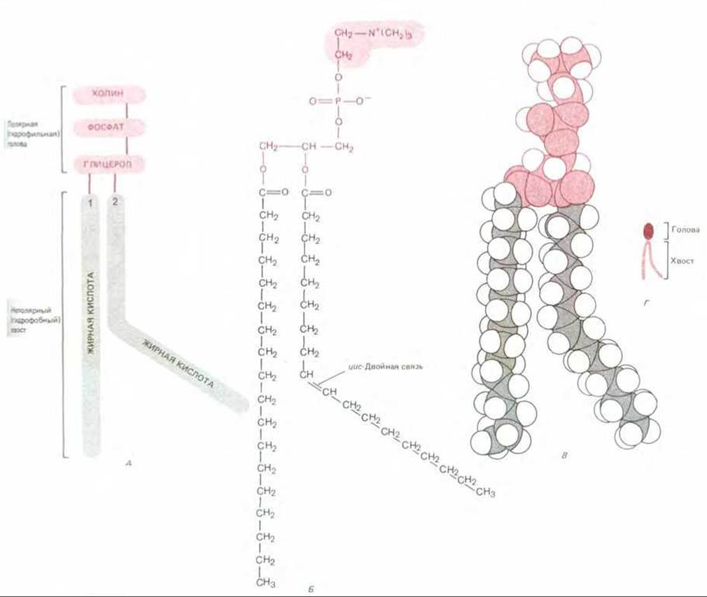
Fig. 6-2. A phospholipid molecule of phosphatidylcholine, represented schematically (A), by its chemical formula (B), as a space-filling model (C), and as a symbol (D). To distinguish the unsaturated fatty acid chain from the saturated one, in this figure (and throughout the book) the unsaturated chain is shown with a distinct kink. In reality, however, only the double bond in an unsaturated fatty acid is rigid. Free rotation is possible around all single bonds, so in each lipid monolayer, both fatty acid chains—both saturated and unsaturated—will tend to pack parallel to each other.
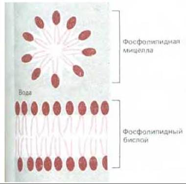
Fig. 6-3. A phospholipid micelle and a phospholipid bilayer in cross section. Phospholipid molecules spontaneously form such structures in water.
It is the amphipathic nature of lipid molecules that drives them to spontaneously form bilayers In aqueous solutions. When amphipathic molecules are in an aqueous environment, they tend to aggregate in such a way that their hydrophobic tails are shielded from water molecules, while their hydrophilic heads are in contact with water. This type of aggregation occurs in one of two ways: either by forming spherical micelles with tails pointing inward, or by forming bimolecular sheets, or bilayers, in which the hydrophobic tails are sandwiched between two layers of hydrophilic heads. Both of these possibilities are illustrated in Fig. 6-3.
Most Phospholipids and glycolipids spontaneously form bilayers in an aqueous environment. Moreover, these lipid bilayers tend to close in on themselves, resulting in The formation of sealed compartments. This eliminates free edges where hydrophobic tails would be exposed to water. For the same reason, compartments composed of lipid bilayers tend to self-seal by reuniting the edges of torn areas. In addition to its self-assembling capability, the lipid bilayer possesses other characteristics that make it an ideal material for cell membranes. The most important of these properties is fluidity, which, as we will see later, underlies many Membrane Functions.
6-6
6-7
6.1.2. The lipid bilayer is a two-dimensional fluid [3]
That individual lipid molecules are able to diffuse freely within a lipid bilayer was first discovered, surprisingly enough, only in the early 1970s. This was initially demonstrated using artificial lipid bilayers. Two Types of artificial bilayers have proven highly useful for experimental studies: 1) Liposomes, which are spherical vesicles ranging in diameter from 25 nm to 1 µm depending on how they are prepared (Fig. 6-4), and 2) planar bilayers, called black membranes, which "seal" a hole in a partition between two water-filled compartments (Fig. 6-5).
A variety of methods are used to measure the mobility of individual lipid molecules and their parts. For example, a "spin label," such as a nitroxide group (= N — O) containing an unpaired electron, can be attached to the polar head of a lipid molecule. The spin of this electron produces a paramagnetic signal detected by electron spin Resonance (ESR) (the principles of this method are similar to those of nuclear magnetic resonance, NMR). This technique makes it easy to determine the motion and orientation of such a spin-labeled lipid within the bilayer. Such studies show that lipid molecules in synthetic membranes very rarely migrate from one monolayer of the membrane to the other. Any individual lipid molecule undergoes such a flip-flop less than once every two weeks (Fig. 6-6). On the other hand, lipid molecules readily exchange places with their neighbors within the same monolayer (about 107 times per second), leading to rapid lateral diffusion, with a diffusion coefficient D of about 10-8 cm2 s-1. This means that an average-sized lipid molecule diffuses a distance equal to the length of a large bacterial cell (~ 2 µm) in approximately 1 s. In addition, such studies show that lipid molecules rotate very rapidly about their longitudinal axes, and their hydrocarbon chains are flexible, with the greatest mobility observed near the center of the bilayer and the least near the polar head (Fig. 6-6).
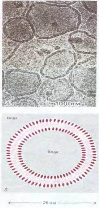
Fig. 6-4, A. Electron micrograph of unfixed, unstained phospholipid vesicles (liposomes) in water. Note that the bilayer Structure OF THE vesicles is easily discernible. B. Schematic cross-sectional view of a small spherical liposome. Liposomes are commonly used in experimental research as model membranes. (A, courtesy of Jean Lepault.)

Fig. 6-5. Schematic cross-sectional view of an artificial lipid bilayer, called a black membrane. The membrane seals a small hole in a partition between two water-filled compartments. Black membranes are used to measure the permeability of Artificial Membranes.
Studies of labeled lipid molecules in isolated biological membranes and relatively simple whole cells, such as Mycoplasmas, Bacteria, and red Blood Cells, have shown that The behavior of lipid molecules in cell membranes is largely similar to their behavior in artificial bilayers. The lipid component of a biological membrane is a two-dimensional fluid in which individual lipid molecules are free to move within the plane of the membrane. As in synthetic bilayers, individual lipid molecules usually do not migrate from one monolayer to the other. However, there are exceptions: in membranes where lipids are actively synthesized (such as the membranes of The Endoplasmic reticulum), rapid flip-flop of specific lipids must occur. To facilitate this process, there are even specialized membrane-bound Enzymes called phospholipid translocators (see Section 8.6.14).
6.1.3. The fluidity of a lipid bilayer depends on its composition [4]
A synthetic lipid bilayer composed of a single type of phospholipid transitions from a liquid state to a crystalline (or gel) state when the Temperature is lowered to a specific value (the freezing point). This change of state is called a phase transition. The transition temperature is lower (i.e., the membrane is harder to freeze) if the hydrocarbon chains are short or contain double bonds. With shorter chain lengths, interactions between the hydrocarbon tails are less likely, and the kinks caused by cis-double bonds prevent the tails from packing tightly together (Fig. 6-7).
In a synthetic bilayer containing a mixture of phospholipids with different degrees of saturation (and thus different phase transition temperatures), phase Separation can occur: as the temperature drops to the freezing points, phospholipid molecules of a specific type spontaneously aggregate within the bilayer, forming "frozen" domains. Because biological membranes typically contain both a saturated and an unsaturated fatty acid chain within the same lipid molecule, such phase separation does not occur in cells.
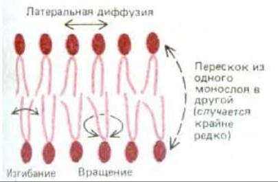
Fig. 6-6. Types of movement of phospholipid molecules in a lipid bilayer.

Figure 6-7. Double bonds in unsaturated hydrocarbon chains increase the fluidity of the lipid bilayer by making it difficult for the hydrophobic tails to pack together.

Figure 6-8. A cholesterol molecule represented as a chemical formula (A), a schematic drawing (B), and interacting with two phospholipid molecules in a monolayer (C).
Another factor influencing membrane fluidity is cholesterol. Eukaryotic Plasma Membranes contain relatively large amounts of cholesterol—approximately one molecule for every phospholipid molecule. Cholesterol molecules orient themselves in the bilayer so that their hydroxyl groups are adjacent to the polar head groups of the phospholipid molecules. Their rigid, planar steroid rings partially immobilize those Regions of the hydrocarbon chains closest to the polar head groups. The remaining PARTS OF THE hydrocarbon chains do not lose their flexibility (Figure 6-8). Although cholesterol makes the lipid bilayer less fluid, at the high concentrations found in most eukaryotic plasma membranes, it prevents the hydrocarbon chains from packing together and crystallizing. Thus, cholesterol also inhibits potential phase transitions.
Cholesterol decreases not only the fluidity of the lipid bilayer, but also its permeability to small water-soluble molecules. In addition, cholesterol increases the elasticity and mechanical strength of the bilayer. It is because of cholesterol that the membrane can change its shape in response to an applied force. This is because, unlike phospholipids, cholesterol can rapidly redistribute between monolayers. This is explained by the fact that the small polar head of cholesterol (the hydroxyl group) passes relatively easily through the center of the bilayer; the energy barrier for the flip-flop of cholesterol molecules is low, and therefore its redistribution occurs rapidly.
The Importance of cholesterol in maintaining the mechanical strength of membranes is demonstrated by studies of mutant animal cell lines unable to synthesize it. In the absence of cholesterol in the culture medium, these cells rapidly lyse (burst, releasing their contents). When cholesterol is added to the medium, it integrates into the plasma membrane, thereby stabilizing the lipid bilayer, and the cells survive.
A certain level of plasma membrane fluidity is of great biological importance. This is evidenced by the fact that bacteria, Yeast, and other poikilothermic organisms, which adapt to the ambient temperature, alter the fatty acid composition of their plasma membranes to keep membrane fluidity relatively constant. For example, when the temperature drops, Fatty acids with more cis-double bonds are synthesized to prevent the decrease in bilayer fluidity that would otherwise occur. It has been shown that some membrane transport processes cease and certain enzymatic activities disappear as soon as the bilayer viscosity exceeds a certain threshold. It remains to be determined which membrane-associated processes are most sensitive to bilayer fluidity.
6.1.4. The Lipid Bilayer Serves as a Solvent for Membrane Proteins [5]
The lipid compositions of various biological membranes are compared in Table 6-1. Bacterial plasma membranes usually contain only one type of phospholipid and lack cholesterol; in the absence of cholesterol, mechanical strength is provided by the surrounding Cell wall (see Figure 5-54). In contrast, most eukaryotic plasma membranes contain not only significant amounts of cholesterol but also A wide variety of phospholipids. For example, four major phospholipids are found in the plasma membrane of many animal cells: phosphatidylcholine, sphingomyelin, phosphatidylserine, and phosphatidylethanolamine. The structures of these molecules are shown in Figure 6-9; note that of these, only phosphatidylserine carries a net negative charge, while the other three are electrically neutral at physiological pH. Together, these four phospholipids constitute more than half of the total lipid mass in most membranes (Table 1-6). Other phospholipids, such as Inositol phospholipids (see Section 12.3.9), are also functionally important but are present in relatively small amounts. The unique role of inositol phospholipids in cell signaling is discussed in Chapter 12.
Table 6-1. Approximate Lipid Composition of various cell membranes
|
Lipids |
Percentage of total lipid (by weight) |
|||||
|
Liver cell plasma membrane |
Red blood cell plasma membrane |
Myelin |
Mitochondrial outer and inner membranes |
ER |
E. coli |
|
|
Cholesterol |
17 |
23 |
22 |
3 |
6 |
0 |
|
Phosphatidylethanolamine |
7 |
18 |
15 |
35 |
17 |
70 |
|
Phosphatidylserine |
4 |
7 |
9 |
2 |
5 |
trace |
|
Phosphatidylcholine |
24 |
17 |
10 |
39 |
40 |
0 |
|
Sphingomyelin |
19 |
18 |
8 |
0 |
5 |
0 |
|
Glycolipids |
7 |
3 |
28 |
trace |
trace |
0 |
|
Others |
22 |
13 |
8 |
21 |
27 |
30 |
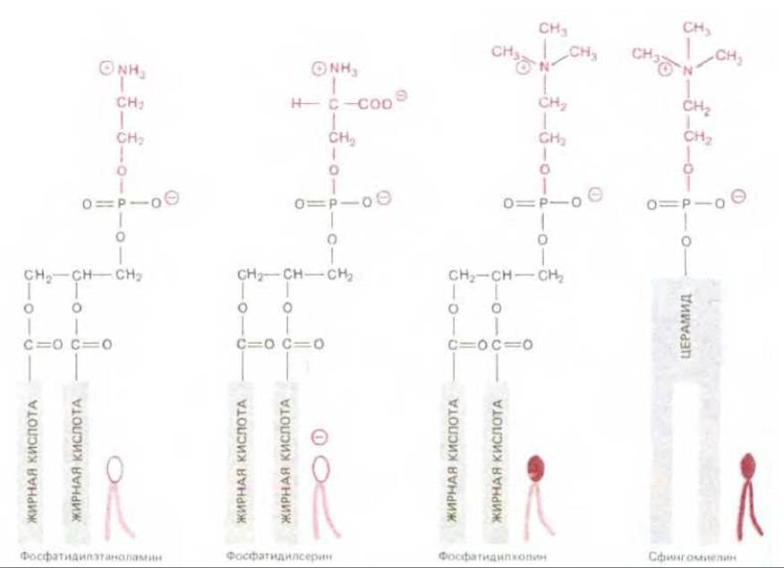
Figure 6-9. Formulas and symbols of the four major phospholipids found in plasma membranes. Note that the different polar head groups are represented by different symbols (in this and the next figure). All the lipid molecules shown are derived from glycerol, except for sphingomyelin, which is derived from Serine. The structure and formula of ceramide are shown in Figure 6-11.
This raises a question: why is there such a diversity of phospholipids in eukaryotic plasma membranes? Perhaps the lipid bilayer serves as a two-dimensional solvent for membrane proteins, just as water serves as a three-dimensional solvent in an aqueous solution. It is also possible that certain membrane proteins function only in the presence of specific phospholipid polar head groups, much like many enzymes that require a specific ion for activation in an aqueous solution. This hypothesis is supported by findings that in artificial lipid bilayers, optimal activity of functioning membrane proteins requires specific phospholipids.
6-5
6-6
6.1.5. The Lipid Bilayer Is Asymmetric [6]
In all plasma membranes studied, the two halves of the bilayer have strikingly different lipid compositions. For example, in the human erythrocyte membrane, most of the lipid molecules that contain Choline in their polar head group—namely, phosphatidylcholine and sphingomyelin—are located in the outer monolayer, whereas most of the phospholipids that contain a terminal amino group (phosphatidylethanolamine and phosphatidylserine) are in the inner monolayer (Figure 6-10). The fatty acid tails of phosphatidylcholine and sphingomyelin are more saturated than those of phosphatidylethanolamine and phosphatidylserine. Consequently, the Asymmetry in the distribution of polar head groups is accompanied by an asymmetry in the distribution of hydrocarbon tails. This may result in the inner monolayer being slightly more fluid than the outer monolayer. It is also known that the negatively charged phosphatidylserine is localized in the inner half of the bilayer; thus, the Two Sides of the bilayer differ significantly in charge as well.
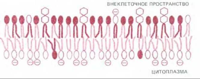
Figure 6-10. Asymmetric distribution of phospholipids and glycolipids in the lipid bilayer of human erythrocytes. The symbols used for phospholipids are the same as in Figure 6-9. The polar head groups of glycolipids are depicted as hexagons. Cholesterol (not shown) is thought to be distributed almost equally between the two monolayers.
Most Eukaryotic Cell membranes, including the plasma membrane, are synthesized in the endoplasmic reticulum (ER). This means that the asymmetry in phospholipid distribution is a consequence of the action of phospholipid translocators in the ER, which transfer specific phospholipid molecules from one monolayer to the other (see Section 8.6.14). Although the Functional Significance of asymmetric lipid distribution is largely unclear, membrane-bound enzymes have been shown to exploit this phenomenon. For example, when protein kinase C is activated, it binds to the cytosolic side of the plasma membrane, where phosphatidylserine—a negatively charged phospholipid required for enzyme activity—is concentrated (see Section 12.3.10). The importance of specific inositol phospholipids concentrated on the cytosolic side of the bilayer, where they can be used to generate intracellular mediators in response to extracellular signals, is discussed in Chapter 12.
6.1.6. Glycolipids Are Found On the surface of All Plasma Membranes, but Their Function Is Unknown [7]
In the plasma membranes of animal cells, the most asymmetrically distributed lipid molecules are those belonging to the class of oligosaccharide-containing lipids called glycolipids. These intriguing molecules are found exclusively in the outer monolayer of the bilayer, with their sugar groups oriented toward the cell surface (Figure 6-10), suggesting that they play a role in the interaction of the cell with its environment. The asymmetric distribution of glycolipids in the bilayer is established when sugar residues are added to lipid molecules in the lumen of the Golgi apparatus.
Glycolipids are probably present in the plasma membranes of all animal cells, typically constituting about 5% of the lipid molecules in the outer monolayer. They vary greatly between species and even among different Tissues of the same species. In bacteria and plants, almost all glycolipids are derivatives of glycerol-based lipids, such as the widespread phosphatidylcholine. In animal cells, glycolipids are based on ceramide, which is also the backbone of the phospholipid sphingomyelin (see Figure 6-9). The overall structure of glycosphingolipids is similar to that of glycerol-derived phospholipids. They also possess a polar head group and two hydrophobic fatty acid chains. However, one of the fatty acid chains is initially linked to serine to form the amino alcohol sphingosine, to which a second fatty acid chain is then attached to form ceramide (Figure 6-11).
All glycolipid molecules differ in the number of sugar residues in their polar head groups. Among the many glycolipids of the plasma membranes of eukaryotic and Prokaryotic Cells, a group of neutral glycolipids can be distinguished. Their polar head groups contain from 1 to 15 or more neutral (uncharged) sugars, depending on the Organism and cell type. An example is galactocerebroside, one of the simplest glycolipids, whose polar head group consists of only a single galactose. Galactocerebroside is the major glycolipid of myelin, the multilayered membranous sheath surrounding axons. Myelin is the plasma membrane of a specialized myelin-forming cell, wrapped in many concentric layers around a nerve fiber. A distinctive feature of these cells is the high content of galactocerebroside in their plasma membrane (up to 40% of the mass of the outer monolayer). In the membranes of other cells, there is little galactocerebroside. This probably indicates that it plays an important role in the specific interaction between the myelin-forming cell and the axon.
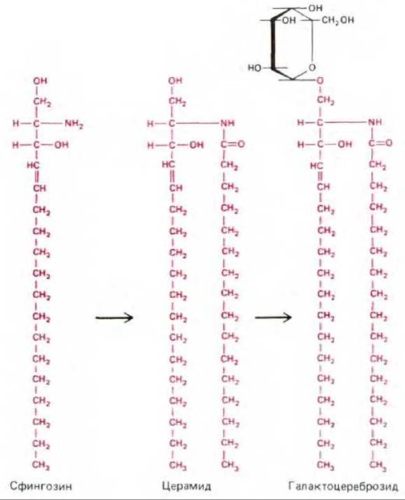
Fig. 6-11. The final steps in The Biosynthesis of a simple glycosphingolipid, galactocerebroside. Sphingosine is formed by the Condensation of The amino acid serine with a single fatty acid molecule. A second fatty acid molecule is then attached, forming ceramide. Ceramide synthesis occurs in the ER, and the carbohydrate is added in the Golgi apparatus. Ceramide is also used in the Synthesis of the major phospholipid, sphingomyelin (see Fig. 6-9).
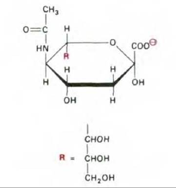
Fig. 6-12. Structure of sialic acid (N-acetylneuraminic acid, or NANA). In cells, it exists, as shown in the figure, in its ionized form (—СОС-).
The most complex glycolipids, gangliosides, contain one or more sialic acid residues (N-acetylneuraminic acid, or NANA), which give ganglioside molecules a negative charge (Fig. 6-12). Gangliosides are particularly abundant in the plasma membrane of nerve cells, where they constitute 5-10% of the total lipid mass; however, they are found in much smaller amounts in most other cell types. More than 40 different gangliosides have now been identified. Some of these are shown in Fig. 6-13, which also presents the nomenclature used to describe these compounds.
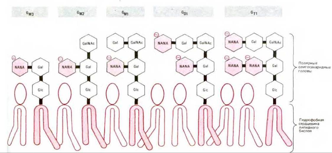
Fig. 6-13. Some typical gangliosides and their structural designations. In GM1, GM2, GM3, GD1, and GT1, the letters M, D, and T denote the number of sialic acid residues—one, two, and three, respectively—and the numbers 1, 2, and 3 represent the difference obtained by subtracting the number of uncharged carbohydrate residues from 5. NANA is N-acetylneuraminic (sialic) acid, Gal is galactose, Glc is glucose, and GalNAc is N-acetylgalactosamine. Gal, Glc, and GalNAc are uncharged, whereas NANA carries a negative charge (see Fig. 6-12).
Little is known about the functions of glycolipids. It has been established, for example, that ganglioside GM1 (Fig. 6-13) acts as a cell-surface receptor for the bacterial toxin that causes the debilitating diarrhea of cholera. Cholera toxin binds to the surface and enters only those cells that have GM1 in their plasma membrane (such as intestinal epithelial cells). The entry of cholera toxin into the cell leads to a prolonged increase in the concentration of intracellular cAMP, which causes a massive efflux of Na+ and water into the intestine. Of course, the binding of Bacterial toxins cannot be the normal function of gangliosides. It is reasonable to suggest that they act as receptors for specific signaling molecules involved in normal cell-to-cell communication.
Summary
Biological membranes consist of a continuous double layer of lipid molecules in which various proteins are embedded. The lipid bilayer is a fluid in which individual lipid molecules can diffuse rapidly within their own monolayer, but extremely rarely migrate spontaneously from one monolayer to the other. Membrane lipids are amphipathic molecules that spontaneously form bilayers in an aqueous environment. These bilayers self-assemble into closed compartments that can spontaneously reseal when damaged. There are three Major Classes of lipid molecules in the plasma membrane—phospholipids, cholesterol, and glycolipids—with the compositions of the inner and outer monolayers differing from each other. Different lipid compositions are characteristic of both the plasma membranes of different cell types and the various membranes of the same eukaryotic cell. In most cases, the functional Significance of the different components in various membranes remains unknown.
Last update: 12/08/2026
Editorial and Educational Adaptation: This material has been compiled based on the primary/original source text. The project team performed an editorial review, corrected technical inaccuracies, structured sections, and adapted the content for an educational format.
What was processed:
- elimination of formatting defects (OCR errors, structural breaks, corrupted characters);
- editorial organization of content;
- standardization of terminology in accordance with academic sources;
- verification of factual statements against the original source text.
All mentions of the author, publication year, and origin of the primary text have been preserved in accordance with the source.