HUMAN MEDICAL BIOLOGY, ANATOMY, PHYSIOLOGY AND PATHOLOGY - Y.I. Fedoniuk 2010
ANATOMY, PHYSIOLOGY, PATHOLOGY
CHAPTER 5. BLOOD AND LYMPH CIRCULATION. PATHOPHYSIOLOGY
THE HEART: STRUCTURE AND TOPOGRAPHY
1. STRUCTURE OF THE HEART WALL. HEART CHAMBERS
The Heart (cor) is an unpaired hollow muscular organ located in the left half of the thoracic cavity (two-thirds of the organ) within the anterior Mediastinum. In shape, the heart resembles a cone (Fig. 5.1). Its narrowed portion, the apex, is directed downward, forward, and to the left, while the expanded portion, the base, is directed upward and backward. The heart has anterior (sternocostal), inferior (diaphragmatic), and mediastinal (pulmonary) surfaces. The coronary sulcus runs transversely across The surface of the heart, forming the boundary between the atria and ventricles. The anterior interventricular sulcus runs along the anterior surface, and the posterior interventricular sulcus runs along the inferior surface. These sulci meet near the apex of the heart.
Class="center">
Fig. 5.1. The heart; right view
The heart wall consists of three layers: the endocardium (inner), the myocardium (middle), and the epicardium (outer).
The endocardium (endocardium) lines all the Chambers of the heart and forms its Valves.
The myocardium (myocardium) is composed of specialized cardiac Striated Muscle tissue. Its contractions are involuntary. The atrial myocardium and ventricular myocardium are distinguished, with their muscle bundles remaining separate. The sequential and rhythmic contraction of the ventricles and atria is driven by the cardiac conduction system, which consists of specialized muscle fibers forming bundles and nodes.
The epicardium (epicardium) is formed by a delicate Connective Tissue membrane and a layer of epithelial-like Cells, representing the visceral layer of the serous Pericardium. In addition to the heart, the epicardium covers the initial segments of the large vessels entering or leaving the heart: the aorta, pulmonary trunk, and the inferior and superior venae cavae.
The pericardium (pericardium) is a thin yet robust fibro-serous sac enclosing the heart (Fig. 5.2). It consists of two layers: an outer fibrous layer and an inner serous layer. The outer fibrous layer merges with the adventitia of the great vessels near the Base of the heart. The serous pericardium has two parts: the parietal layer, which lines the inner surface of the fibrous pericardium, and the visceral layer, which covers The Heart and serves as its outer coat (the epicardium). Between these two layers lies a slit-like cavity containing 1-2 ml of serous fluid, which reduces friction during cardiac contractions.

Fig. 5.2. The heart, pericardial sac opened
The human heart is four-chambered, divided longitudinally by the interatrial and interventricular septa into two separate halves—left and right—which normally do not communicate (Fig. 5.3). The upper portion of each half contains an atrium (right and left), and the lower portion contains the right and left ventricles. Each atrium communicates with its respective ventricle through an atrioventricular opening. Externally, near each atrium, There is a right and left auricle. The muscular wall of the left ventricle is thicker than that of the right ventricle. The internal surface of the ventricles features muscular projections called papillary Muscles (two on the right, three on the left). Four Pulmonary Veins carrying arterial Blood empty into the left atrium
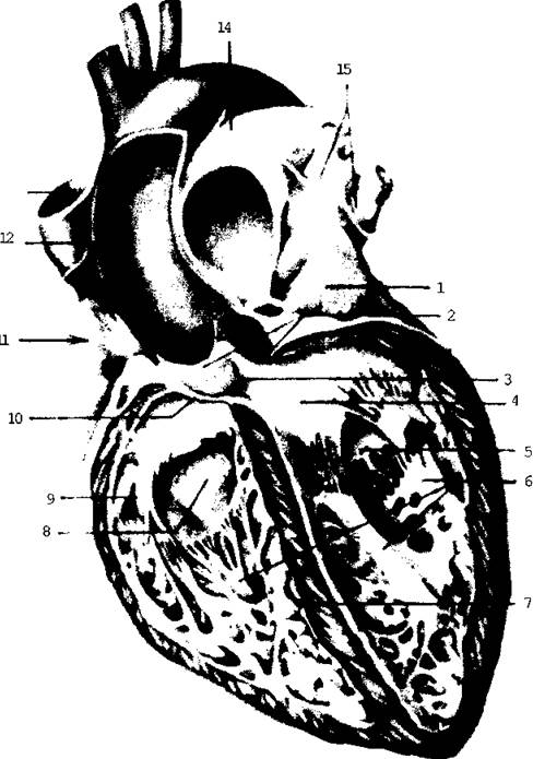
Fig. 5.3. The heart (longitudinal section); anterior view
from the Lungs. The superior and inferior venae cavae, which carry venous blood from all PARTS OF THE body, empty into the right atrium (Fig. 5.1, 5.2).
The aorta emerges from the left ventricle, carrying arterial blood through the systemic Circulation to all Organs and Tissues.
The pulmonary trunk emerges from the right ventricle, initiating the Pulmonary Circulation. It carries venous blood to the lungs.
Thus, the right half of the heart contains venous blood, and the left half contains arterial blood.
The heart valves are formed by folds of the endocardium. The atrioventricular openings are closed by atrioventricular valves. The valve between the right atrium and ventricle is called the tricuspid valve (having three cusps) (Fig. 5.3). The left half of the heart contains a bicuspid valve, known as the mitral valve (Fig. 5.4). The cusps of the valves are connected to the papillary muscles by chordae tendineae (Fig. 5.5), preventing them from everting from the ventricles into the atria during ventricular contraction (systole). Consequently, blood is prevented from flowing backward from the ventricles into the atria during their contraction. When the cusps are damaged (e.g., due to rheumatic fever or Syphilis), the atrioventricular orifices fail to close completely, impairing cardiac function and resulting in heart defects.
The openings of the pulmonary trunk and aorta are also equipped with semilunar valve cusps (Fig. 5.4). During ventricular relaxation (diastole), these cusps fill with blood and seal the vessel openings, preventing the backflow of blood into the ventricles.

Fig. 5.4. Heart valves

Fig. 5.5. Right atrium and ventricle (opened); right lateral view
Last update: 08/08/2026
Editorial and Educational Adaptation: This material has been compiled based on the primary/original source text. The project team performed an editorial review, corrected technical inaccuracies, structured sections, and adapted the content for an educational format.
What was processed:
- elimination of formatting defects (OCR errors, structural breaks, corrupted characters);
- editorial organization of content;
- standardization of terminology in accordance with academic sources;
- verification of factual statements against the original source text.
All mentions of the author, publication year, and origin of the primary text have been preserved in accordance with the source.