Review of Medical Physiology - William F. Ganong 2002
Respiration
Pulmonary Functions
Pulmonary Circulation
Pulmonary Blood Vessels
The Blood vessels of the Lungs are similar to systemic vessels (see Chapter 30), except that the walls of the pulmonary artery and its major branches are about 30% thicker than the wall of the aorta. Unlike systemic arterioles, small pulmonary arterial vessels have relatively few smooth Muscle Cells in their walls. Smooth muscle is also sparsely present in the walls of postcapillary vessels. Pulmonary capillaries are abundant and anastomose extensively, so that every alveolus is surrounded by a capillary basket.
Pressure, Volume, and Blood Flow
Except for two minor exceptions, blood ejected from the left ventricle enters the right atrium and is subsequently pumped out by the right ventricle, making the pulmonary vasculature unique. It accommodates the entire Cardiac Output, ensuring a flow rate comparable to that of all other Organs in the body. One exception involves a portion of the bronchial blood flow. As noted above, anastomoses exist here between bronchial capillaries and pulmonary capillaries and Veins; consequently, part of the bronchial blood drains into bronchial veins, while another part enters pulmonary capillaries and veins, bypassing the right ventricle. The other exception is the blood flowing from the coronary Arteries into the Chambers of the left side of The Heart (see Chapter 32). Because of these two physiological shunts, blood in the systemic arteries has a PO2 that is 2 mm Hg lower than blood equilibrated with alveolar air, resulting in Hemoglobin saturation being lower by 0.5% (see Chapter 35).
Overall, the pulmonary Vascular System is distended by low pressures. Pulmonary arterial pressure is about 24/9 mm Hg, mean pressure is around 15 mm Hg, and left atrial pressure is 8 mm Hg during diastole, yielding a pressure gradient across the pulmonary system of about 7 mm Hg, compared with 90 mm Hg in the systemic Circulation (see Fig. 34-4). Interestingly, the pressure drop from the pulmonary arteries to the capillaries is relatively small, whereas the pressure drop within the veins is quite pronounced.
The blood volume in the pulmonary vessels is constantly about 1 L, of which less than 100 mL is contained within the capillaries. The mean blood velocity at the ROOT of the pulmonary artery is similar to that in the aorta (about 40 cm/s). It decreases rapidly and then increases slightly in the large Pulmonary veins. The transit time of erythrocytes through pulmonary capillaries is about 0.75 s at rest and 0.3 s or less during exercise.
Capillary Pressure
Pressure in the pulmonary capillaries is 10 mm Hg, whereas oncotic pressure is 25 mm Hg, resulting in an inward-directed pressure gradient of 15 mm Hg that keeps the alveoli free of fluid. When pulmonary capillary pressure exceeds 25 mm Hg—which may occur, for instance, in left ventricular "backward failure"—pulmonary congestion ensues, leading to pulmonary edema. Patients with mitral stenosis exhibit a chronic, progressive increase in pulmonary capillary pressure accompanied by widespread fibrotic Changes in the pulmonary vessels.
Gravity also exerts a partial influence on pulmonary circulation. In the upright position, the upper regions of a human's lungs are located above the level of the heart, while the basal regions are at or below this level. This obviously affects the pressure gradient in the pulmonary arteries from the apex to the Base of the lungs due to gravitational pull, ultimately resulting in a linear increase in pulmonary blood flow from the apex to the base (Fig. 34-20). Capillary pressure at the lung apex is close to alveolar atmospheric pressure. Normal pulmonary arterial pressure is generally sufficient to maintain perfusion; however, if it decreases or alveolar pressure increases, some capillaries collapse, halting Gas Exchange in the affected alveoli and turning them into physiological dead space. In the middle portion of the lungs, pulmonary arterial and capillary pressures exceed alveolar pressure, but pressure in the pulmonary venules may fall below alveolar pressure during normal inspiration, causing them to collapse. Consequently, blood flow is determined more by the difference between pulmonary arterial and alveolar pressures than by the arteriovenous difference. Beyond the constriction, blood "falls" into the pulmonary veins, which collect the blood flowing through the narrowed vessels. This is known as the waterfall effect. Naturally, if the degree of constriction decreases and pulmonary blood flow increases, arterial pressure rises relative to the basal Regions of the lungs.
Class="center">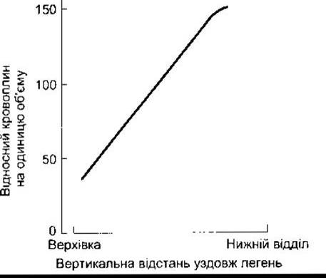
Fig. 34-20. Comparison of blood supply from the apex to the lower PARTS OF THE lung in the upright position. Blood flow values are scaled such that a value of 100 corresponds to steady-state flow.
In the lower regions of the lungs, alveolar pressure is lower than the pressure throughout the pulmonary circulation, and blood flow is determined by the arteriovenous pressure difference.
Ventilation-Perfusion Ratio
The ratio of pulmonary ventilation to pulmonary blood flow for the entire lung at rest is 0.8 (4.2 L/min of ventilation divided by 5.5 L/min of blood flow rate). Obviously, significant regional differences in this ventilation-perfusion ratio across different parts of the normal lung are a consequence of gravity, and local alterations are frequent during disease. If ventilation in an alveolus decreases relative to its perfusion, alveolar PO2 drops because less O2 is delivered, and PCO2 rises because less CO2 is exhaled. Conversely, if perfusion decreases relative to ventilation, PCO2 falls because less CO2 is delivered, and PO2 rises because less O2 enters the blood. These effects are illustrated in Fig. 32-21.
As noted above, both ventilation and perfusion in the upright position decrease linearly from the base to the apex of the lungs. Furthermore, the ventilation-perfusion ratio is high in the upper lung regions. This is evidenced by the predilection of tuberculosis for these areas. The high ventilation-perfusion ratio at the lung apices creates a favorable environment for the growth of tuberculosis Bacteria due to the relatively high alveolar PO2.
Widespread heterogeneity of ventilation and perfusion in the lungs can contribute to CO2 retention and a decrease in systemic arterial PO2. The consequences of such heterogeneity in disease states are described in Chapter 37.
Blood Deposition in the Lungs
Due to their distensibility, the pulmonary veins serve as an important reservoir for blood storage. Normally, when a person lies down, pulmonary blood volume increases by 400 mL, and upon standing, this blood returns to the general circulation. This shift accounts for the decrease in vital capacity in recumbent patients and is responsible for orthopnea in Heart Failure (see Chapter 33).
Regulation of Pulmonary Blood Flow
It remains unclear whether pulmonary veins and arteries are regulated independently, although venous constriction increases pulmonary capillary pressure, and pulmonary arterial constriction increases the workload on the right side of the heart. Small arteries with diameters of several hundred micrometers are the primary site of vascular resistance.
Pulmonary blood flow is influenced by both active and passive factors. The pulmonary vessels possess autonomic innervation, and stimulation of the cervical sympathetic ganglion reduces blood supply by nearly 30%. The vessels also respond to circulating humoral factors. Receptors that mediate and encode these responses are listed in Table 34-5. Many dilator responses are endothelium-dependent and are thought to be mediated through the release of NO (see Chapter 31). In pulmonary Hypertension, endothelial NOS activity is deficient.
Passive factors, such as cardiac output and gravity, also significantly affect pulmonary blood flow. Local matching of perfusion to ventilation is governed by local O2 levels or Hypoxia. During exercise, cardiac output increases, and pulmonary arterial pressure rises proportionally and gradually, though not As a result of vasodilation.
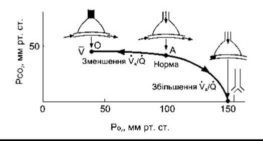
Fig. 34-21. Effect of decreased or increased ventilation-perfusion ratio (VA/Q) on alveolar PCO2 and PO2. Diagrams above the curve represent an alveolus and a pulmonary capillary, with black areas indicating sites of blockage. With complete airway obstruction to the alveolus, PCO2 and PO2 approach mixed venous blood (V) values. With complete perfusion blockade, PCO2 and PO2 approach expired gas values (reproduced with permission from West JB: Ventilation/Blood Flow and Gas Exchange, 3rd ed. Blackwell, 1977).
Table 34-5. Receptors of smooth muscle in pulmonary arteries and veins1
Receptors |
Subtype |
Response |
Endothelium-dependent |
Adrenergic |
а1 |
Contraction |
No |
а2 |
Relaxation |
Yes |
|
ß2 |
Relaxation |
Yes |
|
Muscarinic |
М3 |
Relaxation |
Yes |
Purinergic |
Р2х |
Contraction |
No |
Р2у |
Relaxation |
Yes |
|
Tachykinin |
NK1 |
Relaxation |
Yes |
NK2 |
Contraction |
No |
|
VIP |
? |
Relaxation |
? |
CGRP |
? |
Relaxation |
No |
Humoral |
|||
Adenosine |
А1 |
Contraction |
No |
A2 |
Relaxation |
No |
|
Angiotensin II |
AT1 |
Contraction |
No |
ANP |
АNРA |
Relaxation |
No |
ANPB |
Relaxation |
No |
|
Bradykinin |
В1? |
Relaxation |
Yes |
B2 |
Relaxation |
Yes |
|
Endothelin |
ЕТA |
Contraction |
No |
ЕТВ |
Relaxation |
Yes |
|
Histamine |
H1 |
Relaxation |
Yes |
Н2 |
Relaxation |
No |
|
5-НТ |
5-НТ1 |
Contraction |
No |
5-НТ1C |
Relaxation |
Yes |
|
Thromboxane |
ТР |
Contraction |
No |
Vasopressin |
V1 |
Relaxation |
Yes |
1 Modified and reproduced with permission from Barnes PJ, Lin SF: Regulation of pulmonary vascular tone. Pharmacol Rev 1995;47:88.
In this case, more erythrocytes move through the lungs without any decrease in hemoglobin O2 saturation, and the total amount of O2 entering the systemic circulation evidently increases. Capillaries dilate, and previously unperfused capillaries become recruited to participate in blood supply. The net effect is a marked increase in pulmonary blood flow.
The 133Xe isotope can be used to determine regional pulmonary blood flow by administering a saline solution containing the gas intravenously and monitoring the thoracic region. The gas rapidly enters normally perfused alveoli, whereas it enters poorly perfused ones to a lesser extent. Another technique for identifying regional hypoperfused areas involves the administration of radioiodinated macroaggregated albumin. These aggregates are large enough to block capillaries and small arterioles, and they become deposited only in vessels through which blood is flowing upon reaching the lungs. Although this may seem paradoxical when studying patients with impaired pulmonary blood flow due to vascular obstruction, the technique is safe because relatively few particles are administered, blocking only a small fraction of pulmonary vessels and being rapidly cleared from the body.
Bronchial or bronchiolar obstruction leads to hypoxia in hypoventilated alveoli. In such areas, O2 deficiency undoubtedly exerts a direct effect on vascular smooth muscle, causing constriction and diverting blood away from the hypoxic region. The accumulation of CO2 leads to a local decrease in pH, which consequently promotes vasoconstriction in the lungs, in contrast to the vasodilation observed in other Tissues. Conversely, a reduction in blood flow to a part of the lung decreases alveolar PCO2 in that area, which triggers constriction of the adjacent Bronchi, thereby redirecting ventilation away from the poorly perfused region.
Systemic hypoxia also induces constriction of the pulmonary arterioles and, consequently, an increase in pulmonary arterial pressure.
Pulmonary Hypertension
A documented elevation in pulmonary arterial pressure is observed in neonates (see Chapter 32) and adults. In adults, the Etiology is most often idiopathic. Increased pulmonary arterial pressure may occur in individuals who inhale cocaine and those who use dexfenfluramine and related appetite suppressants that elevate extracellular serotonin levels. Elevated pressure directly leads to The Development of right-sided heart failure and death. Treatment with vasodilators, including prostacyclin (epoprostenol), is indicated. In some cases, the therapeutic efficacy of prostacyclin extends beyond vasodilation to include vascular remodeling. However, its administration is challenging because it requires continuous intravenous infusion.
One of the normal Functions of the lungs is to filter out small blood clots without producing any symptoms. When emboli obstruct a major branch of the pulmonary artery, they provoke an increase in pulmonary arterial pressure and rapid, shallow breathing (tachypnea). The elevation in pulmonary arterial pressure undoubtedly triggers reflexive vasoconstriction mediated by sympathetic nerve fibers; however, an alternative view suggests that reflex vasoconstriction does not occur when major Branches of the pulmonary arteries are obstructed. Tachypnea is a reflex response to the activation of vagally innervated pulmonary receptors that monitor vessel walls (see Table 36-2), mediated by the effects of serotonin released from platelets at the site of the embolus.
Last update: 10/08/2026
Editorial and Educational Adaptation: This material has been compiled based on the primary/original source text. The project team performed an editorial review, corrected technical inaccuracies, structured sections, and adapted the content for an educational format.
What was processed:
- elimination of formatting defects (OCR errors, structural breaks, corrupted characters);
- editorial organization of content;
- standardization of terminology in accordance with academic sources;
- verification of factual statements against the original source text.
All mentions of the author, publication year, and origin of the primary text have been preserved in accordance with the source.