MEDICAL BIOLOGY, HUMAN ANATOMY, PHYSIOLOGY AND PATHOLOGY - Ya.I. Fedonyuk 2010
ANATOMY, PHYSIOLOGY, PATHOLOGY
CHAPTER 5. THE PROCESS OF BLOOD AND LYMPH CIRCULATION. PATHOLOGY THEREOF
VESSELS OF THE SYSTEMIC CIRCULATION
1. THE AORTA
1.2. Branches of the DESCENDING AORTA
As noted above, the descending aorta is divided into the thoracic and abdominal parts, depending on the cavity through which it passes.
The thoracic part of the aorta (pars thoracica aortae) lies to the left of THE Vertebral Column in the posterior Mediastinum (Fig. 5.23). As it traverses the thoracic cavity, its branches supply Blood to the Esophagus, Trachea, Bronchi, Pericardium, Diaphragm, and the walls of the thoracic cavity. Passing through the aortic hiatus in the diaphragm, it enters the Abdominal cavity AND is referred to as the Abdominal Aorta.
Class="center">
Fig. 5.23. The Thoracic Aorta
The abdominal part of the aorta (pars abdominalis aortae) lies on the posterior wall of the abdominal cavity, anterior to the vertebral column (Fig. 5.24). The INFERIOR VENA CAVA is situated to its right. The Branches of the Abdominal Aorta are subdivided into visceral and parietal branches, with the visceral branches further categorized into paired and unpaired.

Fig. 5.24. The abdominal aorta
Unpaired Visceral Branches of the abdominal aorta include:
- the celiac trunk (tr. coeliacus), which is a short trunk originating from the aorta at the level of the 12th thoracic vertebra and dividing into three relatively large branches: the left gastric artery, the common hepatic artery, and the splenic artery, supplying blood to The Stomach, duodenum, Liver, Spleen, and the HEAD of the Pancreas;
- the superior mesenteric artery (a. mesenterica superior), which branches off the aorta at the level of the 1st lumbar vertebra. Its branches supply the Small Intestine, the vermiform Appendix with the cecum, the ascending colon, the right half of the transverse colon, and the pancreas;
- the inferior mesenteric artery (a. mesenterica inferior), which arises from the aorta at the level of the 3rd lumbar vertebra and supplies blood to the left half of the transverse colon, the descending colon, the sigmoid colon, and the upper section of the rectum.
Paired Visceral Branches of the abdominal aorta extend to the paired abdominal Organs:
- middle suprarenal Arteries, supplying the Adrenal Glands;
- renal arteries, supplying the Kidneys;
- testicular (ovarian) arteries, supplying the Gonads.
The Parietal branches of the abdominal aorta are typically paired. They supply blood to the diaphragm and the Muscles of the posterior abdominal wall (via four pairs of lumbar arteries).
The median sacral artery originates from the aorta, descends into the lesser pelvis, and runs along the pelvic surface of the sacrum.
At the level of the 4th lumbar vertebra, the abdominal aorta bifurcates into the right and left common iliac arteries (a. iliaca communis), which subsequently divide into the internal and external iliac arteries.
The internal iliac artery (a. iliaca interna) descends into the lesser pelvis, and its branches are subdivided into parietal and visceral branches (Fig. 5.25).

Fig. 5.25. Internal iliac artery and its branches
The parietal branches supply the walls of the lesser pelvis, the perineal muscles, the Medial Group of thigh muscles, the Pelvic Girdle muscles, and the hip joint. The visceral branches supply the middle and lower PARTS OF THE rectum, the Urinary Bladder, the Ureter, as well as the Uterus and Vagina (in females), and the Prostate Gland, Seminal Vesicles, Ductus deferens, and Penis (in males).
The external iliac artery (a. iliaca externa) (Fig. 5.26) runs along the medial border of the psoas major Muscle and gives off branches to the anterior abdominal wall. After passing onto the thigh beneath the inguinal ligament, it is referred to as the femoral artery.
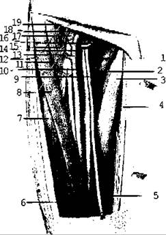
Fig. 5.26. External iliac artery
The femoral artery (a. femoralis) lies lateral to the corresponding vein beneath the inguinal ligament (Fig. 5.27). In the thigh, it runs through the anterior femoral sulcus and then passes through the adductor canal into the popliteal fossa, where it becomes the popliteal artery. In the upper third of the thigh, the femoral artery gives off the deep artery of the thigh, which supplies the Femur, muscles, and Skin of the thigh. In the same region, it gives off branches supplying the external genitalia and the anterior abdominal wall.

Fig. 5.27. Vessels and nerves of the anterior surface of the thigh
The popliteal artery (a. poplitea) gives off branches that form the arterial network of the knee joint together with branches from the femoral and tibial arteries (Fig. 5.28).

Fig. 5.28. Vessels and NERVES OF THE posterior surface of the thigh
At the level of the upper margin of the soleus muscle, the popliteal artery divides into the anterior and posterior tibial arteries.
The anterior tibial artery passes through an opening in the interosseous membrane to the anterior aspect of the leg, supplying the anterior muscle group of the leg. As it continues onto the FOOT, it is called the dorsalis pedis artery (Fig. 5.29).

Fig. 5.29. Nerves of the leg
The posterior tibial artery runs between the superficial and Deep Muscles of the posterior leg compartment and supplies them. It gives rise to the fibular artery, which supplies the Fibula and the Lateral Muscle Group of the leg (Fig. 5.30).
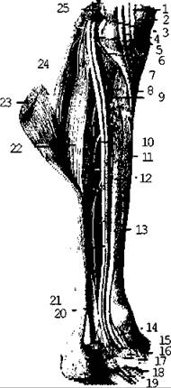
Fig. 5.30. Deep vessels and Muscles of the Leg (posterior view)
Passing behind the lateral malleolus, the posterior tibial artery reaches the plantar surface of the foot, where it divides into the medial and lateral plantar arteries (Fig. 5.31). Together with the dorsalis pedis artery, these supply the bones and soft Tissues of the foot.
In certain areas, arteries lie superficially (such as the dorsalis pedis artery and the radial artery) and can be palpated to determine the pulse or compressed against bones to stop bleeding.

Fig. 5.31. Arteries of the SOLE OF THE foot
Last update: 08/08/2026
Editorial and Educational Adaptation: This material has been compiled based on the primary/original source text. The project team performed an editorial review, corrected technical inaccuracies, structured sections, and adapted the content for an educational format.
What was processed:
- elimination of formatting defects (OCR errors, structural breaks, corrupted characters);
- editorial organization of content;
- standardization of terminology in accordance with academic sources;
- verification of factual statements against the original source text.
All mentions of the author, publication year, and origin of the primary text have been preserved in accordance with the source.