Human Anatomy - Kotsan I. Ya. 2009
Muscles and Fascia of the Trunk
Deep Muscles of the Thoracic Wall
The DEEP Muscles OF the chest are located mainly in the intercostal spaces, act as intrinsic muscles of The thoracic cage, and functionally participate in the act of breathing.
The deep thoracic muscles include the external and internal intercostal muscles, subcostal muscles, transversus thoracis Muscle, and levatores costarum muscles (Figs. 101, 102).
The external intercostal muscles (mm. intercostales externi) fill the intercostal spaces from THE Vertebral Column to the transition of the bony Ribs into their cartilaginous parts. They originate from the lower margin of the rib above, pass obliquely downward and forward, and insert onto the upper margin of the rib below. Function: they elevate the ribs, expand the thoracic cage, and participate in inspiration.
Innervation: intercostal nerves.
Class="center">
Fig. 101. Muscles of the Trunk (anterior view).
On the right: the pectoralis major, external and internal obliques, and rectus abdominis muscles have been removed; on the left: the superficial chest muscles, external and internal oblique Abdominal muscles, and partially the rectus abdominis muscle have been removed.
1 — anterior scalene muscle, 2 — middle scalene muscle, 3 — posterior scalene muscle, 4 — omohyoid muscle (superior belly), 5 — Internal jugular vein, 6 — Subclavian Artery, 7 — subclavian vein, 8 — external intercostal muscles, 9 — internal intercostal muscles, 10 — rectus abdominis muscle, 11 — tendinous intersection, 12 — linea alba, 13 — rectus sheath (posterior lamina), 14 — semilunar line, 15 — rectus sheath (anterior lamina), 16 — transversus abdominis muscle, 17 — inguinal ligament, 18 — Spermatic Cord, 19 — saphenous opening, 20 — pyramidalis muscle, 21 — fascia lata, 22 — adductor longus muscle, 23 — rectus femoris muscle, 24 — pectineus muscle, 25 — iliopsoas muscle, 26 — sartorius muscle, 27 — tensor fasciae latae muscle, 28 — femoral nerve, 29 — femoral vein, 30 — femoral artery, 31 — fascia transversalis, 32 — arcuate line, 33 — aponeurosis of the biceps brachii, 34 — tendon of the biceps brachii, 35 — brachialis muscle, 36 — serratus anterior muscle, 37 — biceps brachii muscle, 38 — latissimus dorsi muscle, 39 — pectoralis major muscle, 40 — coracobrachialis muscle, 41 — deltoid muscle, 42 — pectoralis minor muscle, 43 — subclavius muscle, 44 — trapezius muscle, 45 — omohyoid muscle (inferior belly), 46 — sternocleidomastoid muscle
The internal intercostal muscles (mm. intercostales interni) fill the intercostal spaces from the angles of the ribs to the Sternum. They originate from the upper margin of the rib below, run upward and forward, and insert into the lower margin of the rib above. Function: they depress the ribs, assisting in expiration.
Innervation: intercostal nerves.
The subcostal muscles (mm. subcostales) are located on the inner surface of the lower ribs (X–XII) in the region of their angles. They originate from the inner surface of the lower ribs and, spanning one or two ribs, insert onto the upper ribs. Function: they depress the ribs.
Innervation: intercostal nerves.
The transversus thoracis muscle (m. transversus thoracis) is a flat, thin, fan-shaped muscle located on the inner surface of the anterior thoracic wall. It originates from the posterior surface of the xiphoid process and the lower part of the body of the sternum. The muscle fibers run obliquely upward and laterally, inserting onto the inner surface of the II–VI ribs near the junction of their bony and cartilaginous parts. Function: it depresses the ribs, assisting in expiration.
Innervation: intercostal nerves.
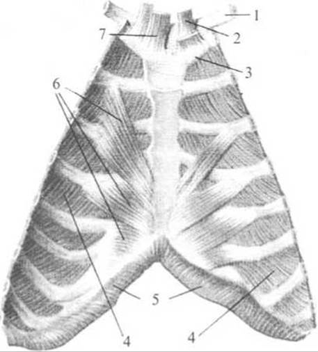
Fig. 102. Muscles of the anterior thoracic wall. Posterior view (from the inside)
1 — clavicle, 2 — sternohyoid muscle, 3 — Cartilage of the first rib, 4 — internal intercostal muscles, 5 — Diaphragm, 6 — transversus thoracis muscle, 7 — sternothyroid muscle
The levatores costarum muscles (mm. levatores costarum) are situated deep to the erector spinae muscle and are present only in the thoracic region of the vertebral column. They are divided into short and long groups. The short levatores costarum muscles (mm. levatores costarum breves) originate from the transverse processes of the seventh cervical and upper eleven thoracic vertebrae, pass obliquely downward and laterally, and insert into the angles of the underlying ribs. The long levatores costarum muscles (mm. levatores costarum longi) are found only in the lower part of the thoracic spine. They originate from the transverse processes of the thoracic vertebrae, run downward and laterally, skip one rib, and insert onto the next rib near its angle. Function: they elevate the ribs.
Innervation: posterior rami of the Spinal Nerves.
Last update: 08/08/2026
Editorial and Educational Adaptation: This material has been compiled based on the primary/original source text. The project team performed an editorial review, corrected technical inaccuracies, structured sections, and adapted the content for an educational format.
What was processed:
- elimination of formatting defects (OCR errors, structural breaks, corrupted characters);
- editorial organization of content;
- standardization of terminology in accordance with academic sources;
- verification of factual statements against the original source text.
All mentions of the author, publication year, and origin of the primary text have been preserved in accordance with the source.