Human Anatomy - Kotsan I. Ya. 2009
Anatomy of the Axial Skeleton
Vertebral Column
The human Skeleton is divided into the axial and Appendicular Skeleton. The Axial Skeleton comprises the trunk skeleton and the Skull, whereas the appendicular skeleton includes the BONES OF THE upper and lower limbs along with their girdles and joints.
The Skeleton of the Trunk consists of THE Vertebral Column and The thoracic cage.
The vertebral column (spine) (columna vertebralis) serves as the body's primary axis and support Structure (Fig. 19). During embryonic development, the spine forms around the Spinal Cord, enclosing it in a protective bony canal. In addition to safeguarding the spinal cord, the vertebral column performs several other vital Functions: it Supports the HEAD, forms part of the walls of the thoracic, abdominal, and pelvic cavities, and facilitates Movements of the trunk and head.
The Development of the key morphological Features of the vertebral column was driven primarily by adaptation to bipedalism and upright posture.
The human vertebral column, much like that of other vertebrates, is segmented in its structure. Its fundamental building blocks are the vertebrae. The Anatomy of the vertebrae clearly illustrates a core principle of the morphophysiology of the Skeletal System: wherever maximum structural strength is required alongside minimal weight and volume, spongy Bone tissue develops. The precise, orderly arrangement of the trabeculae within this tissue—tailored strictly to lines of compression and tension—ensures the structural integrity of individual vertebrae. Furthermore, the overall Stability of the spine relies heavily on its ligamentous apparatus. Despite being remarkably robust, the vertebral column remains exceptionally flexible due to the combination of two TYPES OF JOINTS (diarthroses and synarthroses).
The vertebral column consists of 33–34 vertebrae and is divided into five regions: cervical (7 vertebrae), thoracic (12 vertebrae), lumbar (5 vertebrae), sacral (5 vertebrae), and coccygeal (4–5 vertebrae). The sacral and coccygeal vertebrae fuse together over time to form two distinct bones: the sacrum and the coccyx. While vertebrae from different regions vary in shape and size, they share numerous common features. The thoracic vertebrae are generally considered the most typical in terms of structure.
Each vertebra consists of a body (corpus vertebrae) and a vertebral arch (arcus vertebrae) located posteriorly. The arch attaches to the body via two pedicles (pedunculi arcus vertebrae), which extend into the lamina of the vertebral arch (lamina arcus vertebrae). The space between the vertebral body and the arch is the vertebral foramen (foramen vertebrale). When stacked vertically, the vertebral foramina form the vertebral canal (canalis vertebralis), which houses the spinal cord. Superior and inferior vertebral notches are located on the lateral aspects of the bone where the arch meets the body. The inferior vertebral notch (incisura vertebralis inferior) of an upper vertebra, together with the superior vertebral notch (incisura vertebralis superior) of the vertebra below it, forms the intervertebral foramen (foramen intervertebrale), through which nerves, Blood Vessels, and Lymphatic vessels pass.
Seven processes project from the vertebral arch. A single, unpaired spinous process (processus spinosus) projects posteriorly and serves for the attachment of Ligaments and Muscles. The remaining processes are paired. These include the transverse processes (processus transversi), which extend laterally from the vertebrae, and the superior and lower articular processes (processus articulares superiores et inferiores). The articular processes feature paired superior and inferior articular surfaces (facies articulares). The transverse processes serve as attachment sites for muscles and ligaments, whereas the articular processes articulate with the corresponding processes of adjacent vertebrae above and below.
The vertebrae in different Regions of the spine possess unique Anatomical Features that allow them to be readily distinguished from one another.
Class="center">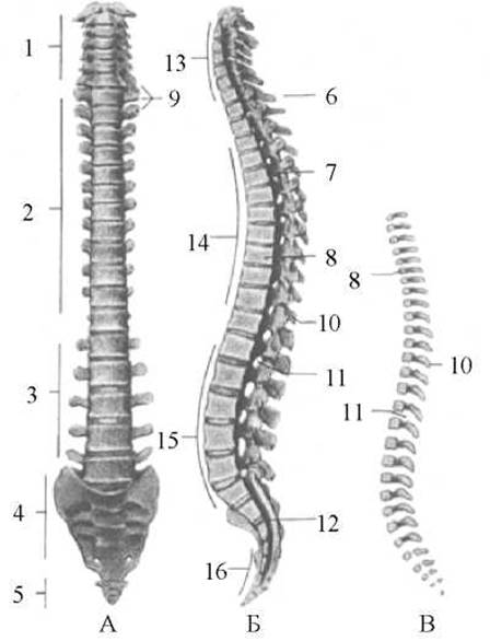
Fig. 19. The vertebral column
A — anterior view, B — lateral view (sagittal section), C — vertebral column of a newborn, 1 — cervical vertebrae, 2 — thoracic vertebrae, 3 — lumbar vertebrae, 4 — sacral vertebrae (sacrum), 5 — coccygeal vertebrae, 6 — vertebra prominens, 7 — vertebral canal, 8 — vertebral bodies, 9 — transverse processes of the vertebrae, 10 — spinous processes of the vertebrae, 11 — intervertebral foramina, 12 — sacral canal, 13 — cervical lordosis, 14 — thoracic Kyphosis, 15 — lumbar lordosis, 16 — sacral kyphosis
Cervical vertebrae (vertebrae cervicales). A characteristic feature of the cervical vertebrae—beginning with the second—is their relatively small, low body, which is roughly rectangular in shape. The size of the vertebral bodies increases progressively down to the seventh (VII) cervical vertebra. The vertebral foramina in this region are relatively large and triangular. The spinous processes are bifid at their tips (except for the VII vertebra). The spinous process of the seventh vertebra is the longest. The articular processes feature rounded, smooth surfaces; on the superior processes, these face backward and upward, while on the inferior processes, they face forward and downward. The transverse processes are short and project laterally. The upper surface of each transverse process is marked by a deep groove for the spinal nerve (sulcus nervi spinalis), which marks the path of the cervical nerve. This groove separates the anterior and posterior tubercles (tuberculum anterius et tuberculum posterius) located at the tip of the transverse process. The transverse processes of the cervical vertebrae are formed from two parts: a posterior and an anterior part. The posterior part constitutes the true transverse process (processus transversus), while the anterior part represents the costal process (processus costalis), which is a rudimentary, underdeveloped rib. Together, these two processes form the costotransverse process (processus costotransversarius) and enclose the transverse foramen (foramen transversum), through which the vertebral artery, Veins, and sympathetic nerve plexus pass.
The first vertebra (the atlas), the second vertebra (the axis), and, to some extent, the sixth and seventh vertebrae depart significantly from the general cervical pattern and exhibit several distinctive characteristics.
The first cervical vertebra, or atlas (atlas), lacks a vertebral body, notches, and a spinous process, consisting instead of a ring formed by an anterior and a posterior arch (Fig. 20A). The anterior arch of the atlas (arcus anterior atlantis) replaces the vertebral body and features the anterior tubercle (tuberculum anterius) on its anterior surface, and the facet for the dens (fovea dentis) on its posterior surface, which articulates with the dens of the axis. The posterior arch of the atlas (arcus posterior atlantis) bears the posterior tubercle (tuberculum posterius) on its outer surface, representing a remnant of the spinous process. On the superior surface of the posterior arch, on either side, lies the groove for the vertebral artery (sulcus arteriae vertebralis). The anterior and posterior arches of the atlas are joined by lateral masses (massae laterales). Short, thick transverse processes (processus transversi) project from these lateral masses, which also bear the superior and inferior articular surfaces (facies articularis superior et facies articularis inferior).
The second cervical vertebra, known as the axis (axis), is named for its primary mechanical function: it acts as a pivot around which the head and the atlas rotate (Fig. 20B). Andreas Vesalius called it the epistropheus, meaning "rotator" (derived from *epistropho* — to turn, hence turning around an axis). Projecting upward from the irregularly shaped body of the axis (corpus axis) is a small, stout process called the dens (odontoid process). Phylogenetically, this process represents the body of the first cervical vertebra that fused with the body of the second during evolution. The dens features an apex, a small anterior articular surface (facies articularis anterior) for articulation with the facet of the atlas, and a posterior articular surface (facies articularis posterior) for articulation with the transverse ligament of the atlas. Flanking the dens on the superior surface of the axis—in place of superior articular processes—are two smooth articular surfaces that articulate with the inferior articular facets of the atlas. The inferior articular processes of the axis are typical in structure, resembling those of other vertebrae. The spinous process of the axis is short, massive, and bifid at the end.

Fig. 20. Cervical vertebrae
A — first cervical vertebra—atlas (superior view): 1 — anterior arch of the atlas, 2 — lateral mass, 3 — costal process, 4 — transverse foramen, 5 — transverse process, 6 — groove for the vertebral artery, 7 — posterior arch, 8 — posterior tubercle, 9 — superior articular fovea (surface), 10 — anterior tubercle, B — second cervical vertebra—axis (superior and posterior view): 1 — superior articular surface, 2 — transverse process, 3 — transverse foramen, 4 — spinous process, 5 — dens of the axis
The sixth cervical vertebra features a somewhat larger anterior tubercle on its transverse process compared to the other cervical vertebrae, known as the carotid tubercle (tuberculum caroticum). This is of significant clinical importance: because the common carotid artery runs directly in front of the carotid tubercle, digital pressure can be applied against this tubercle in the event of Hemorrhage from the artery or its branches to rapidly stem the bleeding.
The seventh cervical vertebra has a long, unbranched spinous process that protrudes prominently beneath the Skin. Consequently, this bone is called the vertebra prominens. The spinous process of the vertebra prominens is easily palpable through the skin, making it a reliable anatomical landmark.
Thoracic vertebrae (vertebrae thoracicae) articulate with the Ribs and therefore feature costal facets (foveae costales) on the lateral surfaces of their bodies near the junctions with the vertebral arches (Fig. 21). Because most ribs articulate with the bodies of two adjacent vertebrae, they typically bear two demifacets (half-facets): one on the superior margin of the vertebral body and another on the inferior margin. Two such demifacets from adjacent vertebrae Complement each other to form a single complete socket that receives the head of the rib. Notable exceptions include the first thoracic vertebra (which features a full facet for the first rib and a demifacet for the second) as well as the eleventh and twelfth thoracic vertebrae (which each feature a single complete facet). The transverse processes of the thoracic vertebrae are robust, thickened at their extremities, and bear a costal facet of the transverse process (fovea costalis processus transversi) on their anterior surfaces for articulation with the tubercle of the rib. The superior and inferior articular processes are flat and oriented nearly in the frontal plane, with their articular facets facing anteriorly and posteriorly, respectively. The spinous processes of the thoracic vertebrae are directed obliquely downward and overlap one another like roof tiles, particularly in the mid-thoracic region. To accommodate the progressively heavier mechanical loads placed upon them, the bodies of the thoracic vertebrae are larger than those of the cervical vertebrae and increase in size from top to bottom. The vertebral foramina of the thoracic region are rounded in shape and smaller than those in the cervical and lumbar regions.
Lumbar vertebrae (vertebrae lumbales) are distinguished by large, oval-shaped bodies and the complete absence of costal facets (Fig. 22). The articular processes of the lumbar vertebrae are massive and oriented almost vertically in the sagittal plane, with the superior facets facing medially and concave, and the inferior facets facing laterally and convex. Projecting upward and dorsally from the base of each superior articular process is a small eminence known as the mammillary process (processus mamillaris), which serves as a Muscle attachment site. The transverse processes of the lumbar vertebrae are thin and flat. At the base of each transverse process lies an accessory process (processus accessorius), which most anatomists consider to be a rudimentary rib fused to the true transverse process (*accessorius* meaning additional or appended). The spinous processes of the lumbar vertebrae are large, long, broad, plate-like, thickened at their tips, and oriented almost horizontally.

Fig. 21. Thoracic vertebra
A — lateral view: 1 — vertebral body, 2 — superior costal facet, 3 — superior vertebral notch, 4 — superior articular process, 5 — transverse process, 6 — spinous process, 7 — inferior articular process, 8 — inferior vertebral notch, 9 — inferior costal facet, B — superior view: 1 — vertebral arch, 2 — transverse process, 3 — vertebral foramen, 4 — superior articular process, 5 — costal facet of the transverse process, 6 — spinous process
The sacral vertebrae (vertebrae sacrales) are separate in children. At the age of 17–25, they fuse to form a single bone — the sacrum (os sacrum) (Fig. 23). This fusion results from the adaptation of the sacrum to the heavy load placed upon it in bipedal humans due to an upright posture (in quadrupedal animals, the sacral vertebrae do not fuse). The sacrum is triangular in shape, with its base (basis ossis sacri) directed superiorly and its apex (apex ossis sacri) directed inferiorly and curved slightly forward. The Base of the sacrum articulates with the body of the last lumbar vertebra at an angle, forming the promontory (promontorium). Extending upward from the posterior part of the base are two small, flat superior articular processes (processus articulares superiores), which, together with the inferior articular processes of the last lumbar vertebra, form the lumbosacral articulation. The anterior or pelvic surface (facies pelvica) of the sacrum is concave, while the posterior or dorsal surface (facies dorsalis) is rough. Five crests, formed by the fusion of processes, are distinguishable on it. The fusion of the spinous processes forms the unpaired median sacral crest (crista sacralis mediana); the fusion of the transverse processes forms the lateral sacral crests (cristae sacrales laterales); and the fusion of the articular processes forms the intermediate sacral crests (cristae sacrales intermediae). On the lateral PARTS OF THE sacrum (partes laterales), formed by the merger of transverse processes and rudimentary sacral ribs, there are auricular surfaces (facies auriculares) for articulation with the iliac bones. Posterior to the auricular surfaces lie the sacral tuberosities (tuberositates sacrales), to which ligaments attach. The anterior (pelvic) and posterior (dorsal) surfaces of the sacrum feature the anterior and posterior sacral foramina (foramina sacralia anteriora et foramina sacralia posteriora), through which Spinal Nerves and blood vessels pass. Between these foramina on the pelvic surface of the sacrum are transverse lines (lineae transversae), which represent remnants of the sacral intervertebral discs. Inside the sacrum runs the sacral canal (canalis sacralis), which is part of the general vertebral canal. The sacral canal contains the filum terminale of the spinal cord and the roots of the lumbar and sacral spinal nerves. The inferior opening of the sacral canal is called the sacral hiatus (hiatus sacralis). On either side of the sacral hiatus are the sacral horns (cornua sacralia), which represent rudiments of the articular processes of the last sacral vertebra. The apex of the sacrum bears a small, oval-shaped articular surface for articulation with the coccyx. The sacrum exhibits well-defined Sexual Dimorphism: in males, it is longer, narrower, and more curved than in females.

Fig. 22. Lumbar vertebra (lateral and superior views)
1 — vertebral body, 2 — transverse process, 3 — superior articular process, 4 — spinous process, 5 — mammillary process, 6 — inferior articular process, 7 — accessory process
The coccygeal vertebrae (vertebrae coccygeae) fuse between the ages of 12 and 25 to form a single coccygeal bone (os coccygis), or coccyx (coccyx) (Fig. 23). The fusion process proceeds in a caudo-cranial direction and occurs earlier in males than in females. Comparative anatomical studies demonstrate that human coccygeal vertebrae are remnants of the caudal spine in animals. They consist almost entirely of vertebral bodies and therefore lack a vertebral canal. The first coccygeal vertebra is larger than the others and bears an articular facet on its superior surface for articulation with the sacrum. Remnants of articular processes can still be distinguished only on the first coccygeal vertebra, transformed into the coccygeal cornua (cornua coccygea), which project superiorly toward the cornua of the sacrum.

Fig. 23. Sacrum and coccyx
A — anterior view: 1 — anterior (pelvic) sacral foramina, 2 — lateral part, 3 — superior articular process, 4 — base of the sacrum, 5 — transverse lines, 6 — apex of the sacrum, 7 — coccygeal bone, B — posterior view: 1 — posterior sacral foramina, 2 — auricular surface, 3 — intermediate sacral crest, 4 — sacral hiatus, 5 — coccygeal cornu, 6 — sacral cornu, 7 — median sacral crest, 8 — posterior surface, 9 — lateral sacral crest, 10 — sacral tuberosity
Last update: 08/08/2026
Editorial and Educational Adaptation: This material has been compiled based on the primary/original source text. The project team performed an editorial review, corrected technical inaccuracies, structured sections, and adapted the content for an educational format.
What was processed:
- elimination of formatting defects (OCR errors, structural breaks, corrupted characters);
- editorial organization of content;
- standardization of terminology in accordance with academic sources;
- verification of factual statements against the original source text.
All mentions of the author, publication year, and origin of the primary text have been preserved in accordance with the source.