Review of Medical Physiology - William F. Ganong 2002
Blood Circulation
Hemodynamics and Lymph Dynamics
Anatomical Features
Arteries and arterioles
The characteristics of various types of Blood Vessels are summarized in Table 30–1. The wall of all arteries consists of three distinct layers: an outer Connective Tissue coat (adventitia); a middle smooth Muscle layer (media); and an inner layer containing endothelium and connective tissue (intima) (Fig. 30–2). The walls of the aorta and other large arteries contain a relatively high proportion of elastic fibers, which stretch during systole and recoil during diastole. The walls of arterioles contain fewer elastic fibers and significantly more smooth muscle. These muscle Cells are innervated by noradrenergic nerve fibers, which act as vasoconstrictors, and to some extent by cholinergic fibers, which mediate vasodilation. Arterioles are the primary resistance Vessels of the Circulation, and even minor changes in their caliber can induce marked alterations in total peripheral resistance.
Class="center">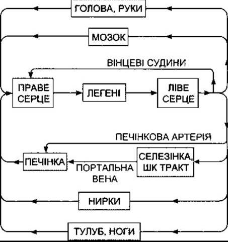
Fig. 30–1. Diagram of the adult human Circulatory system; GI, gastrointestinal.
Capillaries
Arterioles branch into smaller vessels with muscular walls, sometimes referred to as metarterioles, which in turn lead into capillaries (Fig. 30–3). In certain vascular beds that have been studied in detail, metarterioles connect directly with venules through thoroughfare channels, with true capillaries forming an anastomosing network of side branches off this main channel. The entrance of true capillaries is encircled on the arterial side by small bands of smooth muscle known as precapillary sphincters. While it remains unclear whether metarterioles receive innervation, precapillary sphincters definitely do not. Nevertheless, they readily respond to local or circulating vasoconstrictor substances. The diameter of true capillaries is approximately 5 µm at the arterial end and 9 µm at the venous end. When the sphincters are open, the capillary diameter is just sufficient to permit the passage of single erythrocytes. As they traverse the capillaries, red Blood Cells become parachute-shaped because the blood flow forces their central portion forward. This shape change is driven solely by the hydrodynamic pressure within the center of the vessel, regardless of whether the edges of The Cell make contact with the Capillary Wall.
Table 30–1. Characteristics of various human blood vessel types
Vessel |
Lumen diameter |
Wall thickness |
All vessels of each type combined |
|
Approximate total cross-sectional area, cm2 |
Blood volume1, % |
|||
Aorta |
2.5 cm |
2 mm |
4.5 |
2 |
Arteries |
0.4 cm |
1 mm |
20 |
8 |
Arterioles |
30 µm |
20 µm |
400 |
1 |
Capillaries |
5 µm |
1 µm |
4500 |
5 |
Venule |
20 µm |
2 µm |
4000 |
54 |
Vein |
0.5 cm |
0.5 mm |
40 |
|
Vena cava |
3 cm |
1.5 mm |
18 |
|
1 In the systemic circulation. An additional 12% is contained within The Heart and 18% in the Pulmonary Circulation.
The total surface area of the body's capillary walls in an adult exceeds 6,300 m2. These walls, measuring 1 µm in thickness, are composed of a single layer of endothelial cells. Capillary wall Structure varies significantly among different Organs. In many vascular beds, including Skeletal Muscle, cardiac muscle, and smooth muscle, Intercellular junctions between endothelial cells (Fig. 30–4) permit the passage of molecules up to 10 nm in diameter. Plasma and dissolved Proteins appear to be taken up by endocytosis, transported across the endothelial cells, and released by exocytosis (Vesicular Transport; see Chapter 1). However, this mechanism accounts for only a minor fraction of transendothelial transport. In the Brain, capillaries resemble those in skeletal muscle, but the intercellular junctions are tighter, restricting transport primarily to small molecules. In most Endocrine glands, the intestinal microvilli, and PARTS OF THE Kidney, the Cytoplasm of the endothelial cells thinned out to form perforations known as fenestrations. These fenestrations measure 2–100 nm in diameter, allowing relatively large molecules to cross the wall and rendering the capillaries highly permeable (porous). In all structures except the renal glomeruli, the fenestrations are closed by a delicate membrane. In certain Tissues, however, this membrane appears discontinuous, as demonstrated by the freeze-fracture technique. In such cases, the membrane features a central density connected to the margins of the fenestration by spokes (Fig. 30–5).
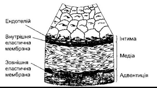
Fig. 30–2. Structure of a muscular artery (reproduced with permission from Ross R, Glomset JA: The Pathogenesis of atherosclerosis. N Engl J Med 1976;295:369).
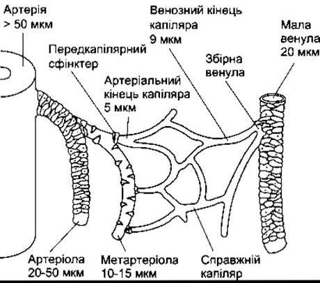
Fig. 30–3. The microvasculature. Arterioles give rise to metarterioles, which in turn branch into capillaries. Capillaries drain blood through short collecting venules into venules. The walls of arteries, arterioles, and small venules contain a relatively large amount of smooth muscle. Isolated smooth muscle cells are found in the walls of metarterioles, and a muscular precapillary sphincter is located at THE ORIGIN OF each capillary. Diameters of the various vessels are also indicated (courtesy of JN Diana).

Fig. 30–4. Cross-section of a capillary. Left: Continuous capillary found in skeletal muscle. Right: Fenestrated capillary (reproduced with permission from Fawcett DW: Bloom and Fawcett, Textbook of Histology, 11th ed. Saunders, 1986).
In the Liver, where sinusoidal capillaries are highly permeable, the endothelium is discontinuous, featuring large intercellular gaps that lack membranes (see Fig. 26–20). Some of these gaps reach 600 nm in diameter, while others may exceed 3,000 nm. Capillary permeability in various parts of the body, expressed in terms of hydraulic conductivity, is listed in Table 30–2.
Pericytes are located on the abluminal surface of capillaries and postcapillary venules, external to the endothelial cells (see Fig. 30–4). These cells possess long processes that wrap around the vessel wall. They are capable of contraction, secrete a variety of vasoactive compounds, and synthesize basement membrane and Extracellular matrix components. One of their
physiological Functions is to regulate fluid flux through interendothelial junctions, particularly during inflammatory responses. They are closely related to the mesangial cells found in the renal glomerulus (see Chapter 38).
Lymphatic drainage from the Lungs and other Regions of the body occurs through a system of converging vessels that empty into the right and left subclavian Veins at their junction with the respective internal jugular veins. Lymphatic vessels contain Valves and routinely pass through Lymph Nodes along their course. The ultrastructure of initial lymphatic vessels differs from that of blood capillaries in several key features: the lymphatic endothelium lacks fenestrations; the basement membrane, if present at all, is extremely thin; and the intercellular junctions between endothelial cells are open, lacking tight intercellular contacts.
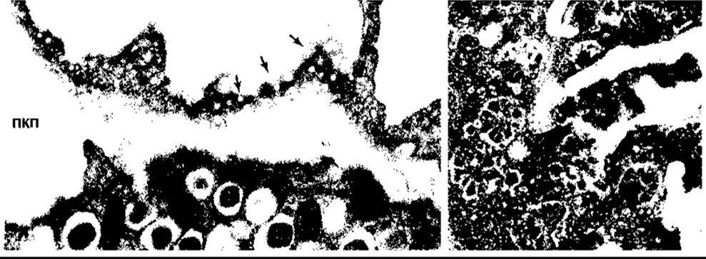
Fig. 30–5. Fenestrations in pancreatic islet capillaries. Left: Arrows indicate fenestrations in a tangential section of the endothelium (×15,000); ECS, extracellular space. Right: Fenestrations visible in capillary endothelium prepared by the freeze-fracture technique (×64,000). Note the centrally located material and the radial spokes attaching it to the margin of each fenestration (reproduced with permission from Orci L: The Insulin cell: Its cellular environment and how it processes (pro)insulin. Diabetes Metab Rev 1986;2:71).
Table 30–2. Hydraulic conductivity of capillaries in various body regions1
Organ |
Conductivity2 |
Endothelial type |
Brain (excluding circumventricular organs) |
3 |
Continuous |
100 |
||
Skeletal muscle |
250 |
|
Lungs |
340 |
|
Heart |
860 |
|
Gastrointestinal tract (intestinal mucosa) |
13,000 |
Fenestrated |
Renal glomeruli |
15,000 |
1 Data provided with the permission of JN Diana.
2 The unit of conductance is 10-13 cm3s-1dyn-1.
Arteriovenous Anastomoses
The fingers, palms, earlobes of humans, as well as the paws, ears, and other tissues of certain animals, contain short channels that connect arterioles to venules, bypassing the capillaries. Such arteriovenous (A-V) anastomoses, also known as shunts, possess a thick muscular wall and are densely innervated, predominantly by vasoconstrictor nerve fibers.
Venules and Veins
The walls of venules are only slightly thicker than those of capillaries. Vein walls are also thin and easily distensible. They contain a minimal amount of smooth muscle; pronounced vasoconstriction is driven by The activity of noradrenergic nerves supplying these veins and by circulating vasoconstrictor substances in the blood, such as endothelin. Anyone who has ever had blood drawn has likely observed the marked local venospasm that occurs in the superficial forearm veins upon injury. Variations in venous tone play a crucial role in vascular regulation.
At regular intervals, the intima of the limb veins folds to form venous valves that prevent retrograde blood flow. William Harvey was the first to demonstrate how these valves function (Fig. 30-6). Such valves are absent in very small veins, large veins, and the VEINS OF THE brain and Internal Organs.
Endothelium
Endothelial cells are situated between the circulating blood and the other two layers of the vessel wall (the media and adventitia), forming a large and vital "organ." They respond to changes in blood flow, vessel wall stretch, and circulating substances, particularly inflammatory Transmitters. Endothelial cells secrete growth regulators and vasoactive compounds (see below and Chapter 31).
Angiogenesis
During tissue growth, blood vessels must proliferate to maintain an adequate blood supply. Therefore, angiogenesis is essential both for fetal life and development, as well as in the adult Organism. In adults, this process plays a vital role in wound healing, The formation of the corpus luteum following ovulation, and the regeneration of the endometrium after menstruation. Pathologically, it is involved in tumor growth; without an adequate blood supply, a tumor cannot expand.
During embryonic development, some blood vessels originate from angioblasts that form tubes, which subsequently fuse. This process is sometimes referred to as vasculogenesis. Other vessels develop via sprouting from preexisting embryonic vessels, a process known as angiogenesis. This is the predominant mechanism of new blood vessel formation in adults.
Numerous growth factors and related compounds stimulate angiogenesis, whereas other substances inhibit it (Table 30-3). Tyrosine Kinases act as receptors for many of these growth factors. Vascular endothelial growth factor (VEGF) is widely considered the most critical stimulatory factor. The Gene encoding VEGF gives rise to three spliced isoforms: VEGF120, VEGF164, and VEGF188. Mice expressing only VEGF120 develop ischemic cardiomyopathy and impaired myocardial angiogenesis. At least three distinct types of VEGF receptors have been identified. Targeted knockout of the gene encoding any of these receptors leads to severe cardiovascular and lymphatic defects. Nonetheless, further research is required to fully elucidate the factors involved in angiogenesis and the mechanisms governing new vessel formation.
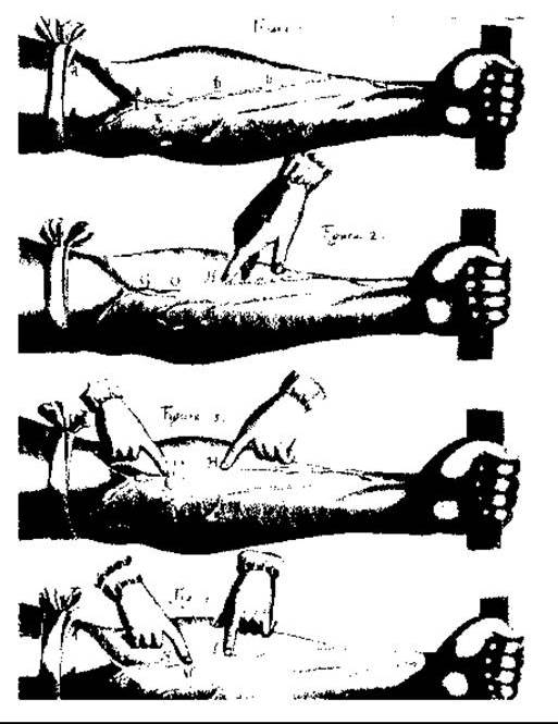
Fig. 30-6. Demonstration of venous valve function in the human forearm, from William Harvey's *Exercitatio Anatomica de Motu Cordis et Sanguinis in Animalibus*, published in 1628; A — a tourniquet applied above the elbow stops venous outflow. If the vein is emptied by stroking from G to H (Fig. 2), it refills from above only as far as the valve O. If it is emptied from K to O with another finger (Fig. 3), it fills above O, while remaining empty from O to H. If the vein is compressed at L (Fig. 4) and stroked with another finger (M) from L to the valve N, it remains empty between L and N. Upon releasing the pressure at L, the vein fills from below.
Last update: 10/08/2026
Editorial and Educational Adaptation: This material has been compiled based on the primary/original source text. The project team performed an editorial review, corrected technical inaccuracies, structured sections, and adapted the content for an educational format.
What was processed:
- elimination of formatting defects (OCR errors, structural breaks, corrupted characters);
- editorial organization of content;
- standardization of terminology in accordance with academic sources;
- verification of factual statements against the original source text.
All mentions of the author, publication year, and origin of the primary text have been preserved in accordance with the source.