Human Anatomy and Physiology (with Age-Related Features of the Child's Body) - Sapin M.R., Sivoglazov V.I. 2002
Splanchnology (The Study of the Viscera)
Angiology (The Study of the Vessels)
Veins of the Systemic Circulation
Blood drains from all Organs and Tissues of the human body into two large vessels—the superior and inferior venae cavae, which empty into the right atrium. A distinction is made between deep Veins, which usually accompany Arteries, and superficial veins (Fig. 84, see color insert).
SUPERIOR VENA CAVA and its tributaries
The superior vena cava, 5—6 cm in length and 2—2,5 cm in diameter, has no Valves. It is located in the thoracic cavity, in the anterior (superior) Mediastinum. The superior vena cava is formed by the union of the right and left brachiocephalic veins behind the junction of the first right costal Cartilage with the Sternum. The vein then descends to the right and posterior to the ascending aorta and empties into the right atrium. The superior vena cava collects blood from the walls and Organs of the thoracic cavity, HEAD, neck, and upper limbs.
Blood drains from the head via the external and internal jugular veins.
The External Jugular Vein collects blood from the occipital and posterior auricular regions. It also anastomoses with tributaries of the internal jugular vein (the retromandibular vein). The external jugular vein descends in the subcutaneous tissue on the superficial surface of the sternocleidomastoid Muscle. Along its course, it receives lateral tributaries—the anterior jugular vein, which forms in the anterior neck, the transverse cervical veins (from the lateral neck), and the suprascapular vein. The external jugular vein empties into the terminal portion of the subclavian or internal jugular vein.
The internal jugular vein exits the cranial cavity through the jugular foramen, continuing from the sigmoid sinus of the dura mater. In the neck, this vein lies adjacent to the common carotid artery and the Vagus nerve. The internal jugular vein drains blood from the Brain, whose superficial and deep veins empty into the adjacent sinuses of the dura mater.
The diploic veins (from the BONES OF THE cranial vault), emissary veins (anastomoses with the veins of the scalp), and the superior and inferior ophthalmic veins empty into the internal jugular vein or its tributaries, including the Dural Venous Sinuses (see below). Its so-called extracranial tributaries empty directly into the internal jugular vein; these include the pharyngeal, lingual, superior and middle thyroid, and facial veins, as well as the large retromandibular vein, which collects blood from the auricle, external acoustic meatus, temporomandibular joint, and the walls of the tympanic cavity. The internal jugular vein unites with the subclavian vein to form the brachiocephalic vein.
The subclavian vein, which is the continuation of the axillary vein, passes anterior to the anterior scalene muscle from the first rib to its junction with the internal jugular vein.
Veins of the upper limb
In the upper limb, deep and superficial veins are distinguished; they possess valves and anastomose abundantly with each other. The deep veins, which collect blood from bones, joints, and Muscles, usually run adjacent to the corresponding arteries in pairs, which is why they are called venae comitantes. In the arm, both deep brachial veins merge and empty into the single axillary vein. The superficial veins of the upper limb form a network on the hand, from which the two main subcutaneous veins of the arm—the lateral and medial—arise.
The medial subcutaneous vein of the arm, located along the ulnar side of the forearm, continues in the medial groove of the arm and empties into the brachial vein.
The lateral subcutaneous vein of the arm passes along the lateral border of the anterior surface of the forearm, then along the lateral surface of the arm, and empties into the axillary vein.
Both subcutaneous veins—both medial and lateral—have numerous lateral tributaries and anastomose with each other. In the cubital fossa, both veins are connected by a short intermediate (median) cubital vein.
The axillary vein, lying adjacent to the axillary artery, becomes the subclavian vein at the level of the first rib. The axillary vein drains blood not only from the Free part of the upper limb; it also receives some veins of the shoulder girdle and thoracic wall—the lateral thoracic, thoracoepigastric, and other veins.
Veins of the Thorax
Venous drainage from the thoracic walls and organs of the thoracic cavity occurs via the azygos and hemiazygos veins, as well as via visceral veins that empty into the brachiocephalic veins and the superior vena cava.
The right and left brachiocephalic veins, which merge to form the superior vena cava, collect blood from the head, neck, and upper limbs. The brachiocephalic veins have no valves; the left brachiocephalic vein is longer (5— 6 cm) than the right (about 3 cm). Tributaries of the brachiocephalic veins include the inferior thyroid, thymic, pericardial, bronchial, esophageal, mediastinal, vertebral, and other veins.
Blood drains from the walls of the thoracic cavity into the azygos vein and its tributary, the hemiazygos vein.
The azygos vein is located in the posterior mediastinum on the bodies of the thoracic vertebrae to the right of the midline. The azygos vein begins between the muscle bundles of the lumbar part of the Diaphragm as a continuation of the right ascending lumbar vein. At the level of the IV—V thoracic vertebrae, the azygos vein arches posteriorly over the ROOT of the right lung, courses anteriorly, and empties into the superior vena cava. Along its path to the superior vena cava, the azygos vein receives the right posterior intercostal, esophageal, bronchial, pericardial, posterior mediastinal, superior phrenic, and other veins, as well as the veins of the internal and external vertebral plexuses and the hemiazygos vein.
The hemiazygos vein, which is the continuation of the left ascending lumbar vein, lies adjacent to the left side of the spine. At the level of the VII thoracic vertebra, the hemiazygos vein turns sharply to the right, crosses THE Vertebral Column anteriorly, and empties into the azygos vein. Tributaries of the hemiazygos vein are the posterior intercostal veins of the left side, esophageal and posterior mediastinal veins, as well as the veins of the vertebral plexuses, which drain blood not only from the spine but also from the Spinal Cord and its Meninges.
Blood drains from the anterior wall of the thoracic cavity via the internal thoracic veins, which possess valves and run adjacent to the corresponding arteries (along the margins of the sternum). Each internal thoracic vein is a continuation of the superior epigastric vein, which collects blood from the upper PARTS OF THE anterior abdominal wall. Tributaries of the internal thoracic vein include the musculophrenic veins (from the diaphragm), as well as the anterior intercostal veins, which anastomose in the intercostal spaces with the posterior intercostal veins—tributaries of the azygos and hemiazygos veins. The left internal thoracic vein empties into the left brachiocephalic vein, and the right empties into the right brachiocephalic vein or directly into the superior vena cava.
INFERIOR VENA CAVA and its tributaries
The inferior vena cava is the largest vein in The Human Body; its diameter at the site of entry into the right atrium reaches 3—3,5 cm, and it has no valves. The inferior vena cava is formed by the union of the right and left common iliac veins at the level of the intervertebral disc between the IV and V lumbar vertebrae, slightly below the bifurcation (division) of the Abdominal Aorta into the common iliac arteries. The inferior vena cava lies retroperitoneally to the right of the aorta and passes through the caval opening of the diaphragm into the thoracic cavity. The vein then enters the pericardial cavity, where it immediately empties into the right atrium at the level of the intervertebral disc connecting the bodies of the VIII and IX thoracic vertebrae. The inferior vena cava collects blood from the veins of the lower limbs, walls, and Internal Organs of the pelvis and abdomen.
Veins of the abdomen
The tributaries of the inferior vena cava in the Abdominal cavity mostly correspond to the paired Branches of the Abdominal Aorta. These tributaries are divided into parietal veins (lumbar and inferior phrenic) and visceral veins (hepatic, renal, right suprarenal, testicular in men, and ovarian in women; the left veins of these organs drain into the left renal vein).
The lumbar veins (4–5 pairs) drain blood from the soft tissues of the abdominal walls; their tributaries correspond to the branches of the lumbar arteries. Near the vertebral column, the lumbar veins are interconnected by the vertically running right and left ascending lumbar veins, which continue superiorly as the azygos vein (on the right) and the hemiazygos vein (on the left).
The inferior phrenic veins (right and left) and their tributaries run in pairs alongside the corresponding arteries and their branches. The inferior phrenic veins drain into the inferior vena cava near the posterior border of the Liver.
The hepatic veins (2–3) drain into the inferior vena cava where it lies adjacent to the posterior border of the liver.
The renal veins (right and left) run horizontally from the renal hilum and drain into the inferior vena cava at the level of the intervertebral disc between the bodies of the I and II lumbar vertebrae. The left renal vein passes anterior to the aorta.
The suprarenal vein emerges from the hilum of the Adrenal gland. The right drains into the inferior vena cava, while the left drains into the left renal vein.
The testicular (ovarian) vein enters the abdominal cavity from the corresponding gonad. The right vein drains into the inferior vena cava just below the renal veins, while the left drains into the left renal vein.
Venous blood from the unpaired abdominal viscera (Stomach, Spleen, small and large intestines, Pancreas) drains into the HEPATIC PORTAL VEIN. After passing through the hepatic vasculature, the venous blood from the portal vein flows via the hepatic veins into the inferior vena cava.
The portal vein and its tributaries
The portal vein is formed behind the head of the pancreas by the union of three veins: the splenic, superior mesenteric, and inferior mesenteric veins (Fig. 85). The splenic vein emerges from the splenic hilum and runs alongside the splenic artery. Small veins from the STOMACH AND PANCREAS also drain into this vein. The superior mesenteric vein drains blood from the veins of the mesenteric part of the Small Intestine, the cecum, the ascending colon, and the right half of the transverse colon. The right gastro-omental, pancreaticoduodenal, and other veins also drain into the superior mesenteric vein. The inferior mesenteric vein drains blood from the left half of the transverse colon, the descending and sigmoid colons, and the upper part of the rectum.
Class="center">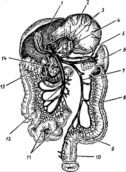
Fig. 85. The portal vein and its tributaries:
1 — liver, 2 — stomach, 3 — gastric veins, 4 — portal vein, 5 — splenic vein, 6 — spleen, 7 — inferior mesenteric vein, 8 — descending colon, 9 — sigmoid colon, 10 — rectum, 11 — loops of the small intestine, 12 — superior mesenteric vein, 13 — transverse colon, 14 — head of the pancreas
After its formation, the portal vein travels within the hepatoduodenal ligament posterior to the hepatic artery proper and the common Bile duct. At the porta hepatis, the portal vein first divides into right and left lobar branches, which then branch into segmental veins, and subsequently into smaller veins and sinusoids (capillaries) that drain into the central veins at the center of the hepatic lobules. The central veins drain into the sublobular veins. These then coalesce into larger veins, eventually forming two to three hepatic veins that empty into the inferior vena cava where it lies adjacent to the posterior surface of the liver.
It should be noted that the tributaries of the portal vein anastomose with each other, as well as with the tributaries of the superior and inferior venae cavae (at the gastroesophageal junction, in the walls of the rectum, and in the periumbilical region of the anterior abdominal wall).
Veins of the pelvis
The pelvic cavity contains tributaries of the inferior vena cava: the right and left common iliac veins, as well as the internal and external iliac veins of the right and left sides, which drain into each common iliac vein.
The common iliac vein is a paired vessel that runs along the medial border of the psoas major muscle on each side of the pelvis. It is formed at the level of the sacroiliac joint by the union of the external and internal iliac veins.
The internal iliac vein has no valves and is one of the tributaries of the common iliac vein. It lies on the lateral wall of the pelvis posterior to the corresponding artery and drains blood from the pelvic walls and pelvic organs. Consequently, the tributaries of the internal iliac vein are divided into two groups: parietal and visceral.
The parietal tributaries of the internal iliac vein are the paired superior and inferior gluteal, obturator, and lateral sacral veins, and the unpaired iliolumbar vein. These veins contain valves and are formed within the branching areas of the corresponding arteries.
The visceral tributaries of the internal iliac vein include the vesical, uterine, middle rectal (from the middle parts of the rectum), and internal pudendal veins. The inferior rectal veins, which drain the lower parts of the rectum, are tributaries of the internal pudendal vein. Veins from the external genitalia—namely, the veins of the Penis (Clitoris) and Scrotum (labia)—also drain into the internal iliac vein.
These visceral veins drain blood from the corresponding organs and the surrounding venous plexuses. Due to the extensive anastomoses among the veins forming these plexuses, they ensure unobstructed venous outflow during the regular filling and emptying of these organs.
Within the pelvic cavity, surrounding the internal organs and in their walls, lie the rectal, vesical, prostatic, uterine, and vaginal venous plexuses.
The external iliac vein is the second major tributary of the common iliac vein and the direct continuation of the femoral vein, which receives blood from all the veins of the lower limb. The external iliac vein has no valves and lies medial to the psoas major muscle. Just superior to the inguinal ligament, the inferior epigastric vein drains into the external iliac vein, carrying blood from the organs and tissues of the lower anterior abdominal wall and anastomosing with the superior epigastric vein in the periumbilical region. Another tributary of the external iliac vein is the deep circumflex iliac vein, whose tributaries correspond to the branches of the corresponding artery.
Veins of the lower limb
Blood is drained from the organs and tissues of the lower limb by the femoral vein, whose tributaries include both superficial and deep veins, which contain valves. The superficial veins of the lower limb lie in the subcutaneous tissue, draining blood from the Skin and underlying tissues, including the superficial fascia. These superficial veins originate on the sole and dorsum of the FOOT. The great saphenous vein is formed from the medially located veins of the foot near the medial malleolus. This vein ascends along the medial side of the leg and thigh. Receiving lateral tributaries, it reaches the saphenous opening, pierces the Superficial layer of the fascia lata, and empties into the femoral vein.
The small saphenous vein forms in the lateral region of the foot. Initially, this vein lies lateral to the calcaneal (Achilles) tendon of the triceps surae muscle, and then ascends along the posterior aspect of the leg to the popliteal fossa, where it drains into the popliteal vein.
The deep veins of the lower limb run in pairs alongside their corresponding arteries, draining blood from deep organs and tissues—bones, joints, muscles, and fasciae. The deep veins of the sole and dorsum of the foot continue onto the leg as the anterior and posterior tibial veins, which run alongside the corresponding arteries. The tibial veins merge to form the single popliteal vein, which receives the genicular veins (veins of the knee joint). The popliteal vein, located adjacent to the popliteal artery, ascends and continues as the femoral vein.
The femoral vein passes through the adductor canal and then into the femoral triangle, lying medial to the femoral artery. After passing under the inguinal ligament through the vascular lacuna, the femoral vein becomes the external iliac vein. Along with the great saphenous vein, the femoral vein receives the deep vein of the thigh, the medial and lateral circumflex femoral veins, the superficial circumflex iliac vein, the superficial epigastric vein, and the external pudendal veins (veins of the penis or clitoris, anterior scrotal or labial veins).
Age-related Features of the Veins of the systemic Circulation
With age, the diameter, cross-sectional area, and length of veins increase. For example, the superior vena cava is short in children due to the high position of The Heart. During the first year of life, in children aged 8—12 years, and in adolescents, both the length and cross-sectional area of the superior vena cava increase. In mature adults, these parameters remain almost unchanged, whereas in elderly and senile individuals, an increase in its diameter is observed due to age-related changes in The Structure of the vein's walls. The inferior vena cava in newborns is short and relatively wide (with a diameter of about 6 mm). By the end of the first year of life, its diameter increases slightly, and subsequently grows faster than the diameter of the superior vena cava. In adults, the diameter of the inferior vena cava (at the level of the renal veins) is approximately 25—28 mm. Along with the increase in the length of the venae cavae, THE POSITION OF their tributaries changes. In newborns, the inferior vena cava is formed at the level of the III—IV lumbar vertebrae. Subsequently, the level of formation gradually descends, and by Puberty (13—16 years), it is located at the level of the IV—V lumbar vertebrae. The angle of Formation of the inferior vena cava in newborns averages 63° (ranging from 45 to 75°). After birth, it gradually increases, reaching about 93° (ranging from 70 to 110°) in adults.
The length of the abdominal part of the inferior vena cava in children during the first year of life increases from 76 to 100 mm, while its intrapericardial part remains virtually unchanged (3.6—4.1 mm). The walls of the inferior vena cava are thicker than those of the tributaries of the superior vena cava. They feature well-developed elastic membranes that more clearly demarcate the tunics. In the tunica media, there are distinct layers of circularly and longitudinally arranged myocytes.
The portal vein in newborns is subject to significant anatomical variation, manifested in the inconsistency of its formative sources, the number of tributaries, their sites of drainage, and their relationship with other elements of the hepatoduodenal ligament. The Water/144.html">Origin of the vein lies at the level of the inferior border of the XII thoracic vertebra or the I and even II lumbar vertebrae, posterior to the head of the pancreas. In newborns, the portal vein is formed primarily from two trunks—the superior mesenteric and splenic veins. The site of drainage of the inferior mesenteric vein is variable; it most commonly empties into the splenic vein, and less frequently into the superior mesenteric vein.
The length of the portal vein in newborns ranges from 16 to 44 mm, the superior mesenteric vein from 4 to 12 mm, and the splenic vein from 3 to 15 mm. The lumen of the portal vein in newborns is about 2.5 mm. Between 1 and 3 years of age, the lumen size doubles; from 4 to 7 years, it triples; at 8—12 years, it increases fourfold; and in adolescence, it increases fivefold compared to that of newborns. By the age of 16, the wall thickness of the portal vein doubles.
After birth, the Topography of the superficial veins of the body and limbs changes. For instance, newborns have dense subcutaneous venous plexuses, against the Background of which large veins are not clearly visible. By 1—2 years of age, the larger great and small saphenous veins of the leg, and on the upper limb, the cephalic and basilic veins of the arm, clearly stand out from these plexuses. The diameter of the superficial veins of the leg increases rapidly from the neonatal period to 2 years of age: the great saphenous vein almost doubles, and the small saphenous vein increases 2.5 times.
Questions for REVIEW AND SELF-assessment:
1. Name the tributaries of the internal and external jugular veins and their Location in the head and neck.
2. Name the superficial veins of the arm, their position on the forearm and upper arm, and their sites of drainage into larger veins.
3. List the tributaries of the azygos and hemiazygos veins.
4. From which veins and where in the body is the superior vena cava formed?
5. Name the parietal and visceral tributaries of the inferior vena cava.
6. Describe the tributaries of the portal vein, its location, and its branching within the liver.
7. List the parietal and visceral tributaries of the internal iliac vein.
8. What venous plexuses are located near the organs of the pelvic cavity?
9. Name the superficial and deep veins of the lower limb, their locations, and their sites of drainage into larger veins.
Last update: 10/08/2026
Editorial and Educational Adaptation: This material has been compiled based on the primary/original source text. The project team performed an editorial review, corrected technical inaccuracies, structured sections, and adapted the content for an educational format.
What was processed:
- elimination of formatting defects (OCR errors, structural breaks, corrupted characters);
- editorial organization of content;
- standardization of terminology in accordance with academic sources;
- verification of factual statements against the original source text.
All mentions of the author, publication year, and origin of the primary text have been preserved in accordance with the source.