MEDICAL BIOLOGY, HUMAN ANATOMY, PHYSIOLOGY AND PATHOLOGY - Ya.I. Fedoniuk 2010
ANATOMY, PHYSIOLOGY, PATHOLOGY
CHAPTER 2. MUSCULOSKELETAL SYSTEM (THE MECHANISM OF MOVEMENT)
SKELETON OF THE LOWER LIMB
JOINTS OF THE LOWER LIMB BONES
3. BONES OF THE FREE LOWER LIMB AND THEIR CONNECTIONS
The Femur (femur) is the longest and largest bone in our Skeleton (Fig. 2.62 a, b). Its upper end features a HEAD, followed by a neck, with the greater and lesser trochanters located superiorly and inferiorly to it. The posterior surface of the bone shaft bears a linea aspera (rough line), which consists of medial and lateral Lips. The lower end of the bone is thickened due to the presence of medial and lateral condyles, which feature the corresponding epicondyles. Anteriorly between the condyles lies the patellar surface, while posteriorly lies the intercondylar fossa. The Patella (patella) is a rounded bone whose posterior surface contains an articular surface for articulation with the femur (Fig. 2.62 c, d).
Class="center">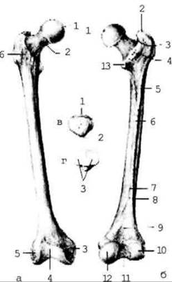
Fig. 2.62. Femur (right)
The Tibia (tibia) (Fig. 2.63) is situated medially in the leg. Its upper end bears lateral and medial condyles, which feature articular surfaces for articulation with the femur. The shaft is triangular in shape with a sharp anterior border.

Fig. 2.63. Tibiae; right
The lower end contains the inferior articular surface (for articulation with the talus) and the medial malleolus.
The Fibula (fibula) (Fig. 2.63) is located laterally in the leg.
Its upper end bears a head with an articular surface (for articulation with the tibia). The lower end features a lateral malleolus, which carries an articular surface for articulation with the talus.
The tarsus consists of the following bones: the talus, calcaneus, cuboid, navicular, and the cuneiforms—medial, intermediate, and lateral (Fig. 2.64).
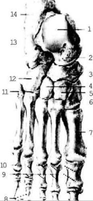
Fig. 2.64. Bones of the FOOT; superior view
The talus features a head with an articular surface (directed anteriorly), followed by a neck, and then the body. The superior surface of the body bears a trochlea for articulation with the tibia, while its inferior surface contains articular surfaces for articulation with the calcaneus.
The calcaneus lies beneath the talus and consists of a body and a rough tuberosity directed posteriorly and inferiorly. The body bears articular surfaces for articulation with the talus and cuboid bones.
The metatarsus consists of five short bones, each comprising a head, a body, and a base.
Phalanges of the digits
Each toe, except for the hallux (big toe), has three phalanges: proximal, middle, and distal. The hallux has only proximal and distal phalanges.
The bones of the free lower limb are connected by both continuous joints (syndesmoses) and discontinuous joints (synovial joints or articulationes). The hip, knee, and ankle (talocrural) joints are of the greatest practical significance.
The hip joint is formed by the articular surface of the femoral head and the acetabulum of the pelvic bone (Fig. 2.65).
Fig. 2.65. Connections of the pelvic bones and the right hip joint (capsule partially removed), posterior and lateral view.
In Structure, it is a simple joint; in shape, it is a cotyloid (cup-shaped) joint; and in function, it is a multiaxial joint. It is a variant of the ball-and-socket joint, allowing movement around all axes, though with a somewhat restricted range of motion. Inside the joint, there is an acetabular labrum (to better grasp the articular surface of the head) and the ligament of the head of the femur, which contains Blood Vessels and nerves supplying the head.
The strongest ligament reinforcing the joint, which can withstand weights of up to 300 kg, is the iliofemoral ligament.
The knee joint is formed by the articulation of three bones: the condyles of the tibia and femur, and the patella (Fig. 2.66). It is a rather complex joint containing the medial and lateral menisci, as well as the anterior and posterior cruciate ligaments, while its capsule forms numerous pouches (bursae). Some of these bursae communicate with the knee joint cavity, which is of clinical importance in inflammatory processes. Structurally, the joint is complex; in shape, it is condylar; and functionally, it is biaxial. It allows flexion and extension, as well as rotation around a vertical axis when the knee is flexed (due to the relaxation of the collateral ligaments that reinforce the capsule).
Fig. 2.66. Right knee joint
Connections between the bones of the leg (crus). The upper ends of the tibia and fibula form a flat, slightly movable joint (tibiofibular joint). The shafts of the leg bones are connected by a syndesmosis (the interosseous membrane), and their lower ends are also connected by a syndesmosis (ligaments).
The ankle joint (talocrural joint, articulatio talocruralis) is formed by the lower ends of the fibula and tibia, whose articular surfaces embrace the talus like a mortise (fork). Structurally, this joint is complex; in shape, it is hinge-like (ginglymus); and functionally, it is uniaxial. It allows flexion and extension of the foot around the frontal axis (Fig. 2.67).
Fig. 2.67. Ligaments and JOINTS OF THE right foot
The tarsal bones articulate with one another through joints whose capsules are reinforced by short ligaments on both the plantar and dorsal surfaces of the foot. Movements in these joints Supplement those of the talocrural joint.
Among the tarsal joints, the talocalcaneonavicular and calcaneocuboid joints are of practical importance; surgeons combine them into a single transverse tarsal joint known as Chopart's joint (Fig. 2.68). Amputation of the foot can be performed along the line of this joint after cutting the interosseous ligament that runs to the navicular and cuboid bones. This ligament is referred to as the "key" to Chopart's joint.
Fig. 2.68. Skeleton of the foot
The tarsal bones articulate with the bases of the metatarsal bones to form flat, slightly movable tarsometatarsal joints. In surgery, they are known as Lisfranc's joint. The "key" to this joint is the medial interosseous tarsometatarsal ligament.
The metatarsophalangeal and interphalangeal joints are similar in structure and movement to the corresponding joints of the hand.
The Foot as a Whole
The foot (pes, pedis) serves as a support during standing and walking. Due to the vertical posture of The Human Body and the increased load on the foot associated with bipedalism, its bones have formed an elastic arch structure. There are five longitudinal arches (corresponding to the five rays of the foot) and one transverse arch. All longitudinal arches originate at the calcaneus and extend forward from there along the metatarsal bones. In the transverse direction, all five arches vary in height. Consequently, the transverse arch is formed in the region of their highest points. The arches are supported by ligaments, Muscles, and fascia. When this supportive apparatus weakens (for example, in older age), the arches flatten, leading to The Development of flat feet (pes planus).
Last update: 08/08/2026
Editorial and Educational Adaptation: This material has been compiled based on the primary/original source text. The project team performed an editorial review, corrected technical inaccuracies, structured sections, and adapted the content for an educational format.
What was processed:
- elimination of formatting defects (OCR errors, structural breaks, corrupted characters);
- editorial organization of content;
- standardization of terminology in accordance with academic sources;
- verification of factual statements against the original source text.
All mentions of the author, publication year, and origin of the primary text have been preserved in accordance with the source.