HUMAN MEDICAL BIOLOGY, ANATOMY, PHYSIOLOGY AND PATHOLOGY - Ya.I. Fedonyuk 2010
ANATOMY, PHYSIOLOGY, PATHOLOGY
CHAPTER 2. THE MUSCULOSKELETAL SYSTEM (THE MECHANICS OF MOVEMENT)
GENERAL ISSUES IN THE ANATOMY AND PHYSIOLOGY OF THE HUMAN MUSCULAR SYSTEM
GENERAL MYOLOGY
The Muscle as an organ. Each Skeletal Muscle (musculus) consists of bundles of striated muscle fibers bound together by loose Connective Tissue into primary bundles, which in turn form secondary bundles, and so on, ultimately constituting the muscle belly. At the extremities of each belly, these connective tissue layers transition into the muscle tendon. Thus, the muscle belly serves as the active component of the muscle, whereas the tendon acts as the passive component through which it attaches to the bones.
The primary function of Muscles is their ability to contract. Muscle contraction brings the two points of attachment closer together. Conventionally, the fixed point of attachment is termed the Water/144.html">Origin of the muscle, while the moving point towards which contraction occurs is called the insertion. These points can interchange, thereby altering the muscle's function. Every movement in any joint results from the coordinated action of antagonistic muscles—that is, muscles performing opposing Functions (for example, when a flexor contracts, the extensor also exhibits controlled tone to prevent excessive movement). Muscles that perform the same function are referred to as synergists.
Classification of Muscles. Based on their shape, muscles are categorized as long, short, and broad (flat). Some long muscles possess multiple bellies, giving rise to biceps, triceps, and quadriceps muscles (Fig. 2.69). They may also feature multiple tendons. For instance, the finger flexors and extensors of the upper and lower extremities have four tendons each; consequently, the contraction of a single belly produces movement across multiple digits, ensuring muscular efficiency.
Class="center">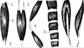
Fig. 2.69. Muscle shapes
The broad Muscles of the Trunk feature wide, flat tendons known as aponeuroses.
Other muscle shapes include quadrate, circular, triangular, pyramidal, deltoid, serratus, soleus, and so forth.
According to the fiber orientation, muscles are classified as straight (parallel), oblique, transverse, circular, etc.
Based on their function, muscles are divided into flexors and extensors; pronators and supinators (which rotate, for example, the forearm and hand inward and outward); adductors and abductors.
Depending on their relation to joints—spanning across one, two, or multiple joints—muscles are designated as uniarticular, biarticular, or multiarticular.
Topographically, muscles are classified as superficial and deep, external and internal, lateral and medial, anterior and posterior.
The Accessory structures of muscles include their Fibrous connective tissue sheaths, known as fasciae. Fasciae provide structural support for Organs, muscles, Blood Vessels, and nerves, protecting them from injury. They are subdivided into superficial and deep fasciae.
Last update: 08/08/2026
Editorial and Educational Adaptation: This material has been compiled based on the primary/original source text. The project team performed an editorial review, corrected technical inaccuracies, structured sections, and adapted the content for an educational format.
What was processed:
- elimination of formatting defects (OCR errors, structural breaks, corrupted characters);
- editorial organization of content;
- standardization of terminology in accordance with academic sources;
- verification of factual statements against the original source text.
All mentions of the author, publication year, and origin of the primary text have been preserved in accordance with the source.