Human Anatomy - H. I. Koliadenko 2009
The Doctrine of Muscles (Myology)
Muscles of the Trunk
This is the largest Muscle group, encompassing the Muscles of the chest, abdomen, and back (Fig. 70, 71).
Muscles of the chest. The thoracic region contains two groups of muscles: deep (proper muscles of the chest cage) and superficial. The deep muscles include the external and internal intercostals, the transversus thoracis, and the Diaphragm, while the superficial (or extrinsic) muscles include the pectoralis major, pectoralis minor, and serratus anterior.
The external intercostal muscles (mm. intercostales externi) fill all the intercostal spaces from the costal cartilages to THE Vertebral Column; their fibers run downward and forward from the upper rib to the rib below. Upon contraction, these muscles elevate the Ribs, thereby increasing the volume of the thoracic cavity in the anteroposterior and transverse directions. These muscles assist in inspiration.
Class="center">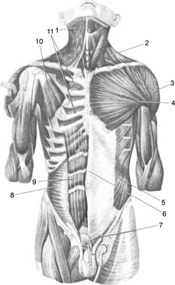
Fig. 70. Muscles of the trunk and neck (anterior view):
1 — platysma; 2 — sternocleidomastoid muscle; 3 — deltoid muscle; 4 — pectoralis major muscle; 5 — external oblique muscle of the abdomen; 6 — linea alba; 7 — superficial inguinal ring; 8 — transversus abdominis muscle; 9 — rectus abdominis muscle; 10 — pectoralis minor muscle; 11 — internal intercostal muscles
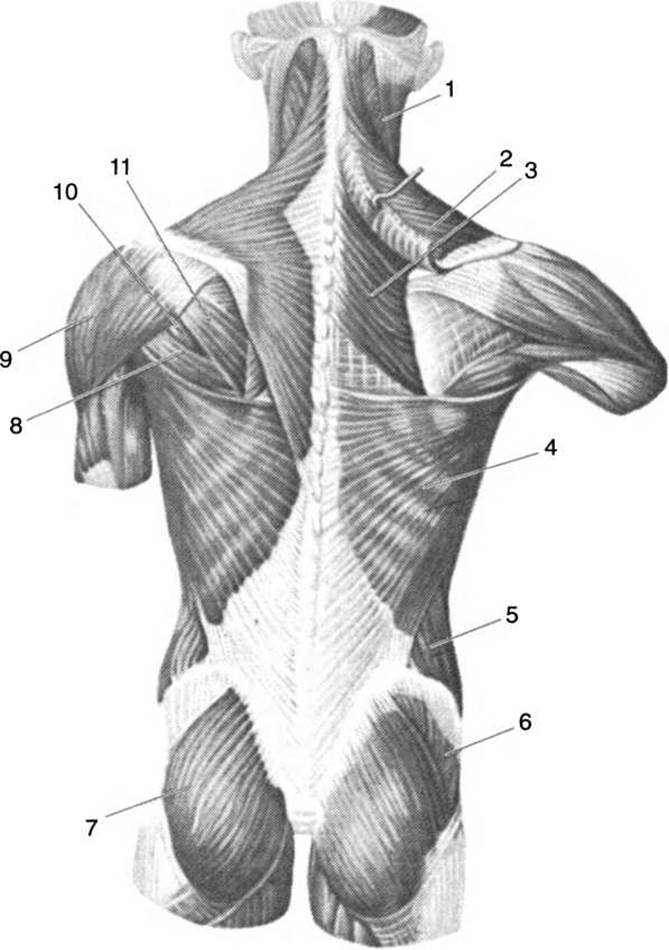
Fig. 71. SUPERFICIAL MUSCLES OF the back:
1 — sternocleidomastoid muscle; 2 — trapezius muscle; 3 — rhomboid muscle; 4 — latissimus dorsi muscle; 5 — external oblique muscle of the abdomen; 6 — gluteus medius muscle; 7 — gluteus maximus muscle; 8 — levator scapulae muscle; 9 — deltoid muscle; 10 — teres minor muscle; 11 — infraspinatus muscle
The internal intercostal muscles (mm. intercostales interni) originate from the upper margin of the rib below, run upward and forward, and insert into the lower margin of the rib above. On the posterior surface of the Thorax, extending from the angles of the ribs to the spine, the internal intercostal muscles are absent. Upon contraction, these muscles depress the ribs, narrow the chest cavity, decrease its volume, and assist in expiration.
The transversus thoracis muscle (m. transversus thoracis) lies on the posterior surface of the anterior thoracic wall. By contracting, it decreases the volume of the thoracic cavity and AIDS in expiration.
The diaphragm (diaphragma), or the thoracoabdominal partition, separates the thoracic cavity from the Abdominal cavity. It has a dome-shaped configuration, with the dome facing into the thoracic cavity. The muscle fibers of the diaphragm originate from the Sternum, ribs, and lumbar vertebrae along a circular line and converge centrally to form the central tendon. The Heart lies just to the left of the central tendon. The diaphragm consists of three parts: sternal, costal, and lumbar.
The sternal part is located in the region of the xiphoid process. The costal part originates with digitations from the inner surface of the lower six costal bones and costal cartilages.
The lumbar part originates with separate bundles from the I–IV lumbar vertebrae and is referred to as the crura of the diaphragm. There are 3 pairs of crura: medial, lateral, and intermediate. Between the medial crura lie two openings — anterior and posterior. The anterior esophageal hiatus transmits the Esophagus and vagus nerves, whereas the posterior aortic hiatus transmits the aorta and the thoracic lymphatic duct. Between the medial and intermediate, as well as between the intermediate and lateral crura, there are openings through which Blood Vessels and nerves pass. In the posterior part of the central tendon on the right side lies the opening for the INFERIOR VENA CAVA. Upon contraction, the diaphragm flattens, increasing the volume of the thoracic cavity, which facilitates inspiration. For this reason, the diaphragm is considered one of the primary Respiratory Muscles (Fig. 72).
The pectoralis major muscle (m. pectoralis major) originates from the sternal portion of the clavicle, the edge of the manubrium, the body of the sternum, the 5th–6th costal cartilages, and the upper region of the rectus sheath. All fibers converge into a strong tendon that inserts into the crest of the greater tubercle of the humerus. Function: upon contraction, the muscle lowers the raised arm, pulls the shoulder forward and inward, and pronates it.
The pectoralis minor muscle (m. pectoralis minor) lies beneath the pectoralis major, originating from the anterior surface of the 2nd–5th ribs and inserting into the coracoid process of the scapula. Upon contraction, the muscle pulls the scapula forward and downward; when the scapula is fixed, it elevates the ribs and participates in the act of inspiration.

Fig. 72. Diaphragm and muscles of the posterior abdominal wall:
1 — sternal part of the diaphragm; 2 — costal part of the diaphragm; 3 — opening for the inferior vena cava; 4 — esophageal hiatus; 5 — aortic hiatus; 6 — lateral crus of the diaphragm; 7 — quadratus lumborum muscle; 8 — psoas minor muscle; 9 — psoas major muscle; 10 — iliacus muscle; 11 — obturator externus muscle; 12 — iliopsoas muscle; 13 — medial crus of the diaphragm; 14 — intermediate crus of the diaphragm; 15 — lateral arcuate ligament; 16 — medial arcuate ligament; 17 — lumbar part of the diaphragm; 18 — central tendon
The serratus anterior muscle (m. serratus anterior) (Fig. 73) lies on the lateral surface of the thoracic wall, originating with digitations from the external surface of the 8th–9th ribs, and inserts into the inferior angle and Medial surface of the scapula. Function: pulls the scapula forward and its inferior angle outward, thereby causing the scapula to rotate around the sagittal axis.
All three muscles described above elevate the ribs upon contraction and serve as accessory respiratory muscles.
The fascias of the thoracic wall are weakly developed.
The Abdominal muscles form the external and internal abdominal walls. Their fibers run in various directions: longitudinal, transverse, and oblique. Muscles with straight fibers include the rectus abdominis and pyramidalis. Horizontally arranged fibers are characteristic of the transversus abdominis, while oblique fiber orientation is found in the external and internal oblique abdominal muscles and the quadratus lumborum.
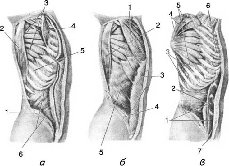
Fig. 73. Muscles of the trunk (lateral view):
a: 1 — iliac crest; 2 — latissimus dorsi; 3 — serratus anterior; 4 — pectoralis minor; 5 — external intercostal muscles; 6 — internal oblique muscle of the abdomen; b: 1 — pectoralis major; 2 — latissimus dorsi; 3 — aponeurosis of the external oblique muscle of the abdomen; 4 — iliac crest; 5 — external oblique muscle of the abdomen; c: 1 — rectus abdominis; 2 — walls of the rectus sheath; 3 — external intercostal muscles; 4 — scapula; 5 — serratus anterior; 6 — internal intercostal muscles; 7 — pyramidalis muscle
The abdominal muscles perform the following Functions: they form the anterior and partially posterior walls of the abdominal cavity, and through their tonus, they hold the abdominal Organs in place; they pull the ribs downward, thereby reducing the size of the thoracic cavity and participating in expiration.
The abdominal press muscles facilitate the evacuation of the bowels and bladder, coughing, and childbirth. In addition, they flex the trunk forward and laterally and rotate it around its vertical axis.
The rectus abdominis muscle (m. rectus abdominis) originates from the xiphoid process and the cartilages of ribs V–VII, and inserts into the pubic bone. It features 3–4 tendinous intersections that strengthen both the muscle and the abdominal wall. The rectus abdominis lies within a fibrous sheath formed by the aponeuroses of the abdominal oblique muscles. This muscle participates in flexing the trunk forward.
The pyramidalis muscle (m. pyramidalis) is a small, triangular muscle that originates by one end in the region of the Pubic Symphysis and inserts by the other end into the lower part of the linea alba, which it tightens upon contraction.
The external oblique muscle of the abdomen (m. obliquus externus abdominis). Its fibers are oriented similarly to those of the external intercostal muscles, running downward and forward. It originates by digitations from the eight lower ribs and attaches to the iliac crest. Anteriorly, the muscle fibers transition into an aponeurosis. The fibers of the aponeurosis intersect with the fibers of the contralateral muscle of the same name to form the anterior wall of the rectus sheath, as well as the linea alba. The lower margin of the aponeurosis thickens to form the inguinal, or Poupart's, ligament, which attaches by its anterior end to the pubic tubercle and by its posterior end to the anterior superior iliac spine. In the region of the pubic bone, the fibers of the aponeurosis diverge to form the superficial inguinal ring.
Upon contraction, the external oblique muscle pulls the thorax downward, flexes the trunk to its side, and, when the thorax is fixed, acts together with the contralateral muscle of the same name to pull the pelvis upward.
The internal oblique muscle of the abdomen (m. obliquus internus abdominis). Its fibers mirror the direction of the fibers of the internal intercostal muscles of the thorax. The muscle originates from the thoracolumbar fascia, the iliac crest, and the lateral two-thirds of the inguinal ligament. It attaches by one part of its fibers to the lower margins of ribs XII, XI, and X, while the other part forms an aponeurosis that splits into two laminae (anterior and posterior), which participate in forming the rectus sheath in women. In men, fibers originating from its lower part, along with the lower fascicles of the internal oblique, form the cremaster muscle (m. cremaster). The anterior aponeurosis of the internal oblique muscle contributes to The formation of the linea alba. Upon unilateral contraction, the internal oblique muscle rotates the trunk toward its own side, and when the thorax is fixed, these muscles elevate the pelvis.
The transversus abdominis muscle (m. transversus abdominis) originates from the thoracolumbar fascia, the inner surface of the lower ribs, the iliac crest, and the lateral two-thirds of the inguinal ligament. The muscle fibers are arranged transversely, with most of them transitioning into an aponeurosis whose fibers blend into the linea alba. The muscle participates in the Formation of the rectus sheath, the linea alba, and the cremaster muscle.
The quadratus lumborum muscle (m. quadratus lumborum) originates from the iliac crest and inserts into the body of the twelfth thoracic vertebra (XII) and the transverse processes of the upper four lumbar vertebrae. The muscle pulls the twelfth rib downward, thereby facilitating expiration, and also flexes the lumbar region of the vertebral column.
In addition to the aforementioned functions, the abdominal press, formed by the abdominal muscles, acts as an accessory respiratory muscle. The Inguinal Canal is located in the lower region of the abdominal wall; in men, it contains the Spermatic Cord, and in women, the round ligament of the Uterus. The superficial opening of the inguinal canal is called the superficial inguinal ring, which is formed by two crura: the superior crus, attached to the pubic symphysis, and the inferior crus, attached to the pubic tubercle.
The wall of the abdominal press varies in thickness and strength. Thus, weak areas of the abdominal wall can sometimes include the inguinal ring, the umbilical ring, and the linea alba; consequently, inguinal and umbilical hernias, as well as hernias of the linea alba, may occur in these regions. Through these weak zones, Internal Organs can protrude beneath the Skin together with the parietal Peritoneum or the omentum.
The back muscles (see Fig. 71) are divided into superficial and deep groups. The superficial group includes the trapezius, latissimus dorsi, rhomboid Major and minor, levator scapulae, and the posterior superior and posterior inferior serratus muscles. The deep muscles include the erector spinae, transversospinales, intertransversarii, interspinales, and short suboccipital and craniovertebral muscles.
The superficial back muscles belong to migrated muscles: the first of these is the trapezius, which migrated from the HEAD, and the latissimus dorsi, which migrated from the upper limbs.
The trapezius muscle (m. trapezius) originates from the superior nuchal line of the Occipital bone, the nuchal ligament, the external occipital protuberance, and the spinous processes of the last cervical and all thoracic vertebrae. The muscle inserts into the spine of the scapula, the acromion, and the lateral end of the clavicle. Upon contraction, the upper fibers of the muscle elevate the lateral angle of the lower scapula, while the lower fibers depress the scapula. Contraction of all fibers retracts the scapula toward the vertebral column. When the scapulae are fixed, simultaneous contraction of both muscles extends the cervical region of the vertebral column.
The latissimus dorsi muscle (m. latissimus dorsi) originates from the thoracolumbar fascia, the spinous processes of the lower five to six thoracic (V–VI) and all lumbar vertebrae, and from the iliac crest. The muscle fibers converge into a single tendon that inserts into the crest of the lesser tubercle of the humerus. Upon contraction, the muscle extends the arm, adducts it, and rotates it medially; when the arm is fixed, it draws the trunk toward the arm.
The rhomboid muscles — major and minor (m. rhomboideus major et minor) — originate from the spinous processes of the lower two thoracic and upper four cervical vertebrae. Their fibers run downward and laterally to insert into the medial border of the scapula. Upon contraction, the muscles retract and slightly elevate the scapula.
The levator scapulae muscle (m. levator scapulae) originates from the transverse processes of the upper four cervical vertebrae. Its fibers run downward to insert into the superior angle of the scapula. The muscle elevates the scapula.
The serratus posterior superior muscle (m. serratus posterior superior) originates from the spinous processes of the lower two cervical and upper two thoracic vertebrae; its fibers run downward and laterally, inserting into ribs II–V. The muscle elevates the ribs and participates in Respiration.
The serratus posterior inferior muscle (m. serratus posterior inferior) originates from the spinous processes of the lower two thoracic and upper two lumbar vertebrae. Its fibers attach via four digitations to the external surface of the lower four ribs. By contracting, this muscle depresses the lower ribs and participates in respiration.
The deep long muscles of the back include the erector spinae, transversospinales, and splenius muscles.
The erector spinae muscle (m. erector spinae) originates from the posterior part of the iliac crest, the thoracolumbar fascia, the posterior surface of the sacrum, and the spinous processes of the lumbar vertebrae. The muscle extends the vertebral column. Below the twelfth rib (XII), the erector spinae divides into the iliocostalis, longissimus, and spinalis muscles of the back.
The iliocostalis muscle lies laterally, attaching to the ribs and the transverse processes of the lower cervical vertebrae.
The longissimus dorsi muscle attaches to the transverse processes of all thoracic and cervical vertebrae and extends to the mastoid process of the Temporal bone.
The spinalis dorsi muscle attaches to the spinous processes of the thoracic and cervical vertebrae and terminates on the axis (epistropheus).
The transversospinalis muscle (m. transversospinalis) extends from the sacrum to the occipital bone, with its fibers running from the transverse processes to the spinous processes. This muscle extends the vertebral column, bends it laterally, and also rotates it.
The splenius muscles of the Head and Neck (m. splenii capitis et cervicis) originate from the nuchal ligament in the region of the III–VII cervical vertebrae and from the spinous processes of the upper six thoracic vertebrae. These muscles attach to the transverse processes of the upper cervical vertebrae and the mastoid process of the temporal bone. Unilateral contraction rotates the head to the same side and tilts the face upward, while simultaneous bilateral contraction extends the cervical region of the vertebral column.
The intertransversarii muscles are located between the transverse processes of adjacent vertebrae and, upon contraction, flex the vertebral column laterally.
The interspinales muscles lie between the spinous processes of adjacent vertebrae; when contracting, they extend the Vertebral Column and maintain it in an upright position.
The short suboccipital muscles (four in number) are situated between the occipital bone, the atlas, and the axis. These muscles extend and rotate the head.
Among the back fasciae, the thoracolumbar fascia is well-developed, covering the Deep Muscles of the back both anteriorly and posteriorly. It forms the osteofascial canal for the deep muscles.
The muscles of the neck are divided into proper Neck Muscles and extrinsic (migrant) muscles, as well as superficial and deep ones.
The Superficial muscles of the neck include: the platysma, the sternocleidomastoid muscle, the suprahyoid muscles, and the Infrahyoid muscles.
The Deep Muscles of the Neck comprise: the anterior, middle, and posterior scalene muscles, the longus colli, and the longus capitis.
The platysma (m. platysma) is vestigial in humans and well-developed in animals (horses, cows, hedgehogs, etc.). In humans, this muscle appears as a thin sheet covering the lateral and anterior surfaces of the neck. It originates from the pectoral fascia below the clavicle and attaches to the Base of the Mandible and the parotid fascia. Blending with some facial expression muscles, it tightens the skin of the neck and pulls the corner of the Mouth downward.
The sternocleidomastoid muscle (m. sternocleidomastoid-eus) is the largest and strongest, standing out prominently on the lateral and anterior surfaces of the neck. It originates by two tendons: one from the manubrium of the sternum and the other from the sternal end of the clavicle. It inserts into the mastoid process of the temporal bone. Unilateral contraction tilts the head to its side, while bilateral contraction tilts it backward.
The superficial muscles also include those that anchor the Hyoid bone from above and below.
The muscles belonging to the hyoid group are named after the bones to which they attach: the sternohyoid, sternothyroid, thyrohyoid, and omohyoid. When contracting, these muscles pull the hyoid bone, and along with it the Larynx, downward.
The muscles lying superior to the hyoid bone are: the geniohyoid, digastric, mylohyoid, and stylohyoid.
The geniohyoid muscle (m. geniohyoid-eus) originates from the mental spine of the mandible and inserts into the body of the hyoid bone. Upon contraction, it pulls the hyoid bone forward and upward, and when the hyoid bone is fixed, it depresses the mandible.
The digastric muscle (m. digastricus) has its anterior belly originating from the digastric fossa of the mandible, while the posterior belly attaches to the mastoid notch of the temporal bone. A tendon lies between the two bellies, forming a loop by which it attaches to the hyoid bone.
The mylohyoid muscle (m. mylohyoid-eus) forms the floor of the Oral Cavity; it originates from the mylohyoid line on the inner surface of the mandible and inserts into the body of the hyoid bone. At the junction of both muscles in the center of the jaw, a median raphe is formed.
The stylohyoid muscle (m. stylohyoid-eus) attaches with one tendon to the styloid process of the temporal bone and with the other to the greater horn of the hyoid bone.
All these muscles, when contracting, pull the larynx and the hyoid bone upward, participating in the act of swallowing as well as in phonation.
Deep muscles of the neck. The deep muscles (Fig. 74) with obliquely arranged fibers include the anterior, middle, and posterior scalene muscles (m. scalenus anterior, medius, posterior), which originate by separate slips from the transverse processes of the cervical vertebrae and attach to the I–II ribs. The anterior scalene muscle attaches to the scalene tubercle on the first rib, the middle one lies posterior to the anterior, and the posterior attaches to the outer surface of the rib. When contracting, the scalene muscles elevate the ribs to which they attach, thereby facilitating respiration. With a fixed rib cage and contraction of these muscles on one side, they bend the cervical region of the vertebral column to their side and forward, whereas bilateral contraction flexes the neck and head forward.
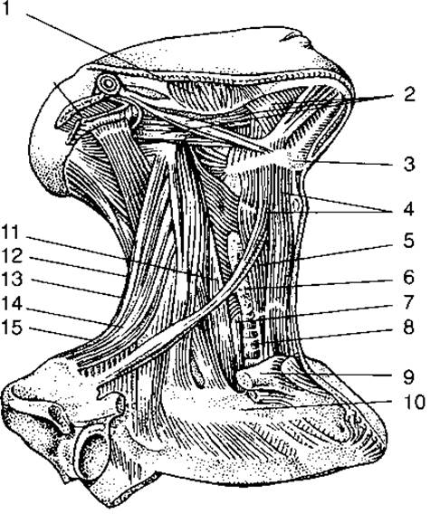
Fig. 74. Muscles of the neck:
1 — masseter muscle; 2 — suprahyoid muscle group; 3 — hyoid bone; 4 — infrahyoid muscle group; 5 — Pharynx; 6 — Thyroid Gland; 7 — esophagus; 8 — first rib; 9 — clavicle; 10 — Trachea; 11 — longus colli muscle; 12 — anterior scalene muscle; 13 — middle scalene muscle; 14 — levator scapulae muscle; 15 — posterior scalene muscle
The longus colli and longus capitis muscles lie on the anterior surface of the cervical region of the vertebral column. The longus colli flexes the neck, while the longus capitis tilts the head.
Muscles of the head. The muscles of the head comprise two major groups: masticatory muscles and Muscles of facial expression.
The masticatory muscles (Fig. 75) are responsible for the active Movements of the mandible during mastication and articulated speech. The mandible is the only movable bone to which all four pairs of masticatory muscles attach: the temporal, masseter, and two pterygoid muscles (lateral and medial).
The temporalis muscle (m. temporalis) fills the temporal fossa of the Skull, originating in a fan-like shape from the temporal line. The fibers of this muscle converge into a strong tendon that passes beneath the zygomatic arch and inserts into the coronoid process of the mandible. Upon contraction, the muscle elevates the mandible, while its posterior fascicles pull it backward.
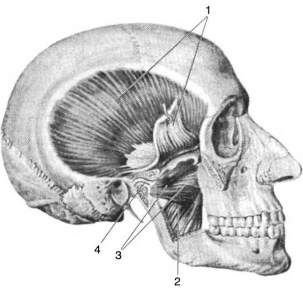
Fig. 75. Masticatory muscles:
1 — temporalis muscle; 2 — medial pterygoid muscle; 3 — lateral pterygoid muscle; 4 — articular disc
The masseter muscle (m. masseter) originates from the inferior border of the Zygomatic bone and the zygomatic arch, and inserts into the lateral surface of the mandibular angle. It is the most powerful masticatory muscle. Upon contraction, it elevates the mandible, presses the Teeth against the Maxilla, and pulls the jaw forward.
The lateral pterygoid muscle (m. pterygoideus lateralis) originates from the Inferior surface of the greater wing and the lateral plate of the pterygoid process of the Sphenoid bone, inserting into the lower capsule of the TEMPOROMANDIBULAR JOINT AND the pterygoid fovea of the mandible. When contracted unilaterally, it pulls the jaw to the opposite side; bilateral contraction protrudes the jaw forward.
The medial pterygoid muscle (m. pterygoideus medialis) originates from the pterygoid fossa of the sphenoid bone and inserts into the medial surface of the angle of the mandible. Upon contraction, the muscle elevates the mandible and pulls it toward its own side.
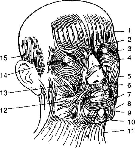
Fig. 76. Facial muscles (muscles of facial expression):
1 — frontal belly of the epicranius muscle; 2 — corrugator supercilii muscle; 3 — procerus muscle; 4 — orbicularis oculi muscle; 5 — zygomaticus major muscle; 6 — levator anguli oris muscle; 7 — orbicularis oris muscle; 8 — depressor anguli oris muscle; 9 — depressor labii inferioris muscle; 10 — mentalis muscle; 11 — platysma; 12 — risorius muscle; 13 — levator labii superioris muscle; 14 — anterior auricular muscle; 15 — superior auricular muscle
The muscles of facial expression (mimetic muscles, Fig. 76) differ from the masticatory muscles in that they originate by one tendon from various bones of The Skull and insert by another tendon directly into the skin. Some facial muscles lie entirely within the soft Tissues of the face. Typically, these muscles surround facial orifices; thus, upon contraction, they close or open these openings, thereby forming folds and wrinkles, primarily on the facial surface of the head. All of this contributes to the expression of various human feelings, emotions, and thoughts.
The epicranius muscle (m. epicranius) covers the top of the cranium and consists of three parts: an anterior part represented by two frontal bellies, a middle part — the epicranial aponeurosis (galea aponeurotica), and a posterior part — the two occipital bellies. The epicranial aponeurosis is a tough fibrous plate covering the entire periosteum of the calvaria. The frontal bellies attach to the facial skin in the region of the supraorbital margins of the frontal bones. The occipital bellies attach to the epicranial aponeurosis, the superior nuchal line of the occipital bone, and the mastoid process of the temporal bone. Contraction of the frontal bellies forms horizontal wrinkles on the forehead, whereas contraction of the occipital bellies tenses the skin of the forehead and forms folds in the occipital region.
The epicranial aponeurosis performs a protective function.
The orbicularis oris muscle (m. orbicularis oris) is located beneath the skin surrounding the oral fissure. Together with other muscles, it forms the upper and lower Lips. Other facial muscles blend into the orbicularis oris. Upon contraction, the muscle narrows the oral aperture.
The orbicularis oculi muscle (m. orbicularis oculi) is located around the orbital opening, beneath the skin of the upper and lower eyelids, as well as near the lacrimal sac. Accordingly, it is subdivided into orbital, palpebral, and lacrimal parts. The orbital part is located along the margin of the Orbit and tightly closes the eye (squinting); the palpebral part lies beneath the skin of the eyelids and gently closes the eye; the lacrimal part dilates the lacrimal sac during tear drainage into the lacrimal canaliculi.
The procerus muscle (m. procerus) lies in the region of the bones forming the bridge of the Nose and produces transverse wrinkles at the ROOT of the nose.
The corrugator supercilii muscle (m. corrugator supercilii) originates from the Frontal bone in the region of the nasal root, passes through the frontal belly of the epicranius, and inserts into the skin of the eyebrow. Upon contraction, the muscle creates vertical wrinkles on the forehead.
The buccinator muscle (m. buccinator) originates with its upper fibers from the outer surface of the alveolar process of the maxilla, with its lower fibers from the body of the mandible below the alveolar process, and with its middle fibers from the pterygomandibular raphe (the junction between the skull base and the mandible). Toward the angle of the mouth, the upper fibers of this muscle blend into the lower lip, the middle fibers into the orbicularis oris, and the lower fibers into the upper lip. The muscle holds food between the masticatory surfaces of the jaws and counteracts intraoral pressure. In young children, adipose tissue accumulates within the buccinator muscle, which is why their Cheeks appear plump and rounded.
The levator anguli oris muscle (m. levator anguli oris) originates from the canine fossa of the maxilla, extends toward the angle of the mouth, and blends into the skin and mucous membrane of the lower lip.
The depressor anguli oris muscle (m. depressor anguli oris) originates from the inferior border of the mandible and blends into the skin and mucous membrane of the upper lip.
Simultaneous contraction of both aforementioned muscles closes the oral fissure.
The nasalis muscle (m. nasalis) originates from the alveoli of the upper canine and incisor teeth; a portion of its muscle fibers ascends along the dorsum of the nose, where they interlace with the fibers of the contralateral muscle of the same name. Upon contraction, these fibers compress the nasal cartilages. The fibers that dilate the nostrils attach to the Cartilage and skin of the alae of the nose.
The zygomaticus major muscle (m. zygomaticus major) originates from the body of the zygomatic bone, and its fibers blend into the skin at the angle of the mouth. Upon contraction, this muscle pulls the angle of the mouth upward and laterally.
The mentalis muscle (m. mentalis) originates from the alveoli of the lower incisors; its fibers course downward and insert into the skin of the chin. The muscle performs several functions: it wrinkles the skin of the chin, creates dimples, and presses the lower lip against the upper lip.
The anterior, superior, and posterior auricular muscles are located around the auricle. In humans, these muscles are poorly developed, and only a few individuals are able to move their auricles.
The risorius muscle (m. risorius) runs between the angle of the mouth and the cheek. When contracted, it forms a dimple on the cheek. It is variable (inconstant).
The levator labii superioris muscle (m. levator labii superioris) originates from three heads: the frontal process and infraorbital margin of the maxilla, and the zygomatic bone. The muscle fibers extend inferiorly and blend into the skin of the nasolabial fold. Upon contraction, the muscle elevates and dilates the upper lip, and pulls the wing of the nose upward and laterally.
The depressor labii inferioris muscle (m. depressor labii inferioris) originates from the base of the mandible and inserts into the skin of the lower lip. When contracted, it pulls the lower lip downward and laterally.
Last update: 08/08/2026
Editorial and Educational Adaptation: This material has been compiled based on the primary/original source text. The project team performed an editorial review, corrected technical inaccuracies, structured sections, and adapted the content for an educational format.
What was processed:
- elimination of formatting defects (OCR errors, structural breaks, corrupted characters);
- editorial organization of content;
- standardization of terminology in accordance with academic sources;
- verification of factual statements against the original source text.
All mentions of the author, publication year, and origin of the primary text have been preserved in accordance with the source.