MEDICAL BIOLOGY, HUMAN ANATOMY, PHYSIOLOGY AND PATHOLOGY - Y.I. Fedoniuk 2010
ANATOMY, PHYSIOLOGY, PATHOLOGY
CHAPTER 2. THE MUSCULOSKELETAL SYSTEM (THE MECHANISM OF MOVEMENT)
MUSCLES AND FASCIAE OF BODY REGIONS
MUSCLES AND FASCIAE OF THE LOWER LIMB
2. MUSCLES OF THE FREE LOWER LIMB
The Muscles of the thigh are divided into three groups: anterior, posterior, and medial.
Anterior Muscle group of the thigh (Fig. 2.101)
Class="center">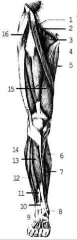
Fig. 2.101. Muscles of the right lower limb; Anterior view
1. The quadriceps femoris Muscle consists of four heads (rectus femoris, vastus lateralis, vastus medialis, and vastus intermedius). It originates from the anterior inferior iliac spine (rectus femoris) and the linea aspera of the Femur (vastus medialis, vastus lateralis, and vastus intermedius), and inserts via a strong tendon into the tibial tuberosity. Function: extends the leg, flexes the thigh.
2. The sartorius muscle runs obliquely downward and medially from the anterior superior iliac spine and inserts into the tibial tuberosity. Function: flexes the leg and the thigh.
Posterior group (Fig. 2.102)
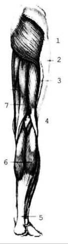
Fig. 2.102. Muscles of the right lower limb; posterior view
1. The semitendinosus and semimembranosus muscles are located medially, originating from the ischial tuberosity and inserting into the Tibia. Function: extend the thigh and flex the leg.
2. The biceps femoris muscle originates by two heads: the long HEAD from the ischial tuberosity and the short head from the femur. It inserts into the head of the Fibula. Function: extends the thigh and flexes the leg.
The medial group includes five muscles: the pectineus, gracilis, and adductors (adductor magnus, adductor longus, and adductor brevis). All of them originate from the pubic and ischial bones and insert into the femur (except for the gracilis, which inserts into the tibia). Function: adduct the thigh.
The Muscles of the Leg are divided into anterior, posterior, and lateral groups. All leg muscles insert into the FOOT.
The anterior group includes the tibialis anterior, extensor hallucis longus, and extensor digitorum longus muscles. Function: extend the toes and the foot.
The posterior group includes the triceps surae muscle (superficial layer), tibialis posterior, flexor digitorum longus, and flexor hallucis longus (deep layer).
The triceps surae inserts via a strong tendon (the Achilles tendon) into the calcaneal tuberosity and participates in plantar flexion of the foot. The deep muscles insert into the tarsal, metatarsal, and phalangeal bones, performing the function of flexing the foot and toes.
The lateral group is formed by the fibularis (peroneus) longus and brevis muscles, which flex the foot and pronate it (lower the medial margin and elevate the lateral margin).
The muscles of the foot are divided into dorsal and plantar groups.
The plantar group comprises the muscles of the great toe eminence, the muscles of the little toe eminence, and the muscles of the middle group (Fig. 2.103).
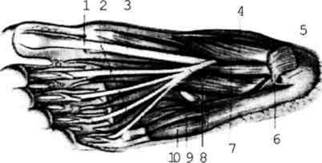
Fig. 2.103. Plantar muscles of the foot
The muscles of the thenar eminence (ball of the big toe) include the abductor hallucis, adductor hallucis, and flexor hallucis brevis.
The muscles of the hypothenar eminence (ball of the little toe) include the abductor digiti minimi, flexor digiti minimi brevis, and opponens digiti minimi.
The middle group comprises the flexor digitorum brevis, quadratus plantae, and four lumbricals, which flex the digits. The interosseous muscles consist of three plantar interosseous muscles, which adduct the digits toward one another, and four dorsal interosseous muscles, which abduct them.
Last update: 08/08/2026
Editorial and Educational Adaptation: This material has been compiled based on the primary/original source text. The project team performed an editorial review, corrected technical inaccuracies, structured sections, and adapted the content for an educational format.
What was processed:
- elimination of formatting defects (OCR errors, structural breaks, corrupted characters);
- editorial organization of content;
- standardization of terminology in accordance with academic sources;
- verification of factual statements against the original source text.
All mentions of the author, publication year, and origin of the primary text have been preserved in accordance with the source.