MEDICAL BIOLOGY, HUMAN ANATOMY, PHYSIOLOGY AND PATHOLOGY - Ya.I. Fedoniuk 2010
ANATOMY, PHYSIOLOGY, PATHOLOGY
CHAPTER 2. LOCOMOTOR SYSTEM (THE MECHANISM OF MOVEMENT)
MUSCLES AND FASCIAE OF BODY PARTS
MUSCLES AND FASCIAE OF THE LOWER LIMB
3. FASCIAE OF THE LOWER LIMB
The internal Muscles of the pelvis are covered by the iliac fascia, which is part of the general intra-abdominal fascia. Passing onto the thigh, the iliac fascia continues into the fascia lata.
The fascia lata is the densest fascia of The Human Body (Fig. 2.104).
Class="center">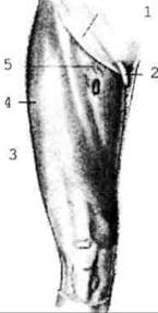
Fig. 2.104. Fascia lata, right thigh: 1 - inguinal ligament; 2 - Spermatic Cord; 3 - fascia lata; 4 - iliotibial tract; 5 - cribriform fascia.
It covers all the muscles of the thigh and gives off three intermuscular septa, which, attaching to the periosteum of the Femur, form strong fascial sheaths for the muscles. On the lateral surface of the thigh, the fascia lata becomes even denser and forms the iliotibial tract. On the anterior surface of the thigh, conversely, the fascia becomes thinner and is pierced by a significant number of Vessels and nerves, which is why this area is called the cribriform fascia. The falciform margin of this fascia delimits the saphenous hiatus (supercutaneous/external ring of the femoral canal).
The Fascia of the leg is a continuation of the fascia lata and covers the Muscles of the Leg, forming sheaths for them. In the region of the ankle joint and above it, the fascia thickens, forming the retinacula of the extensor, flexor, and fibular (peroneal) tendons. Just like in the hand, synovial sheaths are formed here. Anteriorly, there are three synovial sheaths for the extensor tendons; posterior to the medial malleolus, There are also three synovial sheaths for the flexor tendons (Fig. 2.105); and posterior to the lateral malleolus, there are initially two and then one common sheath for the short and long fibular (peroneal) tendons (Fig. 2.106).
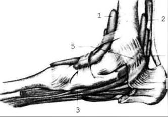
Fig. 2.105. Synovial sheaths of the flexor tendons of the toes and the tibial muscles; medial view
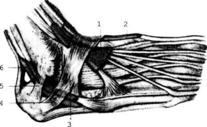
Fig. 2.106. Synovial sheaths of the extensor tendons of the toes and the fibular muscles
The fascia of the dorsum of the FOOT is thin and transparent, whereas on the sole it forms the plantar aponeurosis.
Last update: 08/08/2026
Editorial and Educational Adaptation: This material has been compiled based on the primary/original source text. The project team performed an editorial review, corrected technical inaccuracies, structured sections, and adapted the content for an educational format.
What was processed:
- elimination of formatting defects (OCR errors, structural breaks, corrupted characters);
- editorial organization of content;
- standardization of terminology in accordance with academic sources;
- verification of factual statements against the original source text.
All mentions of the author, publication year, and origin of the primary text have been preserved in accordance with the source.