Human Anatomy - H. I. Koliadenko 2009
Myology (The Study of Muscles)
Muscles of the Upper Limb
The Muscles of the upper limb are divided into two groups: the muscles of the Pectoral Girdle (shoulder girdle) and the Muscles of the Free Upper Limb (Fig. 77).
Muscles of the pectoral girdle. This group includes the deltoid, supraspinatus, infraspinatus, subscapularis, teres major, and teres minor muscles.
The deltoid Muscle (m. deltoideus) originates from the spine of the scapula, the acromion, and the lateral third of the clavicle. Well-developed and consisting of three parts, it covers the proximal epiphysis of the humerus. When contracting, the anterior fibers flex the arm at the shoulder joint, the posterior fibers extend it, and the middle fibers—along with the entire muscle—abduct the arm to a horizontal position. The muscle fibers terminate in a tendon that inserts into the deltoid tuberosity of the humerus.
The supraspinatus muscle (m. supraspinatus) originates from the walls of the supraspinous fossa of the scapula and inserts into the greater tubercle of the humerus. It acts as a synergist to the deltoid muscle, assisting in the abduction of the arm.
The infraspinatus muscle (m. infraspinatus) arises from the entire surface of the infraspinous fossa of the scapula and the overlying fascia, inserting into the greater tubercle of the humerus. It rotates the arm laterally.
The teres minor muscle (m. teres minor) originates from the inferior angle of the scapula and inserts into the greater tubercle of the humerus. It acts as a synergist to the infraspinatus muscle.
The teres major muscle (m. teres major) originates from the dorsal surface of the scapula and its inferior angle, extends laterally and superiorly, and inserts into the crest of the lesser tubercle of the humerus. Upon contraction, it adducts and medially rotates the arm.
The subscapularis muscle (m. subscapularis) originates from the costal surface of the scapula and inserts into the lesser tubercle of the humerus. When contracting, the muscle rotates the arm medially.
The subscapularis, along with the supraspinatus, infraspinatus, and teres minor muscles, blends with the capsule of the shoulder joint. When contracting, these muscles pull the capsule away from the articular surfaces, preventing it from being pinched.
The muscles of the free upper limb are divided into the muscles of the arm (brachium), forearm (antebrachium), and hand. Depending on their action on the Shoulder and Elbow joints, the arm muscles form anterior and posterior groups.
On the anterior aspect of the arm lie three flexor muscles: the biceps brachii, coracobrachialis, and brachialis.
The biceps brachii muscle (m. biceps brachii) arises with its long HEAD from the supraglenoid tubercle of the scapula. The tendon of this muscle passes through the Joint Capsule, enclosed within a synovial sheath (thus preserving joint integrity). Its short head originates from the coracoid process of the scapula. On the anterior surface of the arm, both bellies unite to form a single tendon that inserts into the radial tuberosity. The biceps is a biarticular muscle, as it flexes both the shoulder and elbow joints and supinates the forearm.
Class="center">
Fig. 77. Muscles of the pectoral girdle and free upper limb:
a — anterior view; b — posterior view; 1 — sternocleidomastoid muscle; 2 — pectoralis major muscle; 3 — serratus anterior muscle; 4 — latissimus dorsi muscle; 5 — teres major muscle; 6 — coracobrachialis muscle; 7 — brachialis muscle; 8 — triceps brachii muscle; 9 — brachioradialis muscle; 10 — pronator teres muscle; 11 — bicipital aponeurosis; 12 — flexor carpi ulnaris muscle; 13 — flexor digitorum superficialis muscle; 14 — palmaris longus muscle; 15 — flexor carpi radialis muscle; 16 — biceps brachii muscle; 17 — deltoid muscle; 18 — trapezius muscle; 19 — extensor pollicis longus muscle; 20 — extensor carpi ulnaris muscle; 21 — abductor pollicis longus muscle; 22 — extensor digitorum muscle; 23 — extensor carpi radialis brevis muscle; 24 — extensor carpi radialis longus muscle; 25 — rhomboideus major muscle; 26 — infraspinatus muscle; 27 — teres minor muscle; 28 — extensor pollicis brevis muscle
The coracobrachialis muscle (m. coracobrachialis) originates from the coracoid process of the scapula and inserts into the Medial surface of the humerus. The muscle adducts and flexes the arm.
The brachialis muscle (m. brachialis) originates from the lateral and anterior surfaces of the humerus and inserts into the ulnar tuberosity. It flexes the forearm at the elbow joint.
On the posterior aspect of the arm, There are two extensor muscles: the triceps brachii and the anconeus.
The triceps brachii muscle (m. triceps brachii) has one long head and two short heads. The long head originates from the infraglenoid tubercle of the scapula, while the lateral and medial short heads arise from the posterior surface of the humerus. The lateral head is longer than the medial; it originates from the upper portion of the humerus, whereas the medial head arises from the lower third of the humerus and the intermuscular septa. All three heads converge into a single robust tendon that inserts into the olecranon of the ulna. The muscle extends the forearm, and its long head also extends and adducts the arm.
The anconeus muscle (m. anconeus) is a small, triangular muscle that originates from the lateral epicondyle of the humerus and inserts into the dorsal surface of the proximal ulna. The muscle assists in extending the forearm.
Muscles of the forearm. The forearm contains two muscle groups: anterior and posterior.
The first group comprises superficial and deep muscles, represented by flexors and pronators.
The superficial muscles originate from the medial epicondyle of the humerus, the medial intermuscular septum, and the fascia. They are arranged in the following order: the pronator teres, flexor carpi radialis, palmaris longus, flexor digitorum superficialis, and flexor carpi ulnaris (Fig. 78).
The pronator teres muscle (m. pronator teres) arises from humeral and ulnar heads (from the medial epicondyle of the humerus and the medial margin of the ulnar tuberosity), passes obliquely downward, and inserts into the lateral surface of the middle third of the radius; it pronates the radius and partially flexes the forearm.
The flexor carpi radialis muscle (m. flexor carpi radialis) originates from the medial epicondyle of the humerus and the forearm fascia, passes beneath the flexor retinaculum, and inserts into the Base of the second metacarpal bone. The muscle abducts the forearm, while flexing and abducting the wrist.
The palmaris longus muscle (m. palmaris longus) is rudimentary and sometimes absent. It features a small muscle belly and a long tendon that fans out across the palm, continuing into the palmar aponeurosis. It flexes the wrist and tightens the palmar aponeurosis.
The flexor digitorum superficialis muscle (m. flexor digitorum superficialis) originates with humeroulnar and radial heads (from the medial epicondyle of the humerus and the upper part of the anterior surface of the radius). The common muscle belly ends in four long tendons that attach to the bases of the middle Phalanges of digits II–V. The muscle flexes the middle phalanges of digits II–V and assists in wrist flexion.

Fig. 78. Muscles of the anterior surface of the forearm:
1 — biceps brachii muscle; 2 — brachialis muscle; 3 — pronator teres muscle; 4 — bicipital aponeurosis; 5 — flexor carpi radialis muscle; 6 — palmaris longus muscle; 7 — flexor carpi ulnaris muscle; 8 — flexor digitorum superficialis muscle; 9 — pisiform bone; 10 — palmaris brevis muscle; 11 — palmar aponeurosis; 12 — flexor pollicis brevis muscle; 13 — abductor pollicis brevis muscle; 14 — flexor pollicis longus muscle; 15 — extensor carpi radialis brevis muscle; 16 — extensor carpi radialis longus muscle; 17 — brachioradialis muscle; 18 — brachialis muscle
The flexor carpi ulnaris muscle (m. flexor carpi ulnaris) is located in the forearm, attaching medially to the pisiform bone of the carpus and the base of the fifth metacarpal bone. It flexes and adducts the wrist.
Deep muscles are situated beneath the superficial ones; these include the flexor pollicis longus, flexor digitorum profundus, and pronator quadratus.
Flexor pollicis longus muscle (m. flexor pollicis longus)
originates from the anterior surface of the radius and the medial epicondyle of the humerus, and inserts into the base of the distal phalanx of the thumb. The muscle flexes the distal phalanx of the thumb.
The flexor digitorum profundus muscle (m. flexor digitorum profundus) originates from the upper half of the anterior surface of the ulna and the interosseous membrane of the forearm. Its four long tendons pass beneath the tendons of the flexor digitorum superficialis and attach to the bases of the distal phalanges of digits II–V. It flexes the distal phalanges of digits II–V and partially assists in wrist flexion.
The pronator quadratus muscle (m. pronator quadratus) originates from the anterior surface of the ulna and inserts into the anterior surface of the radius. The muscle acts as a synergist to the pronator teres.
Posterior surface of the forearm. On the posterior surface of the forearm, the muscles are arranged in two layers: superficial and deep.
The SUPERFICIAL MUSCLES OF the forearm include the Lateral Muscle Group running along the radius: the brachioradialis, extensor carpi radialis longus and brevis, extensor digitorum, and extensor carpi ulnaris (Fig. 79).

Fig. 79. Superficial muscles of the forearm (lateral view):
1 — biceps brachii muscle; 2 — flexor digitorum superficialis muscle; 3 — abductor pollicis longus muscle; 4 — extensor pollicis brevis muscle; 5 — extensor pollicis longus muscle; 6 — extensor digitorum muscle; 7 — extensor carpi radialis brevis muscle; 8 — extensor carpi radialis longus muscle; 9 — brachioradialis muscle
The brachioradialis muscle (m. brachioradialis) originates from the lateral supracondylar ridge of the humerus, slightly above the lateral epicondyle, and inserts into the base of the styloid process of the radius. The muscle flexes the elbow joint and participates in both pronation and supination of the radius.
Extensor carpi radialis longus and brevis muscles (m. extensores carpi radialis longus et brevis). The extensor carpi radialis longus originates above the lateral epicondyle of the humerus, while the brevis originates from the lateral epicondyle of the humerus and the radial collateral ligament. The long extensor inserts into the base of the second metacarpal bone, and the short extensor into the base of the third. These muscles extend the wrist.
The extensor digitorum muscle (m. extensor digitorum) originates from the lateral epicondyle of the humerus and the forearm fascia, and by four long tendons inserts into the bases of the distal and middle phalanges of digits II–V. The muscle extends the fingers and the wrist.
The extensor carpi ulnaris muscle (m. extensor carpi ulnaris) originates from the lateral epicondyle of the humerus and inserts into the base of the posterior surface of the fifth metacarpal bone. The muscle extends the wrist (Fig. 80).
The Deep Muscles of the posterior surface include the supinator, the thumb muscle complex, and the extensor indicis muscle.

Fig. 80. Muscles of the posterior surface of the forearm:
1 — extensor digitorum; 2 — extensor carpi radialis brevis; 3 — abductor pollicis longus; 4 — extensor pollicis longus; 5 — extensor retinaculum; 6 — tendons of the extensor carpi radialis longus and brevis; 7 — tendinous connections of the extensor digitorum; 8 — extensor digiti minimi; 9 — extensor carpi ulnaris; 10 — anconeus muscle
The supinator muscle (m. supinator) originates from the lateral epicondyle of the humerus and the ulna, runs obliquely downward, and inserts into the lateral and anterior surfaces of the radius. It supinates the forearm and slightly flexes it.
The abductor pollicis longus muscle originates from the posterior surfaces of the ulna and radius, as well as the interosseous membrane of the forearm. It inserts into the posterior surface of the base of the first metacarpal bone. It abducts the thumb and the hand.
The extensor pollicis brevis muscle lies adjacent to the abductor pollicis longus, slightly inferior to it, and inserts into the base of the posterior surface of the proximal phalanx of the thumb. It abducts the thumb and extends the proximal phalanx.
The extensor pollicis longus muscle originates from the posterior surface of the ulna and the interosseous membrane of the forearm, inserting into the base of the distal phalanx of the thumb.
It extends the thumb and assists in its abduction.
The extensor indicis muscle originates from the posterior surface of the ulna and the interosseous membrane of the forearm, blending with the tendon of the extensor digitorum muscle. It extends the index finger (Fig. 81).
The forearm is enveloped by a deep fascia that gives rise to intermuscular septa, separating the anterior and posterior muscle compartments. Muscles originate from these septa and fasciae to which they attach. At the transition from the forearm to the wrist, the fasciae thicken to form retinacula (ligaments) that retain the flexor and extensor tendons. Distal to these retinacula lies the carpal tunnel, through which the forearm tendons pass.
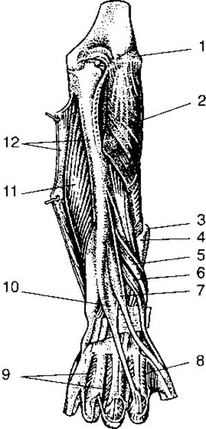
Fig. 81. Deep muscles of the forearm (posterior view):
1 — lateral epicondyle; 2 — supinator muscle; 3 — tendon of the extensor carpi radialis longus muscle; 4 — tendon of the extensor carpi radialis brevis muscle; 5 — abductor pollicis longus muscle; 6 — extensor pollicis brevis muscle; 7 — extensor pollicis longus muscle; 8, 9 — dorsal interosseous muscles; 10 — extensor indicis muscle; 11 — flexor digitorum profundus muscle; 12 — flexor carpi ulnaris muscle

Fig. 82. Muscles of the hand (right):
1 — palmaris longus muscle; 2 — flexor digitorum superficialis muscle; 3 — tendon of the flexor carpi ulnaris; 4 — forearm fascia; 5 — pisiform bone; 6 — abductor digiti minimi muscle; 7 — flexor digiti minimi brevis muscle; 8 — opponens digiti minimi muscle; 9 — lumbrical muscles; 10 — tendons of the flexor digitorum profundus muscle; 11 — tendons of the flexor digitorum superficialis muscle; 12 — adductor pollicis muscle; 13 — flexor pollicis brevis muscle; 14 — abductor pollicis brevis muscle; 15 — transverse carpal ligament
Muscles of the hand. The muscles are divided into those of the dorsal and palmar aspects of the hand.
On the palmar surface of the hand, the palmar aponeurosis is well developed, with the tendons of the superficial and deep digital flexors blending into it.
The Lateral group of thenar muscles includes: the abductor pollicis brevis, flexor pollicis brevis, opponens pollicis, and adductor pollicis. All of these originate from the carpal and Metacarpal bones and insert into the proximal phalanx of the thumb. These muscles provide versatile Movements of the thumb during various manual activities.
The hypothenar eminence on the medial side of the palm is formed by the group of little finger muscles. This group comprises the palmaris brevis, abductor digiti minimi, flexor digiti minimi brevis, and opponens digiti minimi. They originate from the nearby Carpal Bones and insert into the proximal phalanx of the fifth digit and the fifth metacarpal bone.
The central group of palmar muscles comprises four lumbrical muscles, which originate from the tendons of the flexor digitorum profundus and insert into the proximal phalanges of digits II–V into the extensor tendons. These muscles flex the fingers at the metacarpophalangeal joints and extend them at the interphalangeal joints.
The interosseous muscles are located in the spaces between the metacarpal bones. There are seven interosseous muscles in total: three palmar, which adduct the fingers, and four dorsal, which abduct them. On the digits, the palmar aponeurosis forms fibrous sheaths attached to the periosteum. Within these sheaths, the muscle tendons glide freely, surrounded by synovial sheaths that reduce friction, facilitate precise hand movements, and help contain inflammatory processes (Fig. 82).
Last update: 08/08/2026
Editorial and Educational Adaptation: This material has been compiled based on the primary/original source text. The project team performed an editorial review, corrected technical inaccuracies, structured sections, and adapted the content for an educational format.
What was processed:
- elimination of formatting defects (OCR errors, structural breaks, corrupted characters);
- editorial organization of content;
- standardization of terminology in accordance with academic sources;
- verification of factual statements against the original source text.
All mentions of the author, publication year, and origin of the primary text have been preserved in accordance with the source.