Human Anatomy - Kotsan I. Ya. 2009
Muscles and Fascia of the Lower Limb
Muscles of the Leg
Lateral Muscle Group of the Leg
The Lateral group of leg Muscles consists of two fibular (peroneal) muscles: the long and short fibular muscles (Fig. 134).
Class="center">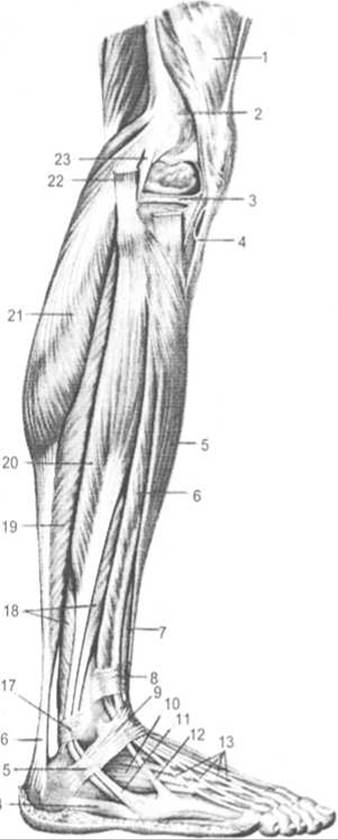
Fig. 134. Muscles of the Leg and FOOT (lateral view)
1 — quadriceps femoris, 2 — lateral epicondyle of Femur, 3 — lateral meniscus, 4 — patellar ligament, 5 — tibialis anterior, 6 — extensor digitorum longus, 7 — extensor hallucis longus, 8 — superior extensor retinaculum, 9 — inferior extensor retinaculum, 10 — extensor digitorum brevis, 11 — tendon of fibularis brevis, 12 — fibularis tertius, 13 — tendons of extensor digitorum longus, 14 — abductor digiti minimi, 15 — inferior fibular retinaculum, 16 — calcaneal tendon, 17 — superior fibular retinaculum, 18 — fibularis brevis, 19 — soleus, 20 — fibularis longus, 21 — lateral HEAD of gastrocnemius, 22 — biceps femoris, 23 — fibular collateral ligament
The fibularis longus Muscle (m. peroneus longus) is located superficially on the lateral aspect of the leg. It originates from the head and proximal third of the lateral surface of the Fibula, as well as from the anterior and posterior intermuscular septa and the crural fascia. At the level of the ankle joint, the muscle tendon hooks behind the lateral malleolus, running first beneath the superior fibular retinaculum within a common synovial sheath with the tendon of the fibularis brevis, and then through the groove on the calcaneus beneath the inferior fibular retinaculum. On the SOLE OF THE foot, the tendon of the fibularis longus courses obliquely forward through the groove of the cuboid bone in its own separate synovial sheath, inserting into the medial cuneiform bone and the bases of the First and Second Metatarsal Bones.
Function: flexes, pronates, and everts the foot, while also supporting the transverse and longitudinal arches of the foot.
Innervation: superficial fibular nerve (LV—SІ).
The fibularis brevis muscle (m. peroneus brevis) lies deep to the fibularis longus. It originates from the middle third of the lateral surface of the fibula and both intermuscular septa of the leg. The tendon passes into the foot posterior to the lateral malleolus within a synovial sheath shared with the preceding muscle beneath the fibular retinacula, and inserts into the tuberosity of the fifth metatarsal bone. Function: flexes, pronates, and everts the foot.
Innervation: superficial fibular nerve (LV—SI).
Last update: 08/08/2026
Editorial and Educational Adaptation: This material has been compiled based on the primary/original source text. The project team performed an editorial review, corrected technical inaccuracies, structured sections, and adapted the content for an educational format.
What was processed:
- elimination of formatting defects (OCR errors, structural breaks, corrupted characters);
- editorial organization of content;
- standardization of terminology in accordance with academic sources;
- verification of factual statements against the original source text.
All mentions of the author, publication year, and origin of the primary text have been preserved in accordance with the source.