Human Anatomy - Kotsan I. Y. 2009
Muscles and fascia of the upper limb
Muscles of the free upper limb
Muscles of the arm
Depending on their Location, the Muscles of the Free Upper Limb are divided into three topographical and anatomical groups: arm muscles, forearm muscles, and hand muscles.
The muscles of the arm, in turn, are divided into two groups: the anterior group (flexors) and the posterior group (extensors). These are separated by Connective Tissue partitions—the medial and lateral intermuscular septa of the arm—which extend from the brachial fascia to the corresponding margins of the humerus.
The Anterior Group of arm muscles consists of three muscles: the coracobrachialis, the brachialis, and the biceps brachii (Fig. 116).
The coracobrachialis Muscle (m. coracobrachialis) originates from the coracoid process of the scapula and inserts below the crest of the lesser tubercle of the humerus, at the level of the insertion of the deltoid tendon. Function: flexes the arm at the shoulder joint and adducts it toward the trunk. If the arm is pronated, it also assists in lateral Rotation of the arm.
Innervation: musculocutaneous nerve (CV—CVIII).
The brachialis muscle (m. brachialis) originates from the lower half of the anterior surface of the humerus and the intermuscular septa of the arm.
It inserts into the tuberosity of the ulna and its coronoid process. Function: flexes the forearm at the elbow joint.
Innervation: musculocutaneous nerve (CV—CVIII).
The biceps brachii muscle (m. biceps brachii) crosses two joints—the shoulder and the elbow—and has two heads: a short HEAD and a long head.
The short head (caput breve) originates from the coracoid process of the scapula. The long head (caput longum) arises from the supraglenoid tubercle of the scapula via a tendon that pierces the capsule of the shoulder joint from top to bottom and descends onto the arm, running within the intertubercular sulcus. At the mid-arm level, both heads merge into a common belly that ends in a tendon attached to the radial tuberosity and the forearm fascia. Function: flexes and supinates the forearm at the elbow joint and flexes the arm at the shoulder joint. However, because the long and short heads originate on the scapula at some distance from each other, their Functions regarding the arm differ: the long head flexes and abducts the arm, while the short head flexes and adducts it.
Innervation: musculocutaneous nerve (GV—CVIII).
Class="center">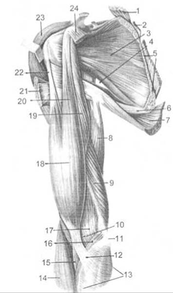
Fig. 116. Muscles of the shoulder girdle and arm (anterior view, right arm)
1 — levator scapulae muscle (cut), 2 — rhomboideus minor muscle (cut), 3 — subscapularis muscle, 4 — triangular space, 5 — serratus anterior muscle (cut), 6 — teres major muscle, 7 — latissimus dorsi muscle, 8 — long head of the triceps brachii muscle, 9 — medial head of the triceps brachii muscle, 10 — cubital fossa, 11 — medial epicondyle, 12 — bicipital aponeurosis, 13 — forearm fascia, 14 — brachioradialis muscle, 15 — tendon of the biceps brachii muscle, 16 — pronator teres muscle, 17 — brachialis muscle, 18 — biceps brachii muscle, 19 — coracobrachialis muscle, 20 — short head of the biceps brachii muscle, 21 — pectoralis major muscle (cut), 22 — long head of the biceps brachii muscle, 23 — deltoid muscle (retracted), 24 — pectoralis minor muscle (cut)
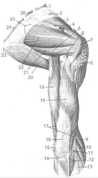
Fig. 117. Muscles of the shoulder girdle and arm (posterior view, right arm)
1 — levator scapulae muscle (cut), 2 — supraspinous fascia, 3 — supraspinatus muscle, 4 — spine of scapula, 5 — triangular space, 6 — quadrangular space, 7 — greater tubercle of the humerus, 8 — deltoid muscle (cut), 9 — brachioradialis muscle, 10 — extensor carpi radialis longus muscle, 11 — lateral epicondyle, 12 — anconeus muscle, 13 — forearm fascia, 14 — olecranon, 15 — medial epicondyle, 16 — tendon of the triceps brachii muscle, 17 — triceps brachii muscle, 18 — lateral head of the triceps brachii muscle, 19 — long head of the triceps brachii muscle, 20 — teres major muscle, 21 — teres minor muscle, 22 — infraspinatus muscle, 23 — inferior angle of scapula, 24 — rhomboideus major muscle (cut), 25 — infraspinous fascia, 26 — rhomboideus minor muscle (cut)
The posterior group of arm muscles consists of two muscles: the triceps brachii and the anconeus (Fig. 117).
The triceps brachii muscle (m. triceps brachii) is quite well developed and runs along the posterior surface of the arm over its entire length. It has three heads: long, medial, and lateral.
The long head (caput longum) originates from the infraglenoid tubercle of the scapula. The medial head (caput mediale) and lateral head (caput laterale) arise from the posterior surface of the arm (the medial head below and the lateral head above the radial nerve sulcus) as well as from the medial and lateral intermuscular septa. All three heads form a common muscle belly that attaches via a flat, broad tendon to the olecranon of the ulna. Some muscle fibers blend into the capsule of the elbow joint and the forearm fascia. Between the medial and lateral heads of the triceps brachii on one side and the humerus on the other lies the humeromuscular canal, through which pass the radial nerve and the deep brachial artery. Function: extends the forearm at the elbow joint, while the long head also extends and adducts the arm at the shoulder joint.
Innervation: radial nerve (CV—СVIII).
The anconeus muscle (m. anconeus) originates from the lateral epicondyle of the humerus and the radial collateral ligament. It inserts into the olecranon, the posterior surface of the proximal ulna, and the forearm fascia. Function: extends the forearm at the elbow joint.
Innervation: radial nerve (СV—СVIII).
Last update: 08/08/2026
Editorial and Educational Adaptation: This material has been compiled based on the primary/original source text. The project team performed an editorial review, corrected technical inaccuracies, structured sections, and adapted the content for an educational format.
What was processed:
- elimination of formatting defects (OCR errors, structural breaks, corrupted characters);
- editorial organization of content;
- standardization of terminology in accordance with academic sources;
- verification of factual statements against the original source text.
All mentions of the author, publication year, and origin of the primary text have been preserved in accordance with the source.