Human Anatomy - Kotsan I. Y. 2009
Brain
Medial surface of the hemispheres
All lobes of the hemisphere, with the exception of the insular lobe, contribute to The formation of its medial surface (Fig. 242). Located above the corpus callosum and separating it from other Regions of the hemisphere is the callosal sulcus (sulcus corporis callosi). Sweeping around the splenium of the corpus callosum posteriorly, this sulcus extends inferiorly and anteriorly, continuing into the hippocampal sulcus (sulcus hippocampi, s. hippocampalis). Positioned superior to the callosal sulcus is the cingulate sulcus (sulcus cinguli). This sulcus begins anteriorly and inferiorly near the rostrum of the corpus callosum, ascends superiorly, then curves backward and runs parallel to the callosal sulcus, terminating superiorly and posteriorly to the splenium of the corpus callosum as the subparietal sulcus (sulcus subparietalis).
Class="center">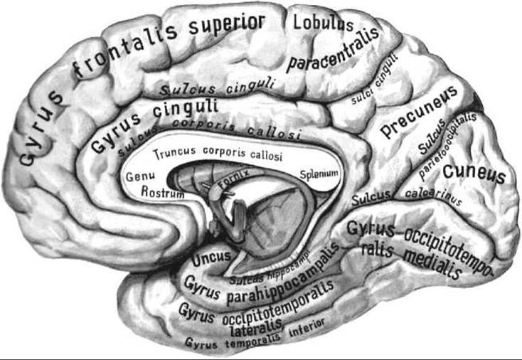
Fig. 242. Brain; right hemisphere; medial surface (after R. D. Sinelnikov)
Between the callosal sulcus and the cingulate sulcus lies the cingulate gyrus (gyrus cinguli), which arches over the corpus callosum anteriorly, superiorly, and posteriorly. Inferior and posterior to the splenium of the corpus callosum, the cingulate gyrus narrows to form the ISTHMUS OF THE cingulate gyrus (isthmus gyri cinguli). Further inferiorly and anteriorly, the isthmus transitions into the broader parahippocampal gyrus (gyrus parahippocampalis), bounded superiorly by the hippocampal sulcus. Deep within the hippocampal sulcus lies a delicate, gray-colored strip divided by small transverse grooves—the dentate gyrus (gyrus dentatus). The region of the medial surface of the hemisphere situated between the cingulate sulcus and the superior margin of the hemisphere belongs to the frontal and parietal lobes.
Anterior to the upper margin of the central sulcus lies the medial surface of the superior frontal gyrus, while directly adjacent to this region of the central sulcus is the paracentral lobule (lobus paracentralis), bounded posteriorly by the marginal ramus of the cingulate sulcus. Situated between the marginal ramus anteriorly and the parieto-occipital sulcus posteriorly is the precuneus (precuneus), which belongs to the parietal lobe.
On the medial surface of the occipital lobe lie two deep sulci that meet at an acute angle open posteriorly. These are the parieto-occipital sulcus, which separates the parietal lobe from the occipital lobe, and the calcarine sulcus (sulcus calcarinus), which originates on the medial surface of the occipital pole and extends anteriorly toward the isthmus of the cingulate gyrus. The triangular area of the occipital lobe situated between the parieto-occipital and calcarine sulci, with its apex pointing toward the junction of these sulci, is called the cuneus (cuneus). Clearly visible on the medial surface of the hemisphere, the calcarine sulcus bounds the lingual gyrus (gyrus lingualis) superiorly, which extends from the occipital pole posteriorly to the lower part of the isthmus of the cingulate gyrus. Inferior to the lingual gyrus is the collateral sulcus (sulcus collateralis), which belongs to the Inferior surface of the hemisphere.
Last update: 08/08/2026
Editorial and Educational Adaptation: This material has been compiled based on the primary/original source text. The project team performed an editorial review, corrected technical inaccuracies, structured sections, and adapted the content for an educational format.
What was processed:
- elimination of formatting defects (OCR errors, structural breaks, corrupted characters);
- editorial organization of content;
- standardization of terminology in accordance with academic sources;
- verification of factual statements against the original source text.
All mentions of the author, publication year, and origin of the primary text have been preserved in accordance with the source.