Operative Surgery and Topographic Anatomy - 2016
Content Module No. 1. Introduction to Topographic Anatomy and Operative Surgery. Topographic Anatomy and Operative Surgery of the Head and Neck Regions
Topic 3. Skull Base: External and Internal. Cranial Fossae and Their Contents. Topography of the Meninges and Dural Venous Sinuses. Kronlein-Brussova-Yegorov Craniocerebral Topography Scheme. Trepanation of the Skull
1. Relevance of the topic: the Treatment of cranial injuries, hematomas, post-traumatic cerebral edema, tumors, and cysts requires thorough knowledge of the Topography of the main cerebral sulci and gyri, craniocerebral topography schemes, as well as the surgical techniques for performing operations and arresting vascular Hemorrhage.
2. Specific objectives:
1. Explain the topography of the external and internal Skull base, cranial fossae, and their contents.
2. Explain the topography of the Meninges and Dural Venous Sinuses (Table 3).
3. Draw the Kronlein-Brussova-Yegorov craniocerebral topography scheme (Fig. 14).
Class="center">
Fig. 14. Kronlein-Brussova craniofacial topography scheme:
ABC - lower horizontal line; DEF - middle horizontal line; GHI - upper horizontal line; ADG - anterior vertical line; BEH - middle vertical line; CFI - posterior vertical line; D-J - PROJECTION OF THE central sulcus; H-J - true length of the central sulcus; DI - projection of the lateral sulcus; A - projection of the main trunk of a. meningea media; D - projection of the anterior branch of a. meningea media; F - projection of the posterior branch of a. meningea media
Projection of the cerebral arterial circle:
rectangle a-b-e-d - projection of a. carotis interna;
line d-g-h-i - projection of a. cerebri anterior;
line B-I - projection of the lateral sulcus and a. cerebri media;
line B-E-F - projection of a. communicans posterior;
line D-E - projection of the cerebral arterial circle.
4. Analyze various Methods of skull trepanation (Craniotomy, craniectomy).
5. Explain the surgical technique for osteoplastic craniotomy in the temporoparietal region.
Table 3. Methods for arresting hemorrhages from various cranial regions:
Vessels of the soft Tissues of the cranial vault |
✵ application of hemostatic clamps followed by placement of transfixing ligatures; ✵ application of hemostatic clamps followed by electrocoagulation. |
|
Diploic Veins and emissary veins |
✵ application of a tampon soaked in a 3% hydrogen peroxide solution; ✵ rubbing bone wax into the bleeding bone surface. |
|
Meningeal vessels |
✵ transfixation and ligation of both ends of the injured vessel; ✵ application of clips. |
|
Dural venous sinuses |
✵ suturing the defect with a vascular suture; ✵ ligation of the sinus on both sides. Ligation of the posterior PARTS OF THE superior sagittal sinus or transverse sinuses drastically impairs venous outflow, and in the area of the confluence of sinuses, it invariably leads to a fatal outcome. |
|
✵ sinus tamponade by inserting gauze packs between the bone and the dura mater on both sides of the sinus injury site for 12-14 days; ✵ application of a galea aponeurotica patch; ✵ Burdenko's plasty → ✵ using a fragment of hemostatic sponge. |
|
|
✵ tamponade with a piece of Muscle excised from the surgical wound area, crushed with scissor blades, and pressed against the bleeding site with a moist gauze ball. If digital compression of the muscle piece fails, the muscle is sutured to the dura mater with several interrupted silk sutures or inserted into the sinus lumen. |
||
Cerebral vessels |
✵ electrocoagulation; ✵ application of a hemostatic sponge; ✵ application of tampons soaked in a 3% hydrogen peroxide solution; ✵ vessel ligation; ✵ application of vascular clips; ✵ endovascular technique (intra-arterial administration of microballoons, spheres, and rapidly Setting substances to occlude injured vessels) |
|

3. Assignments FOR INDEPENDENT work in preparation for the class.
3.1. List of key terms, parameters, and characteristics that the student must master in preparation for the class:
Term |
Definition |
1. Primary surgical debridement of a craniocerebral wound 2. Trepanation of the skull |
1. A surgical Procedure involving The conversion of a contaminated (infected) craniocerebral wound into a clean one and creating proper conditions for primary intention healing 2. Surgical opening of the cranial cavity to provide surgical access to the Brain and its meninges for intervention |
3.2. Theoretical questions for the class:
1. Anesthesia for surgical interventions on the cranial vault.
2. Surgical instruments used for operations in the cranial region.
3. Main methods of arresting hemorrhage from vessels, soft tissues of the HEAD, and BONES OF THE cranial vault.
4. Main methods of arresting hemorrhage from the dural venous sinuses and cerebral vessels.
5. MAIN TYPES OF trepanation of the skull (craniotomy and craniectomy).
6. Indications for decompressive craniectomy.
7. Surgical technique for osteoplastic craniotomy.
8. Indications for osteoplastic craniotomy.
9. Surgical technique for decompressive craniectomy.
3.3. Practical assignments performed during the class:
1. Perform an osteoplastic craniotomy on a cadaver.
2. Be able to arrest hemorrhage from the middle meningeal artery and dural venous sinuses.
3. Perform a decompressive craniectomy on a cadaver.
4. Topic Content
TOPOGRAPHIC AND ANATOMICAL Features of the external and internal cranial base
When reviewing the topography of the internal and external cranial base, students focus on THE CONTRIBUTION OF various bones to The formation of the cranial fossae.
Specifically, the anterior cranial fossa on the internal cranial base is separated from the middle fossa by the posterior margin of the lesser wings of the Sphenoid bone. It is formed by the two orbital surfaces of the Frontal bone along with the cribriform plate (lamina cribrosa) of the Ethmoid bone; posteriorly, the fossa is bounded by the body and lesser wings of the sphenoid bone.
It is noted that the anterior cranial fossa lies above the Nasal cavity and orbits. It lodges the frontal lobes of the brain, while directly beneath them, on either side of the crista galli on the cribriform plate of the ethmoid bone, lie the olfactory bulbs (bulbus olfactorius). About 30 small nerve bundles enter them from the nasal cavity through the openings in the cribriform plate. Passing through these openings into the mucous membrane of the nasal cavity are the anterior and posterior ethmoidal Arteries (aa. ethmoidales anterior et posterior) as well as the ethmoidal nerves (nn. ethmoidales). The anterior meningeal artery (a. meningea anterior) runs from the anterior ethmoidal artery toward the dura mater. It is emphasized that the foramen caecum is located anterior to the crista galli, through which the venous plexus of the nasal cavity communicates with the superior sagittal venous sinus (sinus sagittalis superior).
At the base of the lesser wings of the sphenoid bone lie the paired optic canals, through which the optic nerves (n. opticus) and the ophthalmic artery (a. ophtalmica)—without a corresponding vein—pass from the cranial cavity into the Orbit.
When examining the middle cranial fossa (fossa cranii media), we note that it lies between the lesser wings of the sphenoid bone, the superior border of the petrous part of the Temporal bone (margo petrosus superior), and the dorsum sellae. The middle cranial fossa is formed by the sella turcica, the greater wings of the sphenoid bone, and the anterior surface of the petrous part of the temporal bone. The temporal lobes of the brain occupy the lateral parts of the middle cranial fossa, and the Pituitary Gland rests upon the sella turcica. It should be noted that the cavernous venous sinus (sinus cavernosus) is located on both sides of the sella turcica. The right and left cavernous sinuses communicate via the anterior and posterior intercavernous sinuses (sinus intercavernosus anterior et posterior). The ophthalmic veins drain into the cavernous sinuses bilaterally. Blood flows from the cavernous sinuses into the superior petrosal sinus and subsequently into the sigmoid sinus.
The instructor emphasizes that the facial veins communicate with the cavernous sinuses via vessels passing through the foramen lacerum and foramen ovale.
It is pointed out that the Internal Carotid Artery (a. carotis interna) and the Abducens nerve (n. abducens) pass through the cavernous sinuses. Within the lateral wall of the cavernous sinus, between the layers of the dura mater, lie the III and IV Cranial Nerves and the First Division of the Trigeminal nerve. Attention is drawn to the optic chiasm (chiasma optici), which is situated anterior to the sella turcica and the pituitary gland.
One should pay attention to the openings present in the middle cranial fossa that provide communication with adjacent structures. Thus, the middle cranial fossa communicates with the orbit through the superior orbital fissure (fissura orbitalis superior). Passing through this fissure are the oculomotor (III), trochlear (IV), and abducens (VI) nerves, the ophthalmic nerve (V1) along with its branches (frontal, lacrimal, and nasociliary), and the ophthalmic vein. The maxillary nerve (n. maxillaris) exits the cranial cavity into the pterygopalatine fossa through the foramen rotundum. The mandibular nerve (n. maxillaris is incorrect in original, n. mandibularis intended) (n. mandibularis) exits the cranial cavity through the foramen ovale, which is located posterior to the foramen rotundum. Meanwhile, the middle meningeal artery (a. meningea media) and the meningeal branch of the mandibular nerve (n. spinosus) enter the cranial cavity via the foramen spinosum. On the internal cranial base, between the greater wing of the sphenoid bone and the petrous part of the temporal bone, lies the foramen lacerum. It is noted that the petrosal nerves (nn. petrosus major et minor), the tensor tympani muscle, and its supplying nerve (n. tensor tympani) pass through the fibrous membrane of this foramen. The instructor points out that along with these structures, small-caliber veins pass through the foramen lacerum, connecting the inferior petrosal sinus (sinus petrosus inferior) with the VEINS OF THE external cranial base. Adjacent to the foramen lacerum is the internal carotid opening, through which the internal carotid artery enters the cranial cavity, surrounded by the nerve plexus of the same name.
The posterior cranial fossa (fossa cranii posterior) is separated from the middle fossa by the tentorium cerebelli. This fossa lodges the Cerebellum, Pons, and Medulla Oblongata.
In the posterior cranial fossa, the foramen magnum occupies a central position, connecting the cranial cavity with the vertebral canal. Passing through it are the medulla oblongata, vertebral arteries, and a venous plexus that connects the dural venous sinuses with the internal vertebral venous plexus.
Students identify that the jugular foramina are located on the skull laterally to the clivus of the sphenoid bone. Through them pass the IX, X, and XI cranial nerves, the Internal jugular vein originating from the bulb of the sigmoid sinus, while the posterior meningeal artery (a. meningea posterior), a branch of the ascending pharyngeal artery (a. pharyngea ascendens), enters the cranial cavity. It is noted that on the posterior surface of the petrous part of the temporal bone, the acoustic (n. statoacusticus), facial (n. facialis), and intermediate (n. intermedius) nerves, as well as the internal auditory vessels (a. et v. auditiva interna), pass through the internal acoustic opening (porus acusticus internus).
Somewhat lateral to the foramen magnum lies the hypoglossal canal (canalis hypoglossi), through which the Hypoglossal nerve (n. hypoglossus) exits the cranial cavity onto its external base.
Topographic and Anatomical Features of the external skull base
Students identify the BOUNDARIES OF THE external skull base on the skull specimen. It runs along the line connecting the external occipital protuberance (protuberantia occipitalis externa) with the sphenoidal rostrum (rostrum sphenoidale), which lies between the wings of the Vomer: along the superior nuchal line, across the base of the mastoid process, the posterior and inferior margins of the external acoustic meatus, continuing along the zygomatic process of the temporal bone, its infratemporal crest (crista infratemporalis), and the supraorbital margin (margo supraorbitalis) of the frontal bone.
If a line is drawn through the foramen magnum connecting the apices of the mastoid processes, the external skull base is divided into two parts: anterior and posterior.
Within the posterior part are the occipital condyles (condylus occipitalis), which articulate with the atlas, and the foramen magnum, through which the medulla oblongata passes.
The anterior part of the external skull base encompasses the superior wall of the Pharynx, as well as the superior walls of the orbital and nasal cavities.
The pharyngeal aponeurosis (fascia pharyngealis), atlanto-occipital membrane (membrana atlantooccipitalis), and the fascial sheaths of the Muscles originating from the mastoid process attach to the external skull base.
Meninges of the brain (meninges)
Examining the meninges on a head specimen, it is noted that the outermost layer is the dura mater (dura mater encephali). It is pointed out that it is loosely connected to the bones of the cranial vault, but tightly attached to the internal skull base. By making an incision in the dura mater, one can verify that it consists of two leaflets loosely connected to each other, between which the main neurovascular structures of the meninges run.
The instructor emphasizes that the founder of national neurosurgery, N.N. Burdenko, introduced into clinical practice the repair of dura mater defects using a pedicled flap harvested from the outer layer of the dura mater.
Between the layers of the cranial dura mater within the cranial vault run the anterior, middle, and posterior meningeal arteries. On the bony skull specimen and charts, students determine that the anterior meningeal artery (a. meningea anterior) branches off from the anterior ethmoidal artery (a. ethmoidalis anterior), which is a branch of the ophthalmic artery. The anterior ethmoidal artery enters the anterior cranial fossa through the corresponding foramina and branches out into its own ramifying vessels within the squama of the frontal bone.
The middle meningeal artery (a. meningea media) is one of the largest meningeal branches. It originates from the maxillary artery (a. maxillaris) and enters the cranial cavity through the foramen spinosum. Initially, this artery lies in the groove of the same name as a short common trunk and ascends above the zygomatic arch, where it divides into anterior and posterior branches.
The posterior meningeal artery branches off from the ascending pharyngeal artery (a. pharyngea ascendens) and enters the cranial cavity through the jugular foramen, where, within the squama of the temporal bone, it divides into branches supplying the posterior surface of the tentorium cerebelli along with the falx cerebelli.
The meningeal arteries are accompanied in pairs by veins of the same name, of which the anterior and posterior drain into the superior sagittal sinus, while the middle drain into the pterygoid venous plexus.
Lymph from the dura mater drains into various groups of Lymph Nodes. Thus, from its frontal and parietal regions, it flows into the superficial parotid nodes, and from the temporal and occipital regions, into the retroauricular lymph nodes.
Innervation of the dura mater is provided by the I, II, and III Branches of the trigeminal nerve, branches of the periarterial Nervous system, as well as elements of other cranial nerves.
Beneath the cranial dura mater lies the arachnoid mater (tunica arachnoidea), which evenly covers the cerebral gyri and does not penetrate into its sulci. Projections in the form of villi extend from the arachnoid mater. They pierce the dura mater and are connected to the dural venous sinuses by arachnoid granulations (Pacchionian granulations).
The pia mater of the brain (pia mater cranialis) covers the brain tissue and penetrates into all its sulci and ventricles, forming the choroid plexus (plexus chorioideus) there. This membrane contains a network of Blood Vessels that supply blood to the brain tissue.
If the pia mater is peeled off during dissection of a fresh cadaver, it separates easily from the brain because loose Connective Tissue lies between it and the brain.
Since There is a loose connection between the cranial dura mater and the internal surface of the cranial vault bones, blood can accumulate here during trauma, leading to epidural hematomas.
If a hematoma or pus is localized between the dura mater and the arachnoid mater, subdural conditions occur.
When pathological processes develop between the arachnoid mater (arachnoidea encephali) and the pia mater (pia mater), they are termed subarachnoid.
Three processes extend from the dura mater: the falx cerebri (falx cerebri), the falx cerebelli (falx cerebelli), and the tentorium cerebelli (tentorium cerebelli).
On the bony specimen of the dura mater, students carefully study these processes. They determine that the falx cerebri runs in the sagittal plane, extending from the cribriform plate of the ethmoid bone to the internal occipital protuberance and penetrating between the cerebral hemispheres down to the corpus callosum. At the same time, it is revealed that the falx cerebelli is a continuation of the falx cerebri. It separates the cerebellar hemispheres and extends toward the foramen magnum of the Occipital bone.
At the same time, the specimen clearly shows that the tentorium cerebelli lies in an almost horizontal plane, separating the occipital lobes of the cerebral hemispheres from the cerebellum.
The instructor points out to the students that the falx cerebri and the tentorium cerebelli are formed by a doubled layer of the dura mater. As a result, venous sinuses are formed between the leaves of the dura mater. Their distinguishing feature is the presence of an intima within the sinus lumen and the complete absence of Valves. The walls of these sinuses contain no muscle fibers, making them inelastic. If the wall of a sinus is incised, it remains patent (gapes), resulting in massive hemorrhage. Since blood drains from the dural venous sinuses into the internal jugular vein system, injuries to these areas can lead to air embolism.
Venous sinuses of the dura mater of the brain
Using anatomical charts and specimens featuring the dural reflections and the internal cranial base, the students identify the major sinuses.
The superior sagittal sinus (sinus sagittalis superior) begins at the foramen caecum, gradually widens, and terminates near the internal occipital protuberance. The inferior sagittal sinus (sinus sagittalis inferior) runs along the inferior margin of the falx cerebri of the dura mater. It courses from anterior to posterior, merges with the great cerebral vein (v. cerebri magna), and forms the straight sinus.
In the upper portion of the tentorium cerebelli, near the internal occipital protuberance, the straight sinus connects with the superior sagittal sinus.
The students identify that the occipital sinus (sinus occipitalis) originates at the foramen magnum and extends toward the internal occipital protuberance. It should be noted that in the region of the external occipital protuberance, the sinus occipitalis, together with the superior sagittal and straight sinuses, forms the confluence of the sinuses (confluens sinuum). It is emphasized that injuries to this area can cause life-threatening hemorrhage for the patient.
Using anatomical charts and skeletal specimens, the students determine that the transverse sinus (sinus transversus) lies within the transverse sulcus of the occipital bone, through which venous blood drains into the sigmoid sinus, which then leads to the jugular foramen. The cavernous sinus (sinus cavernosus) is a system of venous sinuses surrounding the sella turcica along with the pituitary gland.
Blood supply to the brain (Circle of Willis)
Using anatomical specimens of the brain, the skull, and charts, the students study the Main sources of its blood supply. Specifically, the internal carotid artery enters the cranial cavity through the carotid canal (canalis caroticus) of the petrous part of the temporal bone. Initially, it enters the cavernous sinus, where it gives off several small branches before dividing into the anterior (a. cerebri anterior) and middle (a. cerebri media) cerebral arteries. The second source of the brain's blood supply is the vertebral arteries (a. a. vertebrales). They enter the cranial cavity through the foramen magnum and unite to form a common trunk, the Basilar artery (a. basilaris), from which several branches initially arise to supply the cerebellum and medulla oblongata. Within the region of the dorsum sellae, the basilar artery gives off its terminal branches—the posterior cerebral arteries.
The cerebral arteries surrounding the sella turcica anastomose with one another. For example, the right and left anterior cerebral arteries (a. a. cerebri anterior) are connected by the anterior communicating branch (r. communicans anterior). They course into the longitudinal fissure between the cerebral hemispheres.
Furthermore, communicating branches (r. communicans posterior) are present bilaterally between the posterior cerebral artery and the internal carotid artery. These play a crucial role in ensuring adequate cerebral blood supply. The instructor draws the students' attention to individual anatomical variations in The Development of the anterior communicating branch (r. communicans anterior), including its occasional absence.
The instructor emphasizes that a key feature of cerebral veins is that they do not parallel the course of the arteries. Venous blood from the cerebral hemispheres drains via cerebral veins into the venous sinuses, primarily the superior sagittal sinus. Blood from the inferior sagittal sinus (sinus sagittalis inferior) and the cerebral ventricles drains into the great cerebral vein (v. cerebri magna).
Craniotomy
Osteoplastic craniotomy involves opening the cranial cavity by temporarily reflecting a soft tissue flap and a bone flap on a periosteal pedicle, returning them to their original position at the end of the procedure. It should be noted that, unlike osteoplastic craniotomy, decompressive surgery involves the final resection of a bone flap. Furthermore, in decompressive craniotomy, the dura mater is not sutured, which contrasts with the osteoplastic approach (Figs. 15, 16).

Fig. 15. Osteoplastic craniotomy in the temporal region:
1 - outlining and incising the scalp-aponeurosis flap; 2 - the periosteum is incised and stripped away from the flap using a raspatory, and three burr holes are made in the bone; 3 - the bone bridges between the holes are cut using a Gigli saw; 4 - the osteoperiosteal flap is reflected, and the dura mater is incised; 5 - the reflected dura mater flap exposes the underlying brain tissue; 6 - continuous sutures are placed on the dura mater.
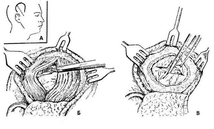
Fig. 16. Cushing's decompressive craniotomy.
The students outline the shape of the potential flap on the cadaveric head. They must ensure that the base of the bone flap is broad and oriented toward the major blood vessels. When reviewing the surgical technique, mention should be made of the single-flap Wagner-Wolf method and the two-flap Olivecrona method. The instructor emphasizes that today this procedure is typically performed using the two-flap technique.
First, a flap comprising the Skin, epicranial aponeurosis, and muscle is outlined and raised. The flap is reflected toward its base and covered with a sponge moistened with warm saline solution. Following this, the surgeon begins creating the bone flap. To do this, an incision is made in the periosteum 1 cm away from the skin wound margin. The periosteum is stripped away on both sides of the incision using a raspatory. Five notches are marked on the bone, ensuring that at the base of the future myo-periosteal flap, the distance between the notches is at least 4 cm. Using a manual hand drill, the assistants create the burr holes. The instructor highlights The Need for a very cautious technique when making the burr holes—especially when thinning the inner table of the skull—to prevent injury to the dura mater and underlying brain tissue. After drilling the five burr holes, a Gigli wire saw attached to a Polenov guide is successively passed through them to cut the bone between the holes. Once the burr holes are connected, an elevator is inserted under the base of the osteoperiosteal flap, and the bone is fractured along the base.
The osteoperiosteal flap is reflected outward, and the dura mater is opened with a cruciate incision. The surgeon then performs the necessary surgical manipulations on the brain tissue, such as removing a tumor, cyst, or vascular aneurysm, among others.
At the Conclusion of the procedure, the dura mater is closed with interrupted sutures (unless contraindicated), the bone flap is returned to its anatomical position, and it is secured with three rows of sutures: the first row is placed in the periosteum, the second in the muscle, and the third in the aponeurosis. Finally, silk sutures are applied to the skin.
The indication for decompressive craniotomy is elevated intracranial pressure caused by large tumors, Hydrocephalus, or other brain pathologies, in cases where the pathological mass cannot be resected, or when progressive Brain Edema and Swelling occur.
The purpose of the surgery is to excise a part of the cranial vault and open the dura mater. Decompressive craniectomy is performed directly over the lesion site (if the Diagnosis is definite) or in the right temporal region (according to Cushing) if the focus localization is unknown.
The surgeon makes an arcuate incision following the attachment of the temporal muscle, with its base directed toward the zygomatic arch. The vessels (superficial temporal artery and its branches) are ligated. The skin flap is reflected toward the base. The temporal fascia and the temporal muscle along the fiber direction are incised. The temporal bone (6x8 cm) is skeletonized. A burr hole is drilled in the center of the bone exposed from the periosteum using a large burr. This opening is enlarged with rongeurs to a size of 6x8 cm. The dura mater is opened with a cruciate incision. The soft tissues, except for the dura mater, are sutured tightly. It is emphasized that before opening the dura mater, a lumbar puncture is usually performed to reduce its tension. This minimizes the risk of acute brain prolapse, hemorrhage, and other complications (Fig. 17, 18).

Fig. 17. Tamponade of the injured sinus with gauze (diagram)

Fig. 18. Skin grafting using asymmetrical Limberg triangles (diagram on a fabric circle)
5. Self-Assessment Materials
A. Self-Assessment Tasks
Test No. 1
A patient exhibits decreased cutaneous sensitivity resulting from a traumatic brain injury. Which area of the Cerebral Cortex is likely to be damaged?
a. Postcentral gyrus
в. Occipital region
c. Parietal cortical area
d. Frontal cortical area
e. Precentral gyrus
Test No. 2
A patient has sustained an injury to the cranial vault. Which sinus may be affected?
а. Superior sagittal sinus
в. Superior petrosal sinus
c. Inferior petrosal sinus
d. Inferior sagittal sinus
e. Cavernous sinus
Test No. 3
A woman is diagnosed with impaired tactile sensation. Which area of the brain is damaged?
а. Postcentral gyrus of the cortex
b. Medulla oblongata
c. Cerebellum
d. Precentral gyrus of the cerebral cortex
e. Temporal region of the cerebral cortex
Test No. 4
A victim has sustained soft tissue and Parietal bone trauma in the area of the sagittal suture, accompanied by severe bleeding. Which of the following structures is most likely damaged?
a. Sinus sagittalis superior
b. Sinus petrosus superior
c. Sinus rectus
d. Sinus sagittalis inferior
e. None of the above
Test No. 5
An injured person was found to have a subdural hematoma in the temporal region. Which artery is damaged?
a. Middle meningeal artery.
b. Middle cerebral artery.
c. Posterior communicating artery.
d. Anterior meningeal artery
e. None of the above
B. Self-Assessment Tasks
Task 1. During a cranioplastic trepanation, the surgeon used a Gigli saw to connect all the burr holes of the bone along with the periosteum. What is the surgeon's mistake?
Task 2. During a cranioplastic trepanation, the surgeon connected the burr holes with a wire saw. When closing the defect, the osteoperiosteal flap sags and lies freely on the dura mater. What is the surgeon's mistake?
Task 3. After incising the dura mater during Cushing's decompressive craniotomy, a sharp herniation (prolapse) of the brain occurred. What did the surgeon fail to do to prevent this complication?
Task 4. During a craniotomy, the surgeon discovered a damaged middle meningeal artery. To stop the bleeding, he ligated the central end of the artery. He is about to suture the wound tightly. What is the surgeon's mistake?
Basic
1. Operative Surgery and Topographic Anatomy, ed. by M.S. Skrypnikov. — K.: Vyshcha Shkola, 2000. — P. 47-64.
2. Operative Surgery and Topographic Anatomy, ed. by M.P. Kovalsky. — K.: Medytsyna, 2010. — P. 58-92.
Supplementary:
1. Kovanov V.V. Operative Surgery and Topographic Anatomy / V.V. Kovanov. — M.: Meditsina, 1978. — P. 276-286.
2. Operative Surgery and Topographic Anatomy, ed. by K.I. Kulchytsky. — K.: Vyshcha Shkola, 1994. — P. 39-45.
3. Operative Surgery and Topographic Anatomy, ed. by G.E. Ostroverkhov. — Rostov-on-Don, 1998. — P. 350-371.
4. Elizarovsky S.I. Operative Surgery and Topographic Anatomy / S.I. Elizarovsky, R.N. Kalashnikov. — M., 1979.
5. Vishnevsky A.V. Local Anesthesia by the Creeping Infiltration Method / A.V. Vishnevsky. — M., 1956.
6. Matyushin I.F. Introduction to the Course of Operative Surgery and Topographic Anatomy / I.F. Matyushin. — Gorky, 1976.
7. Tomashuk I.P. Guide to Surgical Techniques for Beginner Surgeons / I.P. Tomashuk, I.I. Tomashuk. — K.: European University Press, 2001. — 860 p.
8. Bekov D.B. Atlas of the Venous System of the Human Brain / D.B. Bekov. — M., 1965.
9. Zolotko Yu.L. Topographic Atlas of the Head / Yu.L. Zolotko. — M., 1976, part.
10. Zolotareva T.V. Surgical anatomy of the Head / T.V. Zolotareva, G.N. Toporov. — M., 1968.
11. Polenov A.L. Fundamentals of Practical Neurosurgery / A.L. Polenov, I.S. Babchin. — M., 1957.
12. Frautschi V.Kh. Course of Topographic Anatomy and Operative Surgery / V.Kh. Frautschi. — M., 1976.
Last update: 10/08/2026
Editorial and Educational Adaptation: This material has been compiled based on the primary/original source text. The project team performed an editorial review, corrected technical inaccuracies, structured sections, and adapted the content for an educational format.
What was processed:
- elimination of formatting defects (OCR errors, structural breaks, corrupted characters);
- editorial organization of content;
- standardization of terminology in accordance with academic sources;
- verification of factual statements against the original source text.
All mentions of the author, publication year, and origin of the primary text have been preserved in accordance with the source.