Review of Medical Physiology - William F. Ganong 2002
Functions of the Nervous System
Regulation of Posture and Movement
Cerebellum
Structural Division
The cerebellum is located above critical sensory and motor systems of the Brainstem (Fig. 12-13). It is connected to the brainstem on each side by the superior, middle (or pontine), and inferior cerebellar peduncles. The middle part of the cerebellum—the vermis—and the laterally positioned cerebellar hemispheres are much more convoluted and feature deeper sulci than the Cerebral Cortex; the mass of the cerebellum accounts for only about 10% of the cerebral cortex mass, yet its surface area is approximately 75% of that of the cerebral cortex. Anatomically, the cerebellum is divided by two transverse fissures into three main parts. The posterolateral fissure separates the medially located nodulus and the laterally located flocculus on each side from the rest of the cerebellum, while the primary fissure divides the remaining portion of the cerebellum into the anterior and posterior lobes. Smaller fissures divide the vermis into ten primary lobules, numbered from top to bottom from I to X. These lobules are illustrated in Fig. 12-14.
Class="center">
Fig. 12-12. The Basal Ganglia-thalamocortical circuit in Parkinson's disease. Solid arrows denote excitatory influences, and dashed arrows denote inhibitory influences. The strength of each influence is indicated by the thickness of the arrows. The striatum contains D1 dopamine receptors, which enhance the direct pathway to the medial globus pallidus (GPi), and D2 receptors, which reduce the indirect inhibitory influence on the GPi via the putamen, lateral globus pallidus (GPe), and subthalamic nuclei (STN). SNc, substantia nigra pars compacta; Thalam, thalamus; PPN, pedunculopontine Nucleus (reproduced with permission from Grafton SC, DeLong M: Tracing the Brain’s circuitry with functional imaging. Nat Med 1997;3:602).
Functional Division
From a functional standpoint, the cerebellum is also divided into three parts, although this division differs from the anatomical one (Fig. 12-15). The nodulus of the vermis and the flocculus of the hemisphere on each side form the flocculonodular lobe, or vestibulocerebellum. This lobe, which is phylogenetically the oldest part of the cerebellum, is connected with the vestibular system and is important for maintaining balance and for learning-dependent modifications of the vestibulo-ocular reflex (see Chapter 9). The rest of the vermis and the adjacent medial portions of the hemispheres constitute the spinocerebellum, which receives proprioceptive pathways from the body as well as pathways providing information regarding the somatotopic representation of body parts in the motor cortex. By comparing motor planning with the actual execution of movements in various body parts, the cerebellum ensures smooth and coordinated movements. The connections between the cerebellar vermis and brainstem regions are required for The regulation of axial Muscles and proximal limb muscles, whereas similar connections between the cerebellar hemispheres and the brainstem regulate distal limb muscles. The lateral Regions of the cerebellar hemispheres are referred to as the cerebrocerebellum (or neocerebellum). These phylogenetically newest structures of the cerebellum reach their greatest development in humans. Together with the motor cortex, the structures of the neocerebellum are involved in the planning and programming of movements.
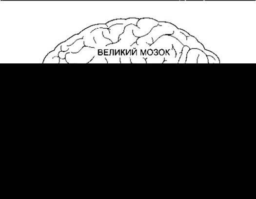
Fig. 12-13. Major PARTS OF THE brain. A slight Displacement of the parts in the figure allows visualization of the cerebellar peduncles, as well as the ring-like Structure formed by the cerebellum, the anterior part of the Pons, and the middle cerebellar peduncle embracing the brainstem (reproduced with permission from Goss CM [editor]: Gray’s Anatomy of the Human Body, 27th ed. Lea & Febiger, 1959).
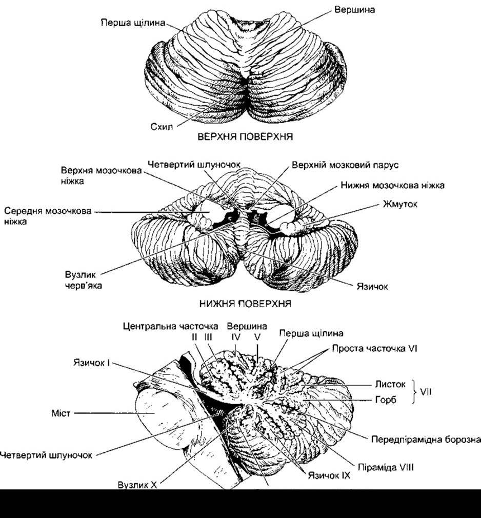
Fig. 12-14. Superior and inferior surfaces and sagittal section of the human cerebellum. The main lobules of the cerebellar vermis are labeled (from I to X) and named.
Organization OF THE Cerebellum
The cerebellum consists of the outer cerebellar cortex, White matter, and deep cerebellar nuclei embedded within it. The primary afferent fibers to the cerebellum, namely mossy and climbing fibers (see below), form collaterals that project to the deep nuclei and continue on to the cortex (Fig. 12-16). There are four pairs of cerebellar nuclei: the dentate, globose, emboliform, and fastigial nuclei.
The globose and emboliform nuclei are sometimes
referred to collectively as the interposed nucleus. Most of the pathways from the vestibulocerebellum pass directly to the brainstem, while the rest run from the cerebellar cortex to its nuclei and thence to the brainstem. Thus, the cerebellar nuclei provide the sole output pathway for impulses from the spinocerebellum and neocerebellum. The medial portion of the pathway from the spinocerebellum projects to the fastigial nucleus and from there to the brainstem. The pathway from adjacent parts of the cerebellar hemisphere, which also belong to the spinocerebellum, runs to the emboliform and globose nuclei, and from them to the brainstem. Fibers from the neocerebellum project to the dentate nucleus, and from there either directly or indirectly to the ventrolateral Nucleus of the thalamus.

Fig. 12-15. Functional subdivisions of the cerebellum (modified from Kandel ER, Schwartz JH, Jessell TM [editors]: Principles of Neural Science, 4th ed. McGraw-Hill, 2000).

Fig. 12-16. Schematic diagram of neural connections within the cerebellum. Plus and minus signs denote excitatory and inhibitory influences, respectively. BC, basket Cell; GC, Golgi cell; GrC, granule cell; CNC, cerebellar nuclear cell; PC, Purkinje cell. Note that PCs and BCs exert an inhibitory influence. The connections of stellate Cells are similar to those of basket cells, but they predominantly terminate on the dendrites of Purkinje cells.
The cerebellar cortex contains five distinct types of Neurons: Purkinje cells, granule cells, basket cells, stellate cells, and Golgi cells. It exhibits a distinct three-layered structure (Fig. 12-17): an outer molecular layer, a ganglionic layer composed of a single layer of Purkinje cell bodies, and an inner granular layer. Purkinje cells are among the largest neurons in the body. Their extensively branched dendrites extend through the molecular layer of the cortex. The axons of Purkinje cells, which form the sole efferent output pathway from the cerebellar cortex, project primarily to the deep cerebellar nuclei. The cerebellar cortex also contains granule cells, which receive inputs via mossy fibers and Relay them to Purkinje cells. The Cell bodies of granule cells reside in the granular layer. The axon of each granule cell ascends into the molecular layer, where it bifurcates in a T-shape. These long, straight branches are called parallel fibers. The dendrites of Purkinje cells form numerous bush-like branches confined to a single plane and oriented at right angles to the parallel fibers. Parallel fibers form synaptic contacts with the dendrites of numerous Purkinje cells, thereby forming an exquisitely regular grid-like network together with the Purkinje cell dendrites.
The other Three types of neurons in the cerebellar cortex are inhibitory interneurons. Basket cells (see Fig. 12-17) are located in the molecular layer. They receive input from parallel fibers, and each basket cell distributes information to numerous Purkinje cells. The axons of these cells form basket-like terminal plexuses around the cell body and axon hillock of Purkinje cells. Stellate cells are similar to basket cells but are located closer to the cortical surface. Golgi cells reside in the granular layer. Their dendrites, which penetrate the molecular layer, receive information from parallel fibers. The cell bodies receive inputs via collaterals from mossy fibers and Purkinje cells. The axons of Golgi cells synapse upon the dendrites of granule cells.
As noted above, the primary pathways mediating excitatory inputs to the cerebellar cortex are formed by climbing and mossy fibers (see Fig. 12-16). Climbing fibers originate from a single source—the inferior olivary nuclei—and project to the primary dendrites of Purkinje cells, winding around them like ivy vines. Proprioceptive impulses from all parts of the body converge on the inferior olivary nuclei. Conversely, mossy fibers convey information directly from all regions of the body, as well as signals from the cerebral cortex via the pontine nuclei to the cerebellar cortex. These fibers terminate on the dendrites of granule cells within complex synaptic structures known as glomeruli. These glomeruli also contain the terminals of inhibitory Golgi cells mentioned previously.
The main circuits for excitation Circulation within the cerebellar cortex are relatively straightforward (see Fig. 12-16). Impulses transmitted by climbing fibers exert a powerful excitatory effect on individual Purkinje cells, whereas mossy fibers, acting via granule cells, provide a weak excitatory drive to a large population of Purkinje cells. Basket and stellate cells are excited by granule cells via parallel fibers, and their influence on Purkinje cells is inhibitory (feedforward inhibition). Golgi cells are excited by inputs arriving via mossy fiber collaterals, Purkinje cell collaterals, and parallel fibers, and they inhibit the transmission of impulses from mossy fibers to granule cells.
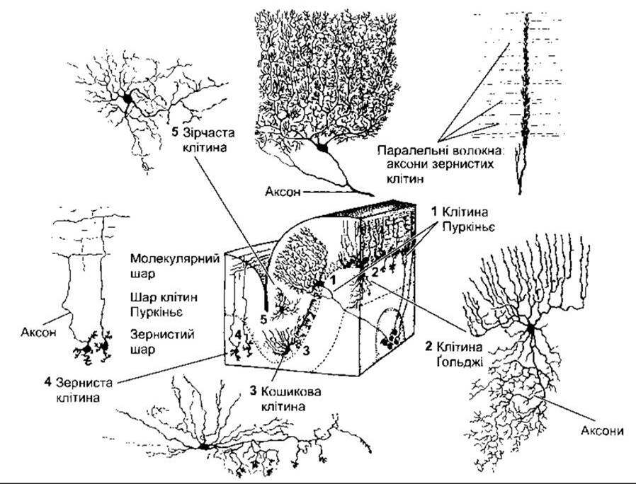
Fig. 12-17. Arrangement and structure of neurons in the cerebellar cortex (reproduced with permission from Kuffler SW, Nicholls JG, Martin AR: From Neuron to Brain, 2nd ed. Sinauer, 1984).
The neurotransmitter likely released by stellate cells, basket cells, Golgi cells, and Purkinje cells is GABA, whereas granule cells release glutamate. GABA acts on GABAA receptors, although the subunit composition of these receptors (see Chapter 4) varies among different cell types. Granule cells are unique in that they are the only class of CNS neurons whose GABAA receptors contain the a6 subunit.
The impulses originating from Purkinje cells exert an inhibitory effect on the cerebellar nuclei. As noted above, these nuclei also receive excitatory inputs via collaterals of mossy and climbing fibers. Interestingly, despite the inhibitory influence of Purkinje cells on the cerebellar nuclei, the output signals transmitted from these nuclei to the brainstem and thalamus are excitatory. Consequently, nearly all circuits mediating excitation within the cerebellum appear to be involved in modulating or regulating the timing of impulse transmission from the cerebellar nuclei to the brainstem and thalamus.
The primary afferent pathways projecting to the cerebellum via mossy and climbing fibers are summarized in Table 12-5.
Flocculonodular Lobe
Animals with lesions of the flocculonodular lobe lose postural stability and walk with a staggering gait on a flat surface. They fail to maintain balance and are prone to falls without external support. Similar motor impairments—which serve as early clinical signs of midline cerebellar tumors originating from the Cells of the nodulus—can be observed in children. In such cases, the structures localized within the flocculonodular lobe are rapidly compromised.
Selective surgical ablation of the flocculonodular lobe in dogs induces motion sickness syndrome (see Chapter 9), whereas extensive damage to other regions of the cerebellum or brain does not produce this effect.
Influence on Stretch Reflexes
Stimulation of cerebellar areas that receive proprioceptive inputs can either inhibit or facilitate movements triggered by stimulation of the cerebral cortex. Experimental lesions involving folia I–VI and medial regions result in limb rigidity on the side corresponding to the damaged cerebellar zones. In humans, however, analogous destructive lesions of the cerebellum lead to muscular hypotonia.
Influence on Movement
Aside from alterations in stretch reflexes, experimental animals and human patients with lesions restricted to the cerebellar hemispheres show no overt abnormalities at rest. However, distinct motor abnormalities become apparent during active movement. These deficits are neither paralytic nor sensory in nature, but manifest as prominent ataxia characterized by dyscoordination resulting from impaired timing, amplitude, force, and direction of movement. This ataxia may be unilateral. When damage is confined to the cerebellar cortex, movement disorders gradually resolve due to compensatory mechanisms. Lesions of the cerebellar nuclei, by contrast, cause more severe and persistent deficits. For this reason, surgical interventions involving the cerebellum must avoid injuring its deep nuclei.
Table 12-5. Functions of the Main Cerebellar Afferent Systems1
Afferent pathway |
Transmitted information / function |
Vestibulocerebellar |
Vestibular impulses from the labyrinths, directly or via vestibular nuclei |
Posterior spinocerebellar |
Proprioceptive and exteroceptive impulses from the body |
Anterior spinocerebellar |
Proprioceptive and exteroceptive impulses from the body |
Cuneocerebellar (dorsal part of external arcuate fibers) |
Proprioceptive impulses, particularly from the HEAD and Neck regions |
Tectocerebellar |
Auditory and visual impulses conveyed via the inferior and superior colliculi of the Midbrain tectum |
Pontocerebellar |
Impulses from the motor and other areas of the cerebral cortex relayed through the pontine nuclei |
Olivocerebellar |
Proprioceptive impulses from the entire body relayed via the olivary nuclei |
1 The olivocerebellar tract projects to the cerebellar cortex as climbing fibers, whereas the other listed pathways terminate as mossy fibers. Additionally, some pathways transmit impulses from brainstem nuclei to the cerebellar cortex and nuclei, notably the serotonergic pathway from the raphe nuclei to the molecular and granular layers, as well as the noradrenergic pathway from the locus coeruleus to all three cortical layers.
Other clinical signs of cerebellar damage in humans underscore the critical role of this brain region in motor control. Ataxia is manifested not only by a crude, staggering, or drunken gait, but also by deficits in fine and dexterous movements, which is particularly evident in speech production. Speech becomes slurred, often referred to as scanning speech. Other voluntary movements are likewise markedly abnormal. For instance, when a patient attempts to reach for an object, the finger oscillates past the target, missing it alternately to one side and then the other. This dysmetria rapidly triggers corrective efforts, but due to overcorrection, the finger overshoots in the opposite direction, resulting in back-and-forth oscillations. This type of tremor associated with cerebellar pathology is termed intention tremor. Unlike the resting tremor seen in Parkinson's disease, intention tremor appears only when the patient attempts a voluntary movement. Another hallmark of cerebellar dysfunction is the inability to "brake" rapidly—that is, to abruptly halt an ongoing movement. Normally, for example, during forearm flexion against resistance, the movement can be stopped instantaneously if the resistance suddenly vanishes. A patient with a cerebellar lesion fails to arrest the limb's trajectory, causing the forearm to swing backward in a wide arc. This abnormal response is known as the rebound phenomenon, and similar deficits manifest during other motor tasks. This is a primary reason why such patients exhibit adiadochokinesia—the inability to perform rapid alternating movements, such as successive pronation and supination of the hand. Cerebellar patients also experience difficulty executing multi-joint movements simultaneously; instead, these movements occur sequentially, joint by joint. This phenomenon is known as decomposition of movement.
The Cerebellum and Learning
Cerebellar function is intimately linked to motor adaptation through learning, such that movement coordination improves with repetition. When a motor task is practiced, neural activity within the brain shifts from the prefrontal cortex to the parietal cortex, motor cortex, and cerebellum. It is widely hypothesized that learning processes within the cerebellum are driven by inputs from the olivary nuclei. In this context, each Purkinje cell receives inputs from 250,000 to 1,000,000 mossy fibers, yet is innervated by only a single climbing fiber originating from the inferior olive. This climbing fiber forms 2,000 to 3,000 synaptic contacts with the Purkinje cell. Activation of a climbing fiber elicits a powerful complex spike in the Purkinje cell, which in turn induces long-term Modification of the signals transmitted to the Purkinje cell via mossy fibers. Climbing fiber activity increases with each newly acquired motor skill, whereas selective damage to the olivary complex abolishes the capacity to form the long-term adaptive plasticity required for executing precise motor responses. The Role of the cerebellum in regulating the vestibulo-ocular reflex and Other forms of procedural memory is discussed in Chapter 16.
Mechanisms
Although the respective functions of the flocculonodular lobe, spinocerebellum, and neocerebellum are relatively well understood, and cerebellar circuitry is less complex than that of the cerebrum, the precise operational mechanisms of its constituent parts remain elusive. Correlating ELECTRICAL PHENOMENA IN the cerebellum with its role as a motor control center is one of the enduring unresolved problems in neurophysiology. The cerebellar cortex exhibits a baseline electrical rhythm of 150–300/s and 200 µV, upon which a lower-amplitude 1000–2000/s component is superimposed. Thus, the frequency of the cerebellar baseline rhythm is more than ten times higher than that of the alpha rhythm recorded from the cerebral cortex using analogous Methods. Incoming impulses to the cerebellum typically modulate the amplitude of this rhythm, much like a broadcast signal modulates the carrier frequency of a radio transmitter. However, the Functional Significance of this bioelectric phenomenon for cerebellar operations remains unexplained.
Last update: 10/08/2026
Editorial and Educational Adaptation: This material has been compiled based on the primary/original source text. The project team performed an editorial review, corrected technical inaccuracies, structured sections, and adapted the content for an educational format.
What was processed:
- elimination of formatting defects (OCR errors, structural breaks, corrupted characters);
- editorial organization of content;
- standardization of terminology in accordance with academic sources;
- verification of factual statements against the original source text.
All mentions of the author, publication year, and origin of the primary text have been preserved in accordance with the source.